Methods, apparatuses and storage mediums for ablation planning and performance

a technology of ablation planning and ablation performance, applied in the field of optical imaging, can solve the problems of a lot of variables, rarely good diagnostic imaging, and a lot of planning for ablation, and achieve the effect of effective planning and reducing the time of ablation procedur

Pending Publication Date: 2020-01-09
CANON USA
View PDF23 Cites 8 Cited by
  • Summary
  • Abstract
  • Description
  • Claims
  • Application Information

AI Technical Summary

Benefits of technology

[0015]Accordingly, it is a broad object of the present disclosure to provide ablation apparatuses and systems, and methods and storage mediums that operate to reduce ablation procedure time through effective planning and performance.
[0016]In accordance with one or more embodiments of the present disclosure, ablation planning and performance apparatuses and systems, and methods and storage mediums may operate to identify target tissue type in addition to providing a morphological image to help an operator's diagnostic / planning or ablation performance decision based on tissue information. On or more embodiments of an ablation planning and performance apparatuses and systems, and methods and storage mediums may include or permit at least one of: simulation of a probe path (including planning the path of the probe with real time simulation of respiratory gating and organ motion and deformation), interaction of a probe and target and surrounding organs, clear definition of a position (e.g., an angle) of a patient (such as with real time gravity simulation), clear communication with an interventional (IR) team, tracking of tools used for better inventory and regulatory control, definition of a number and trajectory of probes based on a patient-specific model (e.g., a 3D model, a CT or MRI scan-derived model, etc.), definition of an ablation geometric zone and equipment setting(s) with real time ablation zone simulation, ability to allow planning viewed and archived in real time (e.g., locally, remotely, etc.) for receiving insight from other clinicians, determination of the treatment target that the probe should reach to and estimation of the ablation zone that covers the target, etc.
[0017]The effective communication in the planning stage and / or the performance stage may permit the IR to connect with multiple stakeholders from a single source. The software equipped with these features will support the IR in the setup of the operating room (OR) environment and will support the communication with the interventional (IR) team more effectively. On a separate channel, the patient may be updated on the schedule and assisted in preparation for the ablation procedure.

Problems solved by technology

However, diagnostic imaging is rarely good enough to plan with, so an IR may conduct initial imaging before developing / finalizing an action plan and starting an ablation procedure.
Ablation takes a lot of planning, and there are a lot of variables.
Even though ablation procedure is very complex, the procedure that is currently performed by clinicians is predominantly done manually and iteratively, which may introduce room for error(s) and may increase the time required to perform ablation (i.e., be inefficient).
In 3D, marking a center position for an object may not be hard, even though many times it may not be accurate due to human visual and motor action inaccuracy / error.
However, a clinician using 3D slice view to figure out a center of a 3D volume which includes a stack of 3D slicers may be difficult and error prone if the center of the volume is the target point, and the clinician may be tricked by image artifacts and / or human limitation in 3D reasoning.
In 3D, marking a center position is much harder because of the intricate limitation of visualization / rendering software.
Relying on clinicians' intuition, experience and visual understanding to define a target point is not optimal (for reliability, repeatability, traceability, etc.), particularly in 3D space.
When the lesion / tumor has a very complicated shape, defining an appropriate target is more or less an art, and it is difficult to achieve consistency.
As discussed above, the common practice of ablation planning is iterative and interactive, and there are many variables to consider such that it is difficult to automate the entire ablation procedure.
If these images do not reveal enough detail(s), clinicians may have to inject a contrast agent into a patient's body to obtain contrast-enhanced images.
This additional step only increases the procedures complexity, cost and time.
Tools that currently exist to help clinicians in ablation planning are not adequate.
However these mechanisms and tools do not alleviate clinicians from crucial steps in planning such as, but not limited to, defining targets, defining target zone(s), and confirming coverage.
Current tools are not integrated with all necessary or preferred components / features for ablation planning and performance, which makes the clinicians still rely on their experience and intuition.
Such reliance inevitably results in inconsistency and guess work, or other inefficiencies, such as, but not limited to, longer procedure times, additional steps, less effective or optimal ablation results, etc.
This not only increases the procedure time, but also wastes a lot of efforts in adjustment / making changes.
Of course, it is also likely having impact(s) on or causing possible damage to nearby structure and tissues.
Considering organ movement and deformation may make ablation planning and performance more complex, and may hamper interaction between clinicians and ablation planning and performance devices.
Such changes may also invalidate the planned insertion.
Respiratory gating, or asking patients to hold their breath, are time consuming monitoring techniques that may be used to assist with insertion.
However, such procedures do not guarantee success or efficiency.
While technologies exist to measure the thermal effect, such technologies do not prove reliable, for example, thermal confirmation is very time sensitive and a measurement may be dramatically different during and after ablation.

Method used


Image

  • Methods, apparatuses and storage mediums for ablation planning and performance
  • Methods, apparatuses and storage mediums for ablation planning and performance
  • Methods, apparatuses and storage mediums for ablation planning and performance

Examples


Embodiment Construction

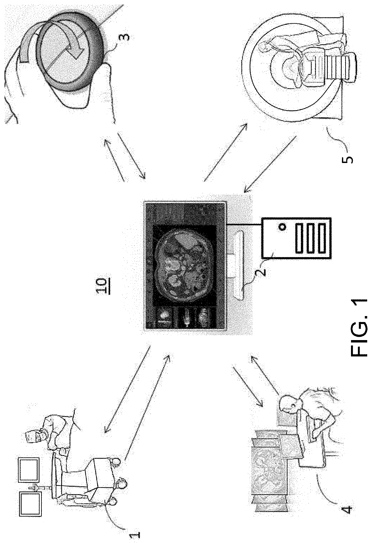
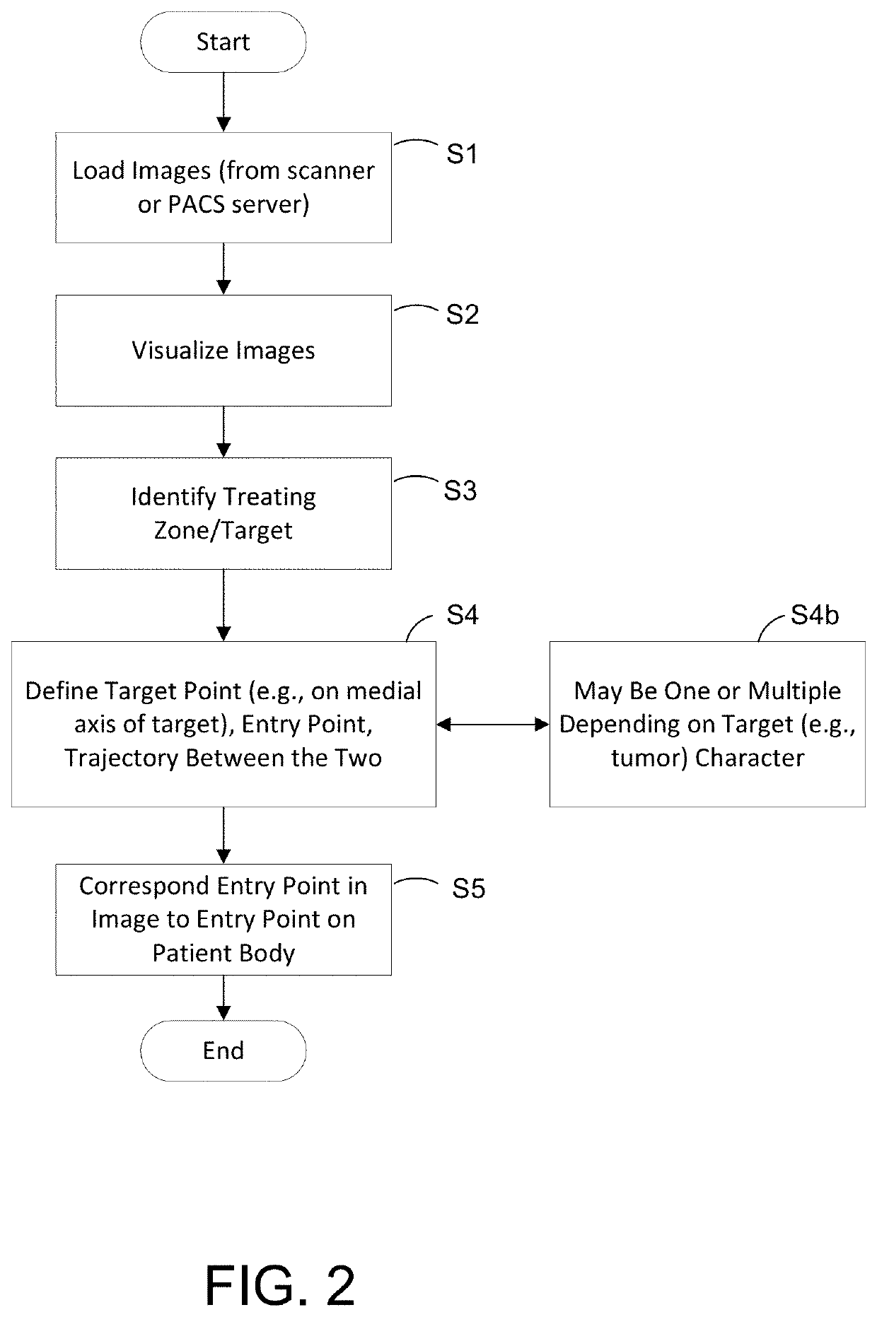
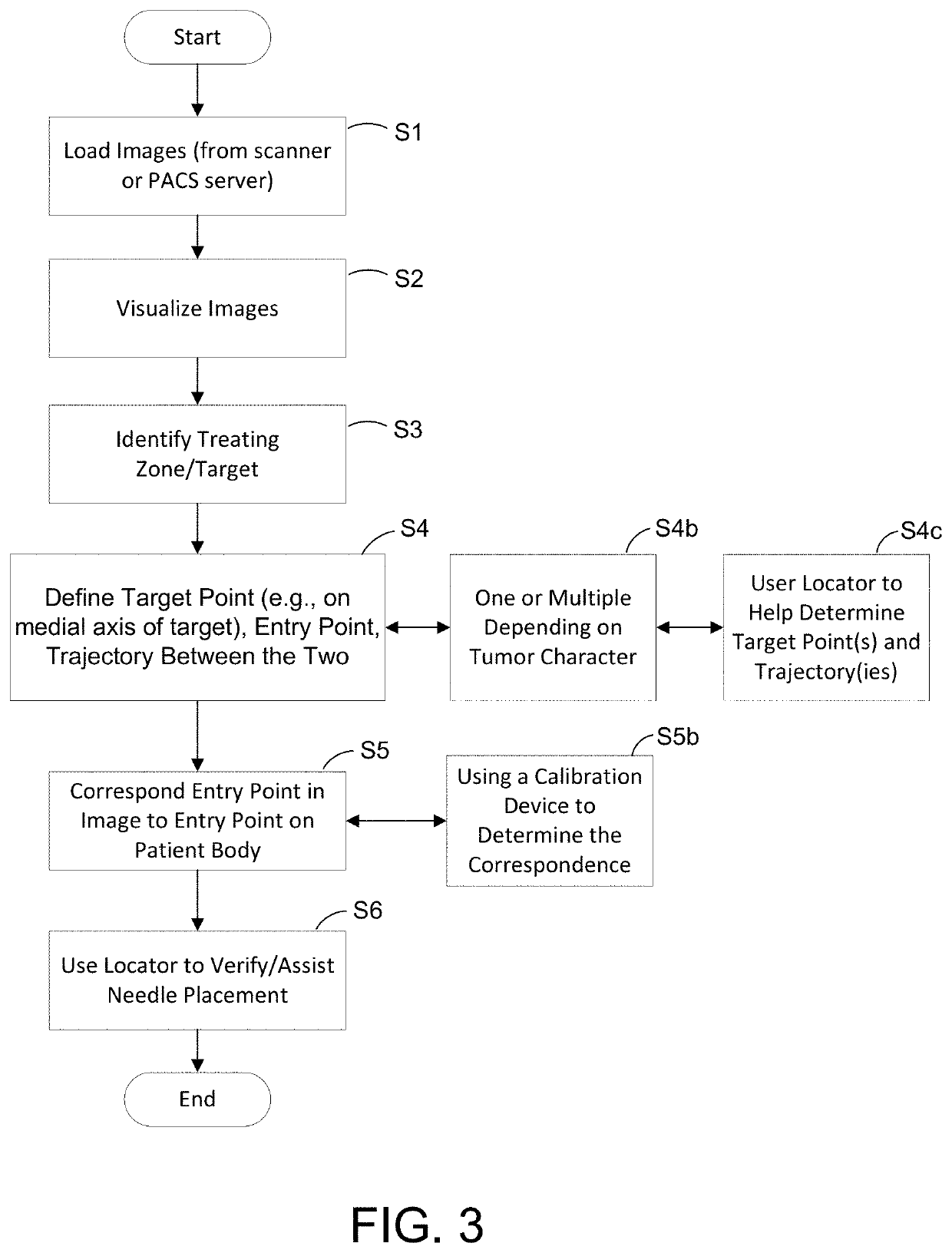
[0046]One or more devices, systems, methods and storage mediums for performing ablation planning and / or performance are disclosed herein. In accordance with at least one aspect of the present disclosure, one or more devices, systems, methods and storage mediums discussed herein perform ablation planning and / or ablation performance using at least one of the following methods: security or credential checking, integrating several steps (e.g., segmentation, registration, differential image view, etc.) to enhance an experience of a user when iteratively and interactively exploring and evaluating the planning and / or performance process, determining a medial axis of a target and performing confirmation with margin view. In one or more embodiments, these methods may be combined to further enhance the effectiveness in planning and ablation performing procedure. Several embodiments of the methods, which may be carried out by the one or more embodiments of an apparatus, system and computer-rea...


PUM


Abstract

One or more devices, systems, methods and storage mediums for performing ablation planning and / or ablation performance are provided. Examples of applications for such devices, systems, methods and storage mediums include imaging, evaluating and diagnosing biological objects, such as, but not limited to, lesions and tumors, and such devices, systems, methods and storage mediums may be used for radiotherapy applications (e.g., to determine whether to place seed(s) for radiotherapy). Preferably, a medial axis or a center line for a predetermined biological object (e.g., a lesion or tumor) is determined / found, one or more target points are picked along the medial axis or center line, and the ablation or radiotherapy zones are defined and optimized. Security checks may be performed in one or more embodiments to ensure proper use of the equipment and patient information.

Description

CROSS-REFERENCE TO RELATED APPLICATION(S)[0001]This application relates, and claims priority, to U.S. Patent Application Ser. No. 62 / 474,265, filed Mar. 21, 2017, the entire disclosure of which is incorporated by reference herein in its entirety.FIELD OF THE INVENTION[0002]The present disclosure relates generally to the field of optical imaging and more particularly to ablation planning and ablation performing apparatuses, systems, methods and storage mediums for use with same. Examples of ablation planning and performance applications include imaging, evaluating and characterizing / identifying biological objects or tissue, such as, but not limited to, for identification, location and treatment of lesions / tumors, operation / procedure planning, simulation and ablation performance.BACKGROUND OF THE INVENTION[0003]There are various forms of ablation, and successful ablation requires good planning. Ablation is normally ordered after diagnosis by oncologists who decide the ablation procedu...

Claims


Application Information

Patent Timeline
Patent Type & Authority Applications(United States)
IPC IPC(8): A61B34/10A61N5/10A61B18/02A61B18/18G06T7/68G16H20/40G16H30/40
CPCG06T2207/20101G06T7/68G06T2207/30004A61N5/1039A61B2018/00577A61B2034/105A61B2090/3735A61B2090/3762A61B2090/367A61B34/10G06T2207/30172A61B2034/107A61B18/02G16H30/40A61B18/1815G16H20/40A61B2034/104A61B2090/374A61B6/03A61N5/103G16H50/50G06V20/64G06V2201/034G06V2201/03G06T7/0012G06T7/11G06T2207/20104G06T2207/30048
Inventor LU, ZHIMIN
Owner CANON USA
Why Eureka
Try Eureka