Intravascular cell therapy device

a cell therapy and intravascular technology, applied in blood vessels, prostheses, coatings, etc., can solve the problems of at least 20% of all vascular disease patients remaining unsuitable for surgical intervention, and achieve the effects of facilitating retention of these cells, reducing vascular disease, and rapid seeding of proangiogenic cells

Active Publication Date: 2020-01-16
UNIV COLLEGE CORK NAT UNIV OF IRELAND CORK
View PDF4 Cites 1 Cited by
  • Summary
  • Abstract
  • Description
  • Claims
  • Application Information

AI Technical Summary

Benefits of technology

[0003]The present invention is in one aspect based on a design of a microbypass intravascular cell therapy device configured to deliver proangiogenic cells to a site of a chronic vascular occlusion for the purpose of developing microvascular bypass networks in continuity from the proximal vessel beyond the occlusion to the distal vessel thereby relieving vascular disease such as ischemia. The device is preferably a biodegradable structure that has a 3-D conformation that allows rapid seeding of proangiogenic cells, facilitates retention of these cells within the device and has a shape that can be altered to allow percutaneous therapy of the device loaded with proangiogenic cells to the vascular occlusion in-vivo by means of an intravascular catheter. The device can be embedded into the vascular wall to allow a microvasculature to develop in continuity from the proximal vessel beyond the occlusion to the distal vessel thus relieving ischemia. The device loaded with proangiogenic cells can be prepared ex-vivo in days—methods of preparation of the cells and seeding of the device is described below and broadly described in Kumar et al. The loaded device can then be delivered to the site of vascular occlusion. In one embodiment, an angiogenic gradient from the site of device placement to the site of distal ischemia facilitates alignment of endothelial cells, pericytes and smooth muscle cells in the surrounding vasculature to create adventitial arterioles that bypass the vascular occlusion and revascularise the distal ischaemic territory. This typically occurs over several weeks. The biodegradable nature of the device reduces the chronic foreign body reaction observed with stainless steel devices and allows angiogenesis and arteriogenesis to progress in-vivo without hindrance by chronic inflammatory processes. The therapy and release mechanism of the device allows rapid deployment into the blunt end of an obstructed artery without the need for a lot of the conventional wires associated with traditional PCI. The device of the invention is for treatment of vascular occlusion, especially chronic vascular occlusion, conditions characterised by vascular occlusion (i.e. peripheral artery disease or atherosclerosis), and symptoms of vascular occlusion (i.e. stroke, myocardial infarction, or ischaemic ulcers).
[0009]In one embodiment, the scaffold is configured to have a distal piercing tip when in a deployed orientation. This allows the scaffold to be at least partly embedded into a plaque and expand within the plaque. This is advantageous because plaque generally comprises microvasculature which is a suitable target for the proangiogenic factors produced by the cells embedded within the device. In addition, embedding the device in the plaque brings the device (and the proangiogenic cells seeded in the device) into close proximity with the plaque microvasculature, as well as the vessel wall microvasculature.
[0010]In one embodiment, the distal piercing tip comprises a helical screw formation. This facilitates the device being inserted / embedded into plaque by means of rotation of the device relative to the plaque.
[0011]In one embodiment, the scaffold is configured to have a conical shape when deployed. Ideally a distal part of the scaffold is conical. This shape facilitates embedding the device at least partially into a plaque.
[0012]In another embodiment, the scaffold is configured to have a partially conical shape when deployed. The term “partially conical” typically means a conical (or frusto-conical) distal part and cylindrical proximal part. This is illustrated in FIGS. 4 and 5. This shape facilitates having a device with a distal end that can be embedded into a plaque, and a proximal end that is exposed proximally of the plaque, whereby the device effects growth of a microbypass network from within the plaque and from within the vessel just proximal of the plaque.
[0037]In another aspect, the invention relates to a method of treating cardiovascular disease, especially coronary artery disease (for example preventing or inhibiting angina, or reducing risk of an ischemic event including a myocardial infarction) in a mammal in need thereof, comprising a step of percutaneous delivery of an intravascular cell delivery device of the invention in a contracted delivery orientation to a site of vascular occlusion, wherein the matrix has been seeded with proangiogenic cells, and deployment of the device at the site of the vascular occlusion, whereby the proangiogenic cells retained within the matrix release proangiogenic factors at the site developing microvascular bypass networks in continuity from the proximal vasculature beyond the occlusion to the distal vasculature thereby increasing regional blood flow.

Problems solved by technology

However, at least 20% of all vascular disease patients remain unsuitable for surgical intervention because of co-morbidities, suboptimal distal artery run off or lack of suitable vascular bypass conduits.

Method used


Image

  • Intravascular cell therapy device
  • Intravascular cell therapy device
  • Intravascular cell therapy device

Examples


example 1

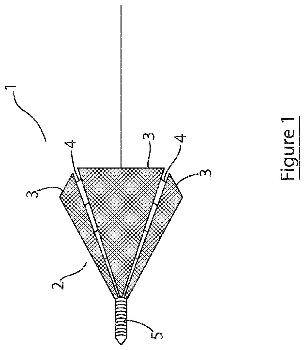
[0091]Referring to FIG. 1, there is illustrated an intravascular cell therapy device (ICTD) of the invention, indicated generally by the reference numerals 1, and comprising a generally conical scaffold body 2 formed by a plurality of sidewall panels 3 of triangular shape and a plurality of adjustable couplings 4 connecting adjacent panels. The scaffold is show in an expanded deployed orientation, with the couplings 4 in an expanded (unfolded) configuration. Although not shown, inward folding of the couplings causes the scaffold to contract and present a smaller profile suitable for percutaneous delivery. The space between adjacent panels when in a deployed orientation will be greater than that illustrated with reference to FIG. 1, with longer couplings, such that when the couplings are folded inwardly, the degree of contraction of the scaffold will be greater. In this embodiment, the couplings are configured to lock when unfolded, thereby locking the scaffold in the deployed config...

example 2

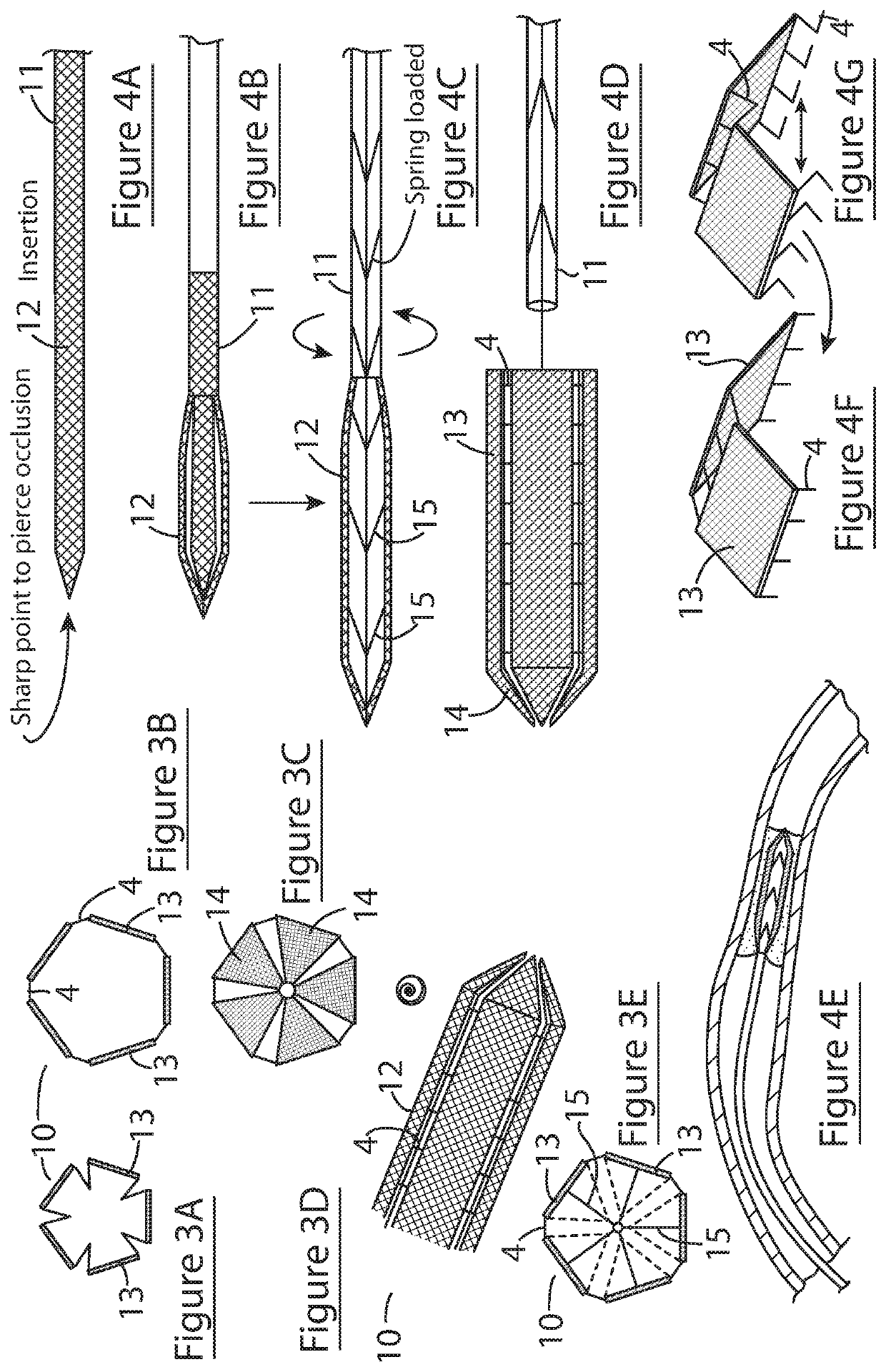
[0094]Referring the FIG. 3, a further embodiment of the ICTD device of the invention, and its use, is illustrated, in which parts identified with reference to the previous embodiments are assigned the same reference numerals. In this embodiment, the device 10 comprises a partially conical scaffold 12 and a delivery catheter 11 coupled to the scaffold by means of a releasable coupling mechanism. Referring to FIGS. 3A to 3D, the scaffold 12 comprises five axially elongated sidewall panels 3 having proximal rectangular sections 13 arranged parallel to an axis of the device (forming the cylindrical distal part of the scaffold) and distal triangular sections 14 that taper inwardly towards a distal end of the device (forming the distal conical part of the scaffold). The distal and proximal sections of each panel are hingedly connected to allow for expansion and contraction of the scaffold. The couplings 4 are foldable struts (as shown in FIG. 3A) that unfold and straighten when the scaffo...

example 3

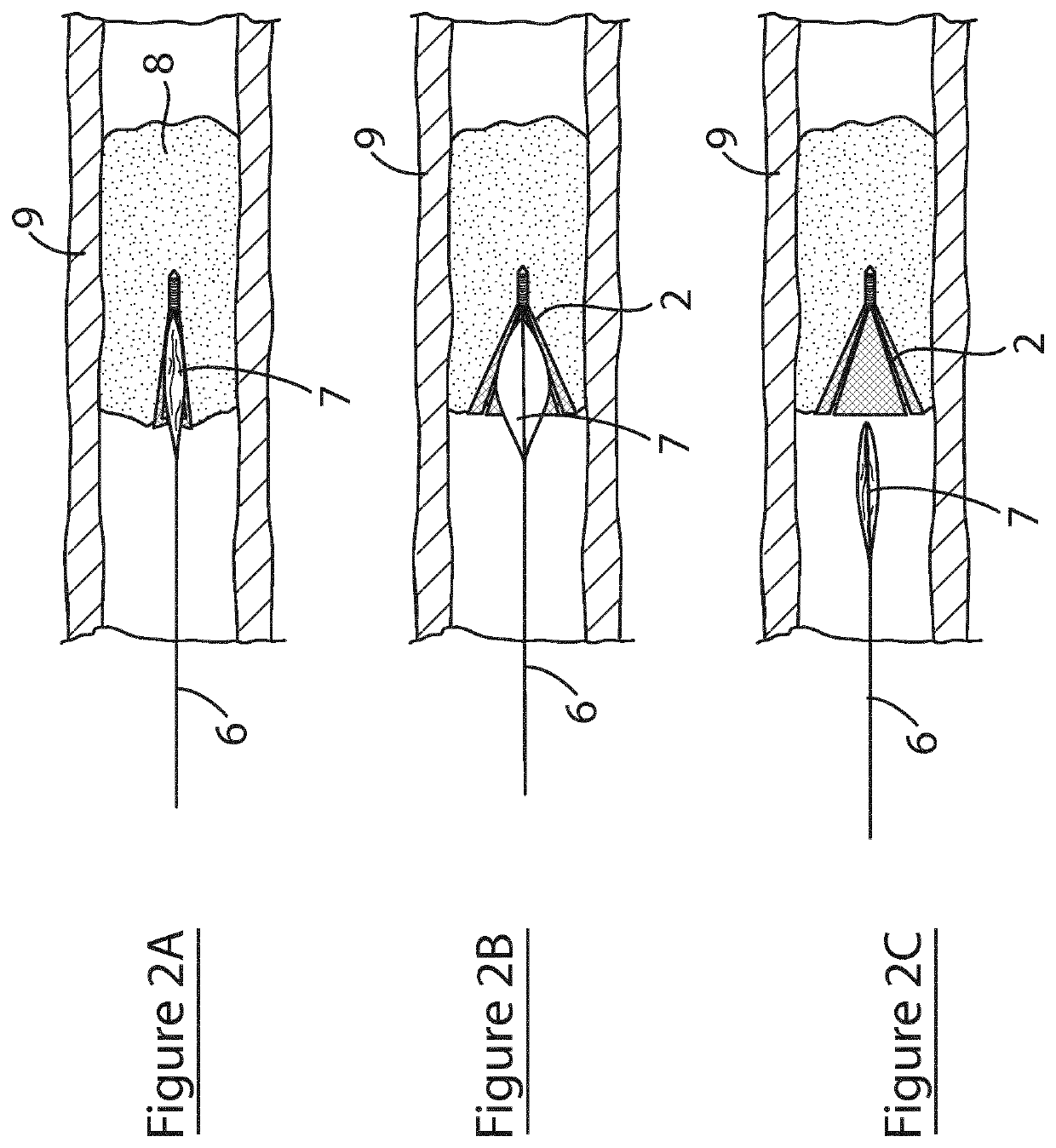
[0097]Referring to FIG. 5, a further embodiment of the ICTD device of the invention, and its use, is illustrated, in which parts identified with reference to the previous embodiments are assigned the same reference numerals. In this embodiment, the device 20 comprises a partially conical scaffold 12 configured for delivery and deployment using a balloon catheter 6. Referring the FIG. 5A, the device 20 is shown mounted on a balloon 7 of a balloon catheter 6. In FIG. 5B, the balloon 6 is inflated thereby deploying the device 20 within the plaque 8. FIG. 5C shows the balloon deflated and retracted away from the plaque 8 leaving the device 20 embedded in the plaque.


PUM

PropertyMeasurementUnit
internal diameteraaaaaaaaaa

Abstract

An intravascular cell therapy device comprises a scaffold (2, 12) that is radially adjustable between a contracted orientation suitable for transluminal delivery to a vascular locus and an expanded orientation, and a biodegradable matrix provided on at least a portion of the scaffold that is suitable for seeding with cells and degrades in a vascular environment. The scaffold is configured to have a distal piercing tip (5) when in a deployed orientation. The scaffold comprises a plurality of sidewall panels (3, 13, 14) arranged around a longitudinal axis of the scaffold, and adjustable couplings (4) between the panels configured for adjustment between an expanded configuration and a contracted orientation, and in which each sidewall panel comprises a matrix suitable for seeding with cells.

Description

FIELD OF THE INVENTION[0001]The present invention relates to an intravascular cell therapy device. Also contemplated are methods of treatment of chronic vascular occlusion of coronary or peripheral vasculature, or symptoms of chronic vascular occlusion, that employs the intravascular cell therapy device.BACKGROUND TO THE INVENTION[0002]Chronic vascular occlusion is currently treated by surgical bypass or less frequently and less successfully by percutaneous coronary intervention (PCI). However, at least 20% of all vascular disease patients remain unsuitable for surgical intervention because of co-morbidities, suboptimal distal artery run off or lack of suitable vascular bypass conduits. These patients may be suitable for a hybrid procedure that can be performed percutaneously (without surgical risks) but also providing microbypass of the occlusion (without the extensive technical, imaging and recurrence limitations of current PCI approaches). This procedure is described in Kumar et ...

Claims


Application Information

Patent Timeline
Patent Type & Authority Applications(United States)
IPC IPC(8): A61F2/02A61F2/06A61L27/44A61L27/50A61L27/54A61L27/58
CPCA61L27/507A61L27/54A61F2230/0067A61F2/022A61F2250/0067A61F2210/0004A61F2/06A61F2230/0069A61L27/44A61L27/58A61L2300/64A61F2002/8486A61F2230/0023A61L27/34A61F2/0077A61F2/062A61F2/07A61F2/82A61F2/848A61B17/3417A61F2/958A61F2002/0086A61F2220/0016A61F2250/0018A61F2250/0031A61F2250/0059C08L89/00
Inventor CAPLICE, NOEL
Owner UNIV COLLEGE CORK NAT UNIV OF IRELAND CORK
Why Eureka
Try Eureka