
Devices and Methods for Vascular Hyperperfusion of Extravascular Space

Active Publication Date: 2020-02-06
ALL VASCULAR PTY LTD
View PDF0 Cites 0 Cited by
  • Summary
  • Abstract
  • Description
  • Claims
  • Application Information

AI Technical Summary

Benefits of technology

The present invention is a method for delivering therapeutic substances to a region of the body through vascular isolation and manipulation. The technique includes restricting vascular inflow, increasing outward oncotic pressure gradient, inducing ischemia, controlling pressure and fluid flow of main blood vessels, and providing the therapeutic substance when fluid flow is controlled. This method avoids the need for drug usage to expel albumins and allow therapeutic substances to cross extravascular space barriers.

Problems solved by technology

The interface between the vein or artery and cannula requires pressure to deliver materials or receive blood and minimise the ability for blood to stagnate around the interface thereby leading to thrombosis.
Dead space also gives rise to an area where blood can pool and stagnate also presenting a situation where thrombosis can occur.
The dead space may also present an area where gas collects giving rise to the risk of a gas embolism forming.
However, when a cannula is connected to the patient's blood vessel at a non-perpendicular angle, the conventional cylindrical shape of the tip of the plunger is not capable of preventing the filling of a small amount of blood into a lower part (called “the dead space”) of the lumen of the cannula unless the tip is slid further through the lumen of the cannula and a leading part of the tip protrudes into the lumen of the vessel.
However, when a cannula with an appropriately chamfered proximal end is connected to the patient's blood vessel at a non-perpendicular angle, the conventional cylindrical shape of the tip of the plunger is not capable of preventing the filling of a small amount of blood into a lower part (called “the dead space”) of the lumen of the cannula unless the tip is slid further through the lumen of the cannula and a leading part of the tip protrudes into the lumen of the vessel.
However, those access devices with multi-access treatment caps have access ports which are such that only a single catheter may be received through a selected access port and then through the lumen of the access device, and therefore each such device can only facilitate either an outflow from the circulatory system to a blood flow pump or an inflow from a blood flow pump in the circulatory system, but not both.
That is, those access devices with multi-access treatment caps cannot facilitate two or more inflow and outflow catheters at any one time because the lumen of those devices is unable to receive two or more catheters.
Additionally, those multi-access treatment caps do not enable a catheter to be directed into specific positions with use of the multi-access treatment cap.
Lymph nodes that are involved are notoriously difficult to treat because of their small size.
Other problems relating to treatment of neoplasia arise from malignant cells residing in small numbers in relatively ischemic tissue, such that systemic treatment will have diminished effect.
However, when a cannula with an appropriately chamfered proximal end is connected to the patient's blood vessel at a non-perpendicular angle, the conventional cylindrical shape of the tip of the plunger is not capable of preventing the filling of a small amount of blood into a lower part (called “the dead space”) of the lumen of the cannula unless the tip is slid further through the lumen of the cannula and a leading part of the tip protrudes into the lumen of the vessel.
However, those access devices with multi-access treatment caps have access ports which are such that only a single catheter may be received through a selected access port and then through the lumen of the access device, and therefore each such device can only facilitate either an outflow from the circulatory system to a blood flow pump or an inflow from a blood flow pump in the circulatory system, but not both.
That is, those access devices with multi-access treatment caps cannot facilitate two or more inflow and outflow catheters at any one time because the lumen of those devices is unable to receive two or more catheters.
There are clear safety issues.
The high flow rates can contribute to congestive cardiac failure due to the high flow rates that manifests itself as peripheral oedema, lethargy, shortness of breath and chest pain.
High flow can also cause venous hypertension.

Method used


Image

  • Devices and Methods for Vascular Hyperperfusion of Extravascular Space
  • Devices and Methods for Vascular Hyperperfusion of Extravascular Space
  • Devices and Methods for Vascular Hyperperfusion of Extravascular Space

Examples


Embodiment Construction

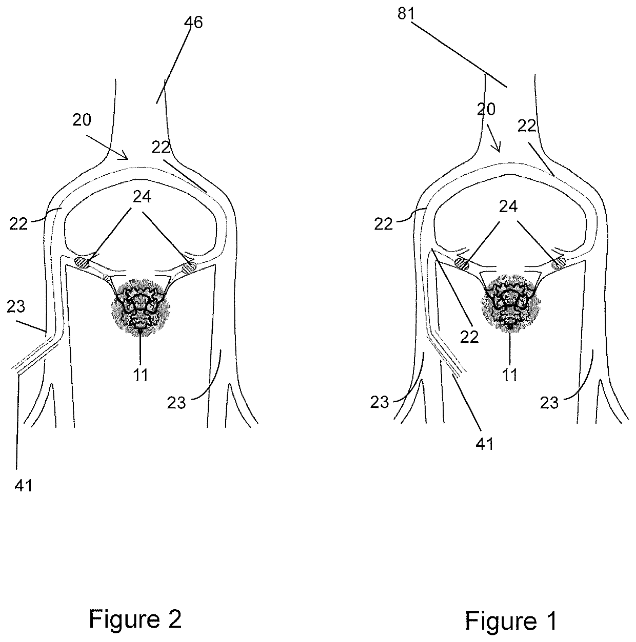
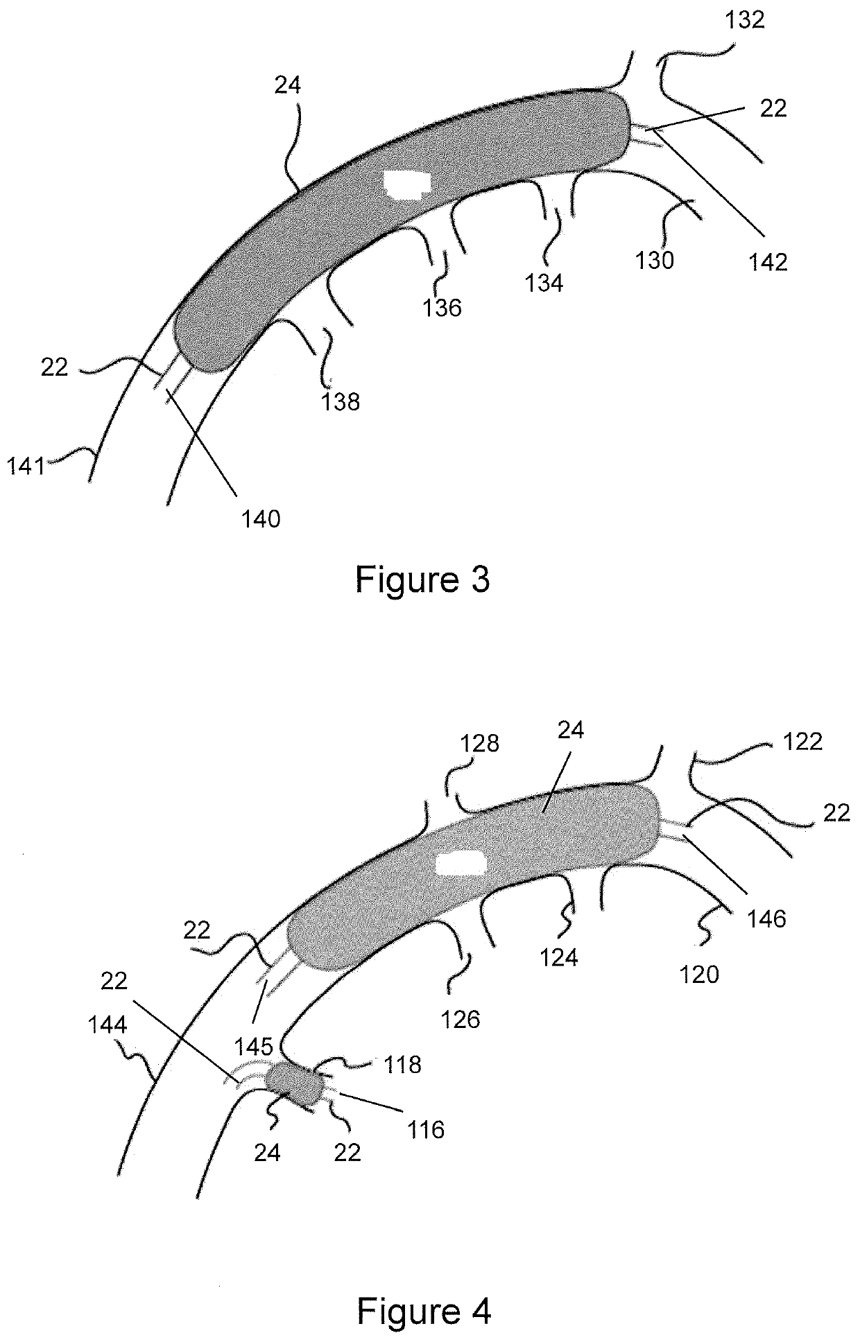
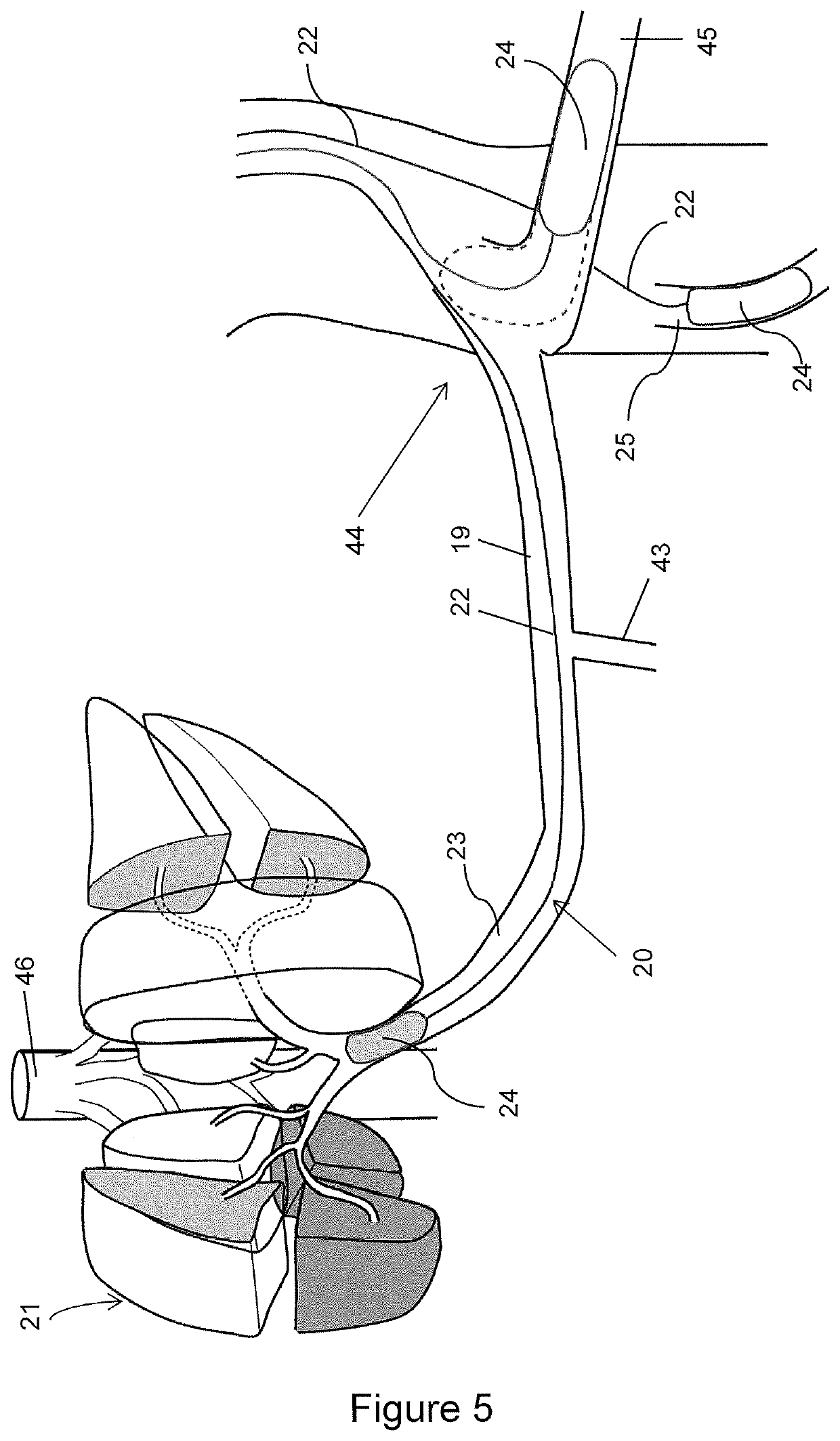
[0124]Broadly, with reference to FIGS. 1 to 13 an embodiment of the present invention relates to a system and devices for improving the delivery of therapeutic substances for therapeutic treatment into the extra vascular, i.e. interstitial space, where the targeted cells or lesions are situated, specifically hyperperfusing the ischemic interstitial space. Hyperperfusion is defined as to cause an above normal amount of fluid (or cells) to pass through a space. The inevitable effect of hyperperfusing therapeutic agents into the interstitial space is to hyperperfuse the target as well as the draining lymphatic channels and lymph nodes. The concept has important ramifications in the treatment of neoplasia as many of the malignant cells invade lymphatics, migrate into the lymphatic nodes, multiply, then embolise via the thoracic duct and then into the venous system and hence is spread systemically. Lymph node involvement is notoriously difficult to treat related to the small size of lymp...


PUM


Abstract

A method of delivering a therapeutic substance for treatment to a region of the body through vascular isolation and manipulation of fluid flux into and from the region of the body including the steps of: restricting vascular inflow to the region of the body; washing out oncotically active plasma proteins from the region of the body by increasing the outward oncotic pressure gradient from the region of the body; inducing ischemia in the region of the body; controlling the pressure and fluid flow of the main blood vessels to and from the region of the body; providing the therapeutic substance to the region of the body when the fluid flow to the region of the body is controlled.

Description

CROSS-REFERENCE AND CLAIM OF PRIORITY TO RELATED APPLICATIONS[0001]This application is a U.S. national phase application of PCT International Patent Application No. PCT / AU2017 / 050266, filed on Mar. 24, 2017, which claims the benefit of and priority to Australian Application No. 2016903834, filed Sep. 22, 2016, Australian Application No. 2016903836, filed Sep. 22, 2016, Australian Application No. 2016904991, filed Dec. 5, 2016, Australian Application No. 2016905067, filed Dec. 8, 2016 and Australian Application No. 2016905068, filed Dec. 8, 2016, each of which are hereby incorporated by reference in their entireties.FIELD OF THE INVENTION[0002]The present invention relates to improvements in vascular isolation of organs and segments thereof and, in particular, to improved devices and methods for vascular isolation of human extravascular spaces in organs and segments thereof, so as to enhance delivery and activity of therapeutic agents, such as chemotherapeutic agents and stem cells, ...

Claims


Application Information

Patent Timeline
IPC IPC(8): A61M1/36A61M25/09A61M25/10A61M39/02A61M5/145
CPCA61M25/1011A61M2025/09008A61M2039/0205A61M1/3613A61M25/09A61M2210/1007A61M1/3615A61M2025/1052A61M2210/0693A61M39/0208A61M1/3655A61M2210/1071A61M5/1452A61M2025/105A61M1/3639A61M39/00A61M39/10A61M39/12
Inventor LANE, RODNEY JAMESHUCKSON, MATTHEW JAMESKYUNG, CHRISLANE, DAVIDKHIN, NYANMURPHY, SCOTT
Owner ALL VASCULAR PTY LTD
Learn More