Method for imaging a biological sample and corresponding probe
a biological sample and probe technology, applied in the direction of fluorescence/phosphorescence, material analysis using wave/particle radiation, instruments, etc., can solve the problems of difficult detection using tem, osmiophilic and therefore visible for tem, and difficult interpretation of images obtained, etc., to achieve the effect of simplifying correlation
- Summary
- Abstract
- Description
- Claims
- Application Information
AI Technical Summary
Benefits of technology
Problems solved by technology
Method used
Image
Examples
example 1
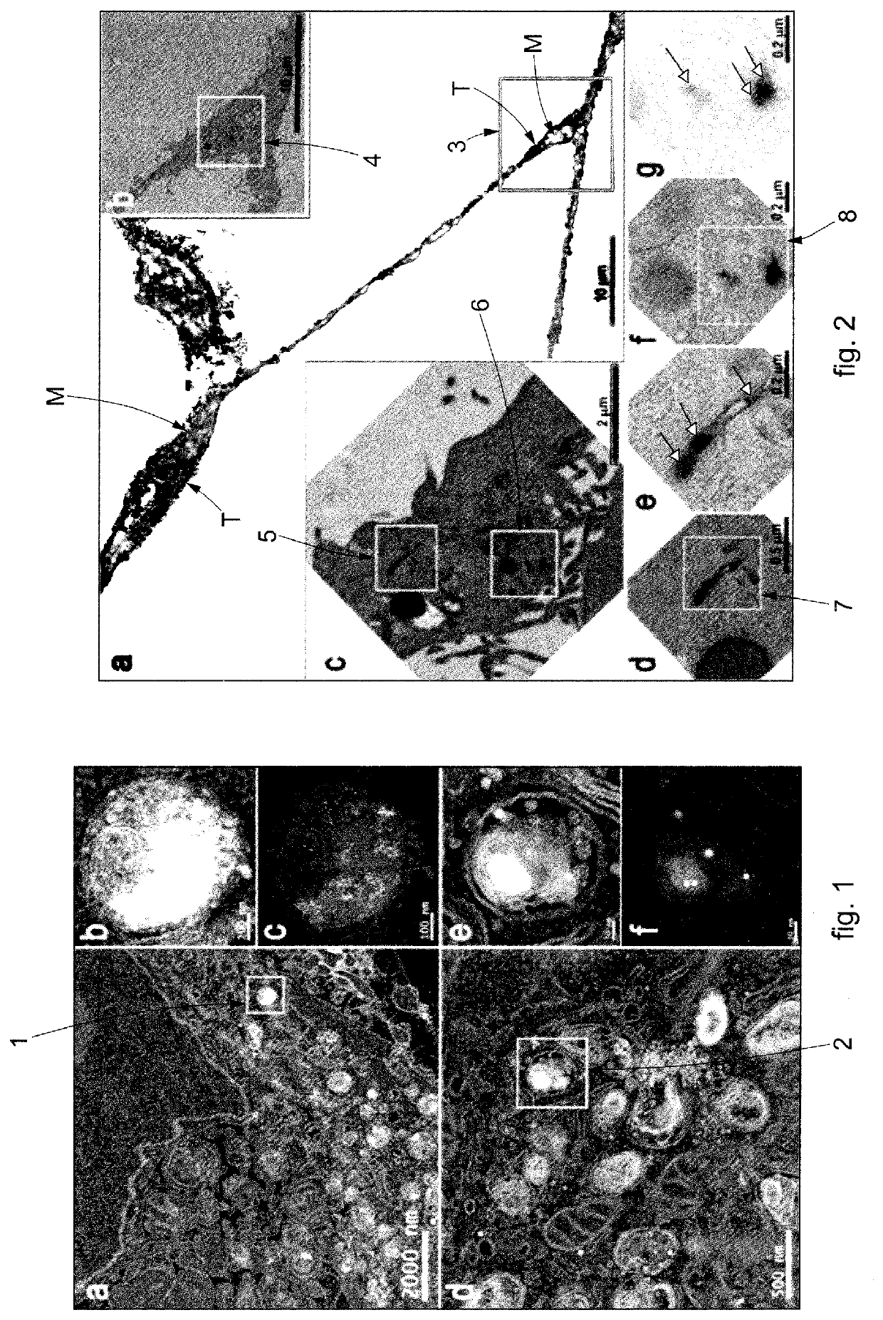
[0097]To test the actual amplification of the electron-dense signal produced by the peroxidase activity of nanoparticles of platinum in a biological sample inside the cells, in the example shown in FIG. 1 a biological sample containing tumor cell lines (HeLa) was incubated with imaging probes having nanoparticles of platinum of 4 nm and 10 nm.
[0098]The images shown in the squares of FIG. 1 were obtained through electron microscopy, in particular acquired by HAADF (high angular annular dark field detector) scanning TEM (STEM), where the squares “b” and “c” are enlargements of the area 1 highlighted in square “a” and squares “e” and “f” are enlargements of the area 2 highlighted in square “d”.
[0099]With reference to FIG. 1, Applicant has observed, for all the dimensional classes of nanoparticles of platinum used, a considerable increase in the electron-dense signal, which made it possible to identify them even at low enlargement (squares “a” and “d”).
[0100]The images of FIG. 1 acquire...
example 2
[0103]In the example of FIG. 2, the efficacy was tested of the imaging probes by CLEM to visualize the endosomal pathway of transferrin inside cells.
[0104]In the example shown, the nanoparticles of platinum were bioconjugated with a specific protein, that is, with fluorescent transferrin, suitably functionalizing the surface of the nanoparticles of platinum.
[0105]In particular, this allowed to create an amide bond between the amino groups present on the surface of the protein and the carboxyl groups present on the surface of the nanoparticles of platinum.
[0106]With reference to the square marked by the letter “a” in FIG. 2, the fluorescent signal of the nanoparticles of platinum bioconjugated with fluorescent transferrin is clearly recognizable for the FOM, indicated in black and with the reference “T”, while the mitochondria are indicated in gray and with the reference “M”.
[0107]The localization observed using FOM was confirmed in TEM using the amplification procedure described abo...
PUM
| Property | Measurement | Unit |
|---|---|---|
| sizes | aaaaa | aaaaa |
| diameter | aaaaa | aaaaa |
| sizes | aaaaa | aaaaa |
Abstract
Description
Claims
Application Information
 - R&D
- Intellectual Property
- Life Sciences
- Materials
- Tech Scout
- Unparalleled Data Quality
- Higher Quality Content
- 60% Fewer Hallucinations
Browse by: Latest US Patents, China's latest patents, Technical Efficacy Thesaurus, Application Domain, Technology Topic, Popular Technical Reports.
© 2025 PatSnap. All rights reserved.Legal|Privacy policy|Modern Slavery Act Transparency Statement|Sitemap|About US| Contact US: help@patsnap.com

