Multiplexed in situ hybridization of tissue sections for spatially resolved transcriptomics with expansion microscopy

a transcriptomics and expansion microscopy technology, applied in the field of imaging, can solve the problems of reducing the accuracy of rna expression level quantification, requiring ultra-thin sections of specimens, and limited imaging of rna

Active Publication Date: 2020-07-30
EXPANSION TECH
View PDF0 Cites 96 Cited by
  • Summary
  • Abstract
  • Description
  • Claims
  • Application Information

AI Technical Summary

Benefits of technology

[0016](HCR) initiator sequence; (g) contacting the sample with the plurality of initiator DNA probes under conditions wherein the sequence complementary to the sequence from one of the nucleic acid targets of interest hybridizes to that sequence from the nucleic acid target of interest; (h) for each initiator DNA probe, providing a pair of fluorophore-labeled DNA hairpins that metastably co-exist in the absence of the initiator DNA probe; (i) contacting the sample with the fluorophore-labeled DNA hairpins under conditions wherein the hairpins self-assemble by HCR, in the presence of their corresponding initiator DNA probe, into fluorescent amplification polymers tethered to that corresponding initiator DNA probe; (j) obtaining an image of the sample; (k) treating the sample with a deoxyribonuclease to remove the initiator DNA probes and amplification polymers; and (l) repeating steps (f)-(k) one or more times for additional RNA targets of interest.
[0017]In another aspect, provided herein are methods of imaging ribonucleic acid (RNA) in a biological sample, said method being performed under RNAse-free conditions and said method comprising: (a) contacting the sample with a gel binding moiety under conditions wherein the gel binding moiety operably links to RNA in the sample; (b) contacting the sample with a solution comprising monomers of a polyelectrolyte gel; (c) by free radical polymerization, polymerizing said monomers to form the polyelectrolyte gel and covalently conjugating the gel binding moiety to the polyelectrolyte gel; (d) proteolytically digesting said sample; (e) dialyzing said sample to expand said polyelectrolyte gel; (f) providing a plurality of initiator deoxyribonucleic acid (DNA) probes targeting a plurality of RNA targets of interest, wherein each of the plurality of initiator DNA probes comprise (A) a sequence complementary to a sequence from one of the RNA targets of interest, and (B) a hybridization chain reaction (HCR) initiator sequence; (g) contacting the sample with the plurality of initiator DNA probes under conditions wherein the sequence complementary to the sequence from one of the RNA targets of interest hybridizes to that sequence from the RNA target of interest; (h) for each initiator DNA probe, providing a pair of fluorophore-labeled DNA hairpins that metastably co-exist in the absence of the initiator DNA probe; (i) contacting the sample with the fluorophore-labeled DNA hairpins under conditions wherein the hairpins self-assemble by HCR, in the presence of their corresponding initiator DNA probe, into fluorescent amplification polymers tethered to that corresponding initiator DNA probe; (j) obtaining an image of the sample; (k) treating the sample with a deoxyribonuclease to remove the initiator DNA probes and amplification polymers; and (l) repeating steps (f)-(k) one or more times for additional RNA targets of interest.

Problems solved by technology

However, the imaging of RNA is still limited due to diffraction-limited resolutions on conventional microscopes which reduce the accuracy of RNA expression level quantification and due to the limited number of transcripts which can be resolved within a single specimen.
Although super-resolution techniques can provide improved spatial resolution, they are slow and require ultra-thin section of specimens.
However, because the number of fluorescent dyes that can be used is small (typically <6), this strategy is limited to imaging only a small number of proteins per sample.
Additionally, the polymerization process dampens the fluorescence of the dyes, which are permanently connected to the gel matrix.
Also, the presence of the DNA on the antibody reduces the extent and the rate at which it binds to the target.
The result is that the current ExM processes are lengthy, and the staining is commonly dim compared to controls, making this approach unsuitable for RNA detection due to the limited number of transcripts which can be resolved within a single specimen.
However, this approach allows only protein detection and is less suitable for RNA detection or imaging both proteins and nucleic acids.
The interrogation of RNA in a highly multiplexed and efficient manner with spatial information remains a challenge in structural biology and medicine.

Method used


Image

  • Multiplexed in situ hybridization of tissue sections for spatially resolved transcriptomics with expansion microscopy
  • Multiplexed in situ hybridization of tissue sections for spatially resolved transcriptomics with expansion microscopy
  • Multiplexed in situ hybridization of tissue sections for spatially resolved transcriptomics with expansion microscopy

Examples


examples

Materials and Methods

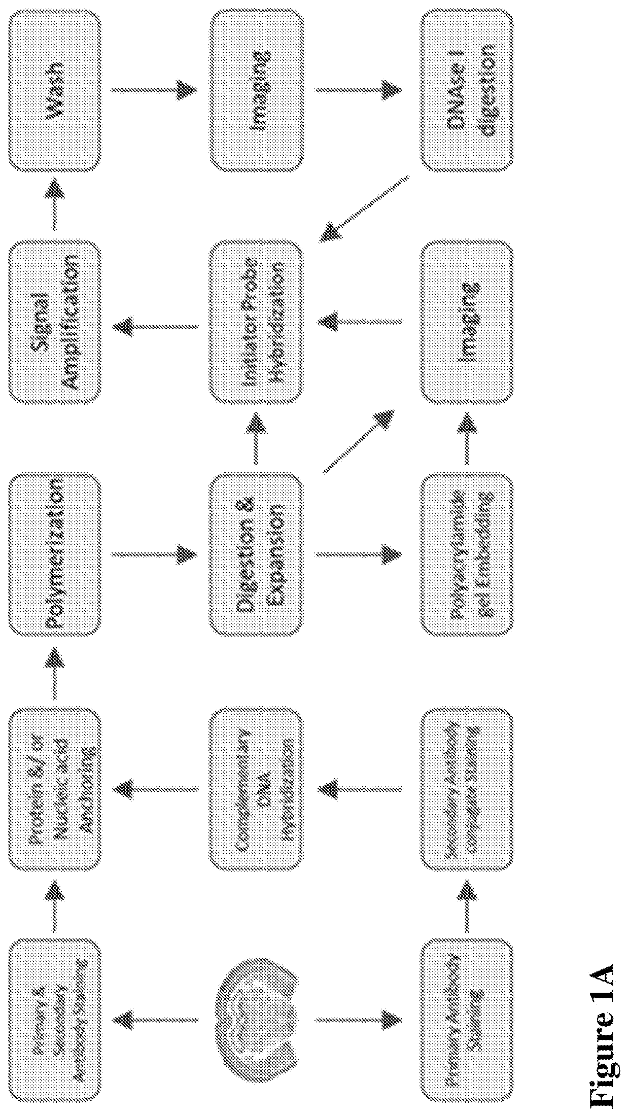
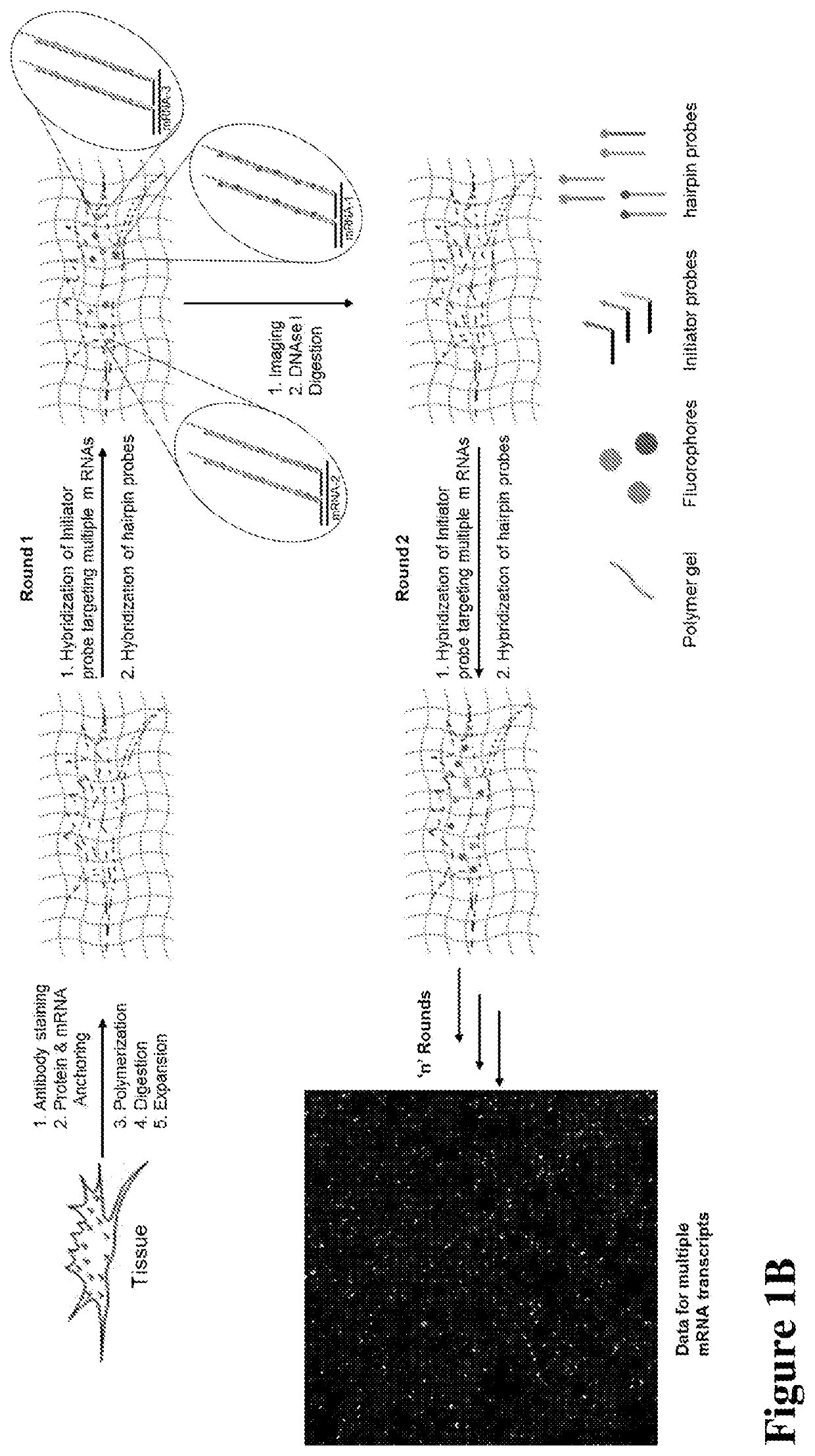
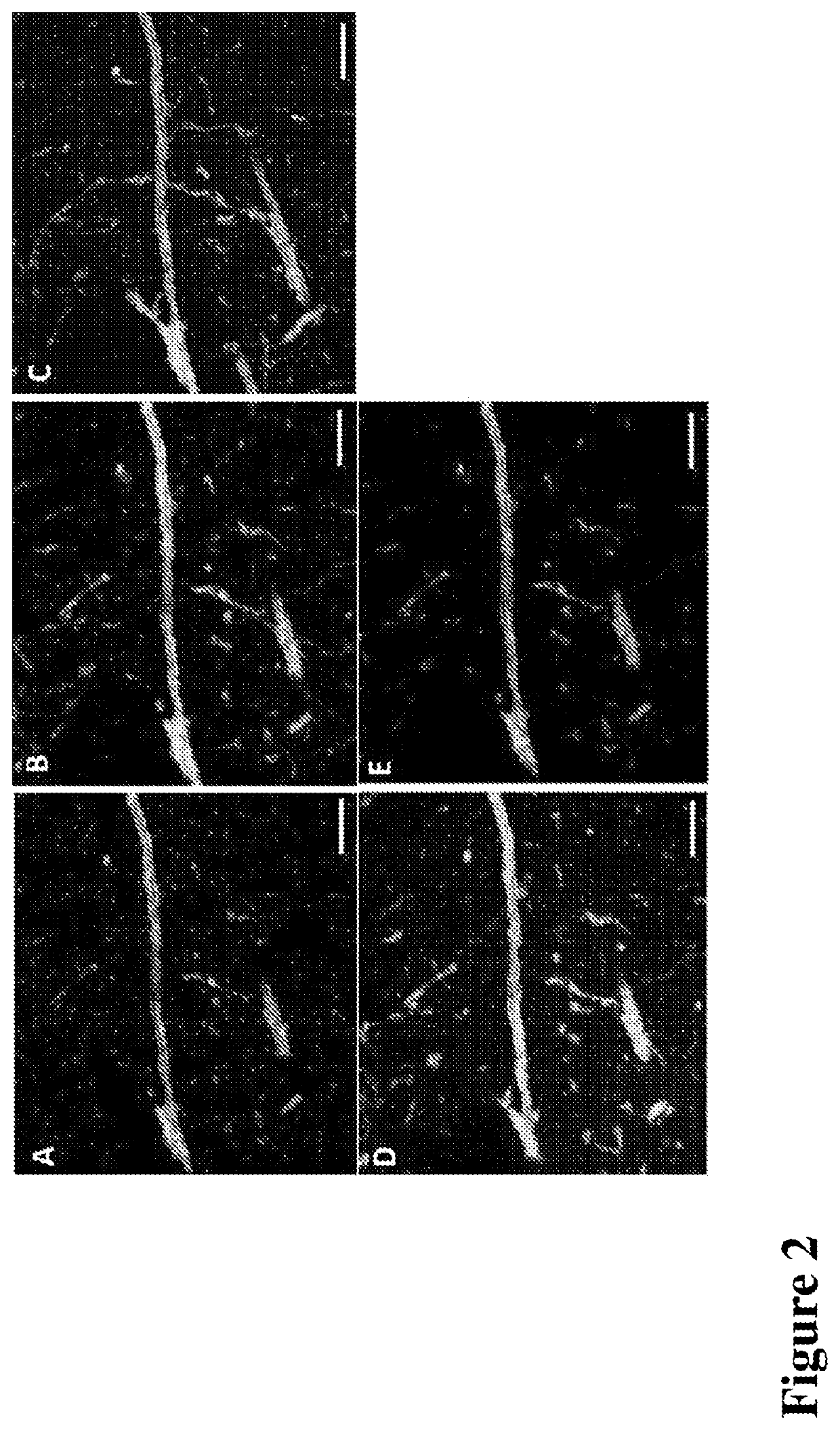
[0149]Antibody staining, Polymerization and Expansion: Brain slices taken from the cryo-protectant solution were washed with 1×PBS and blocked with blocking buffer (5% normal donkey serum and 0.1% TritonX-100 in 1×PBS) for 2 hours at room temperature or overnight at 4 ° C. Slices were incubated with respective primary and secondary antibodies for 6 hours at room temperature (RT) or overnight at 4 ° C. Upon washing with 1×PBS after each antibody incubation, slices were washed with MOPs buffer for 30 minutes, then incubated in a solution of anchoring reagents Acryloyl-X (6-((acryloyl)amino)hexanoic acid succinimidyl ester; 100 μg / mL) and NucliX (FIG. 3A;100 μg / mL) in MOPs buffer for 6 hours or overnight at room temperature. Anchoring reagent solution was removed and slices were washed with 1×PBS three times. Then slices were incubated in monomer solution for 10 minutes with rocking at RT. Polymerization was initiated by adding initiator (10% APS) and accelerator (...


PUM

PropertyMeasurementUnit
dissociation constantaaaaaaaaaa

Abstract

This invention relates to imaging, such as by expansion microscopy, labelling, and analyzing biological samples, such as cells and tissues, as well as reagents and kits for doing so.

Description

CROSS-REFERENCE TO RELATED APPLICATIONS[0001]This application claims priority of U.S. Provisional Application No. 62 / 571,076, filed Oct. 11, 2017, which is hereby incorporated by reference.FIELD OF THE INVENTION[0002]The invention relates to imaging, such as by expansion microscopy, labelling, and analyzing biological samples, such as cells and tissues, as well as reagents and kits for doing so.BACKGROUND OF THE INVENTION[0003]Understanding the transcriptional states and spatial organization of ribonucleic acid (RNA) within tissues is essential to determining the mechanisms of disease and for answering a multitude of scientific questions. However, the imaging of RNA is still limited due to diffraction-limited resolutions on conventional microscopes which reduce the accuracy of RNA expression level quantification and due to the limited number of transcripts which can be resolved within a single specimen. Current imaging technologies for transcriptional profiling of complex tissues in...

Claims


Application Information

Patent Timeline
Patent Type & Authority Applications(United States)
IPC IPC(8): C12Q1/6841
CPCC12Q2525/301C12Q2527/125C12Q2563/107C12Q1/6841
Inventor DEWAL, MAHENDER BABU
Owner EXPANSION TECH
Why Eureka
Try Eureka