
Methods for angiography

a technology of angiography and methods, applied in the field of angiography, can solve the problems of increasing the signal to noise ratio, increasing the risk of the subject, and unsatisfactory images, and achieve the effect of reducing the toxicity of x-ray imaging

Pending Publication Date: 2020-08-06
BUTLER WILLIAM E
View PDF1 Cites 3 Cited by
  • Summary
  • Abstract
  • Description
  • Claims
  • Application Information

AI Technical Summary

Benefits of technology

The patent text discusses methods for reducing the toxicity of imaging mammalian hosts using contrast agents and X-ray angiography. The methods involve extracting specific cardiac frequency angiographic phenomena from the captured angiographic data and using them to generate a diagnostically useful image. By doing so, the amount of contrast agent used and the X-ray dosage required to obtain a diagnostically useful image are significantly reduced, which can help to prevent or reduce contrast nephropathy and other related complications associated with X-ray imaging. Overall, the methods described in the patent text can provide safer and more efficient tools for medical imaging.

Problems solved by technology

Elevating the contrast dose and / or the x-ray dose increases the signal to noise ratio in the produced angiographic images, but also increases the risk to the subject in several ways.
The injected chemical contrast agent has toxic side effects to kidneys and other internal organs, and therefore it often is necessary to lower the dose of contrast agent to reduce the risk of these toxic side effects.
This may produce unsatisfactory images with poor signal to noise ratios which, in turn, may lead to incomplete angiographic studies with inadequately imaged vascular anatomy.
Use of an elevated chemical contrast dose may lead to injury to those organs vulnerable to chemical contrast side effects.
The need to advance the injecting catheter further elevates the risk of complications caused by the catheter injuring ever smaller vessels distal in the vascular tree.

Method used


Image

  • Methods for angiography
  • Methods for angiography
  • Methods for angiography

Examples


example

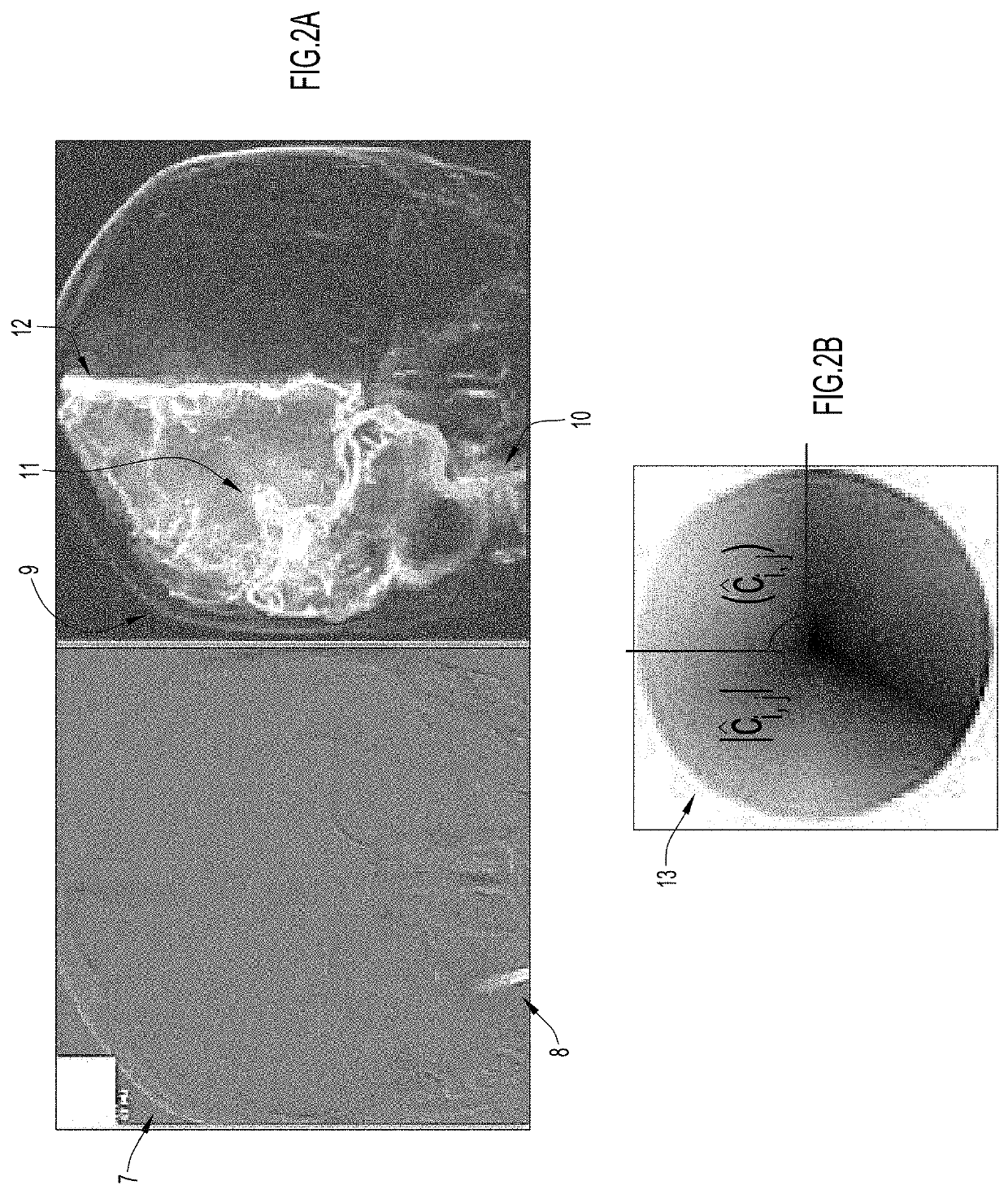
[0070]Two human angiograms were performed in immediate succession in anteroposterior (AP) projection of the right vertebral artery. Since iodinated contrast and x-ray have injurious properties, a so-called “puff” angiogram (preparatory angiogram) was obtained using 10% of the dose of the iodinated contrast agent and 1% of the x-ray dose conventionally used for a diagnostic angiogram.

[0071]For the “puff” injection the chemical agent used was iopamidol (Isovue), at a dose of 1 ml of a formulation of 3 mg / ml, which provides a dose of 3 mg iopamidol for the injection. The x-ray dose area product was 1.968 Gray m2. The “dose area product” is a measure of the absorbed dose per kilogram multiplied by the area irradiated. The x-ray dose is obtained from the image series DICOM metadata.

[0072]For the regular (“full dose”) right vertebral artery injection, the injected contrast dose was also iopamidol, but using 10 ml of the same formulation of 3 mg / ml, providing a dose of 30 mg iopamidol for ...


PUM


Abstract

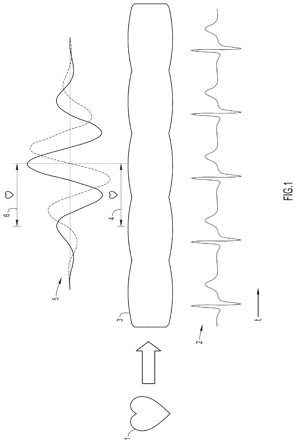
An angiogram is a study of blood vessels where an angiographic chemical contrast agent is injected while a sequence of images (typically x-rays) are obtained. The contrast pattern on the sequence of images provides information about the vascular anatomy and physiology. The discovery that contrast in blood vessels varies at cardiac frequency in magnitude and phase, which may be visualized as a spatiotemporal reconstruction of cardiac frequency angiographic phenomena, enables a set of processes for increasing the signal to noise ratio or equivalently the informational content of an angiogram. In this invention, the organization of cardiac frequency magnitude and phase enables equivalent information on anatomy and physiology to be obtained with less dose of injected chemical contrast agent, less x-ray dose, and / or less navigation of the injecting catheter within blood vessels. The cardiac frequency magnitude and phase is organized so that the arterial and venous subsystems of circulation have coherence at cardiac frequency. This enables processes for diagnosing deficits of circulation that involve alterations in the transit of blood from the arterial to the venous subsystems of circulation. Furthermore, the discovery of cardiac frequency magnitude and phase organization enables the design and manufacture of lighter and more portable angiography equipment.

Description

CROSS-REFERENCE TO RELATED APPLICATIONS[0001]This application claims priority to U.S. Provisional Application No. 62 / 801,780, filed Feb. 6, 2019, the contents of which are hereby incorporated by reference in their entirety.FIELD OF THE INVENTION[0002]Improved methods for angiography are provided. Specifically, methods of obtaining angiographic data are provided that permit use of greatly reduced doses of contrast agent and / or x-ray dosage, while maintaining, or improving the signal to noise ratio of the angiogram.BACKGROUND OF THE INVENTION[0003]In a conventional catheter angiogram, a catheter is placed into an artery and the catheter tip is advanced into the arterial region of interest. A chemical contrast agent is injected and the passage of the contrast to the vascular bed is fluoroscopically imaged and recorded. The contrast agent is opaque to the x-ray, causing a pattern of opacification to appear on the imaging x-ray detector in a sequence of angiographic image frames. Vascula...

Claims


Application Information

Patent Timeline
IPC IPC(8): A61B6/00
CPCA61B6/5217A61B6/5205A61B6/503A61B6/5258A61B6/481A61B6/504A61B6/542A61B6/5211A61B6/4441A61B6/487G06T5/50G06T7/0016G06T2207/10016G06T2207/10116G06T2207/30101G06T5/70A61B6/485A61B6/5288A61K49/04
Inventor BUTLER, WILLIAM E.
Owner BUTLER WILLIAM E
Learn More