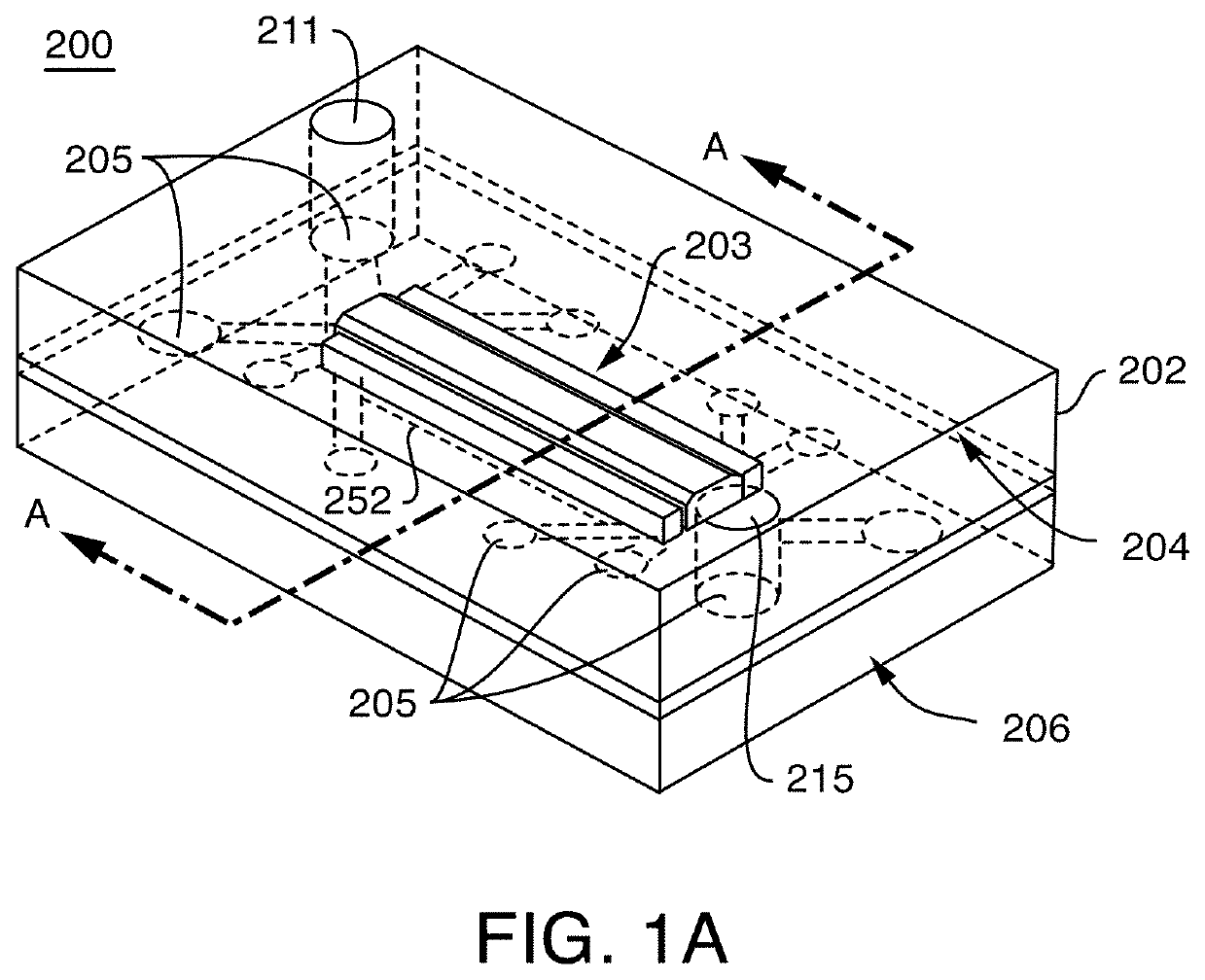
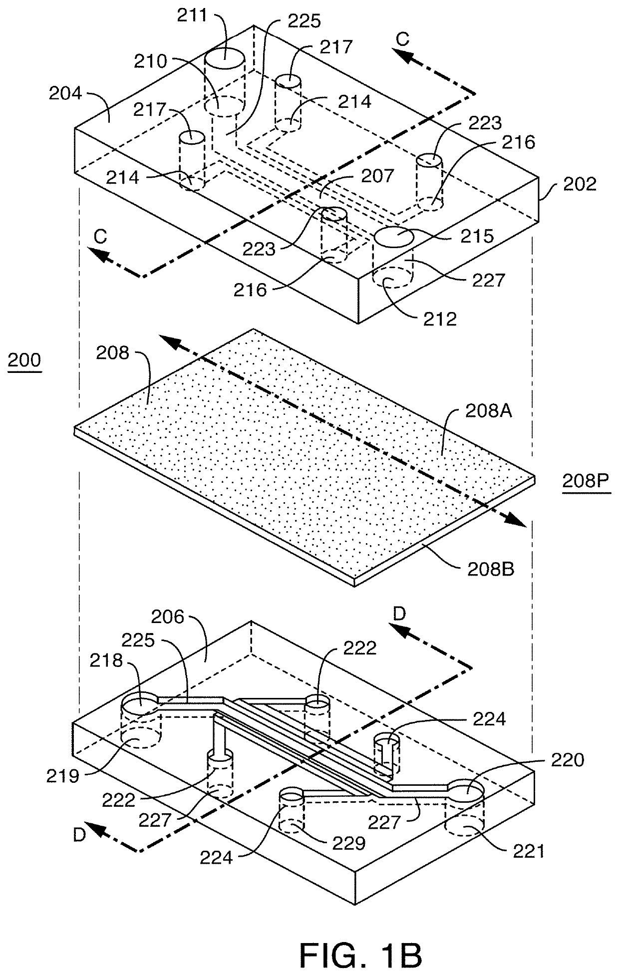
Microfluidic proximal tubule kidney-on-chip
a microfluidic and proximal tubule technology, applied in artificial cell constructs, laboratory glassware, instruments, etc., can solve the problems of not being scalable to a predictive clinical outcome, and based models often fail to predict renal transporter activity
- Summary
- Abstract
- Description
- Claims
- Application Information
AI Technical Summary
Benefits of technology
Problems solved by technology
Method used
Image

Examples
example a-exemplary
Cell Sources and Culture Media
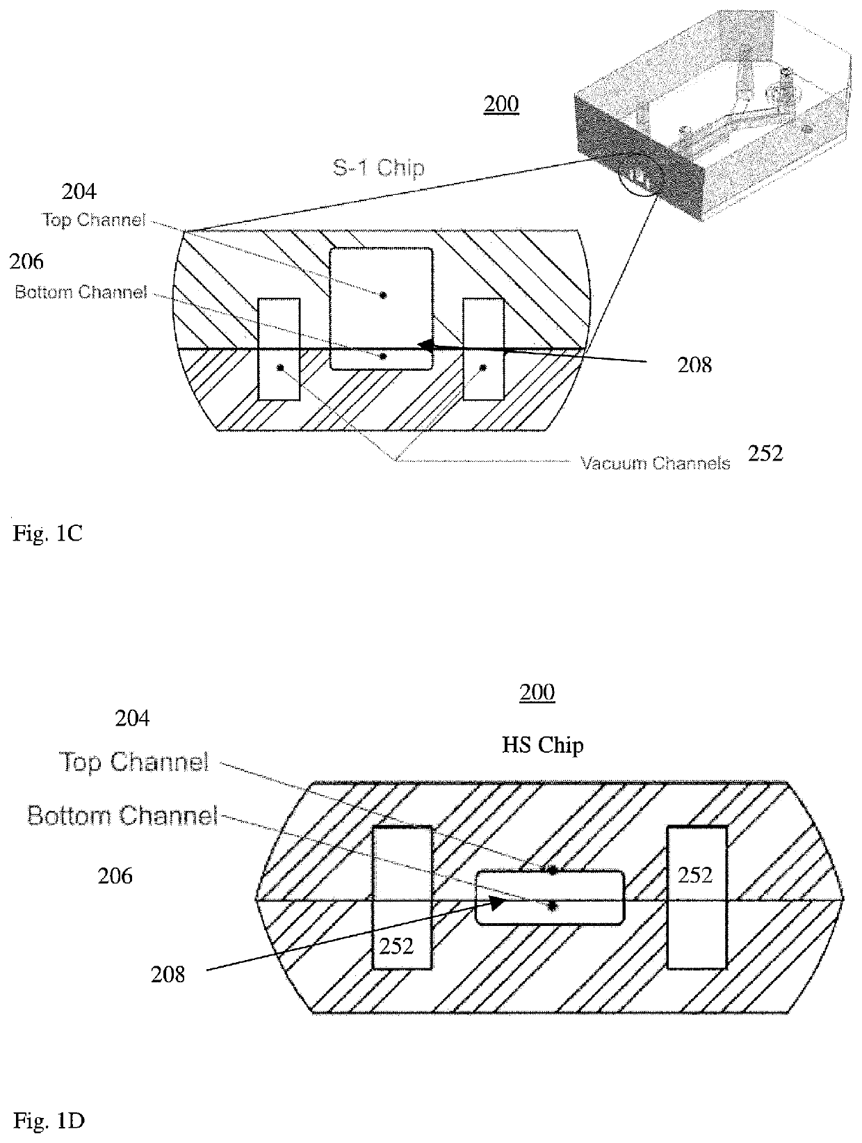
[0148]Exemplary Cells: Top channel-human Renal Proximal Tubule Epithelial Cells (hRPTECs), e.g. Lonza, RPTEC # CC-2553); and Bottom channel-Primary Human Glomerular microvascular Endothelial cells (Cell Systems. ACBRI 128), expand to P7 (e.g. passage 7). Additional examples of commercial cell sources include P3 Proximal Tubular (PT) cells from Biopredic International (www.biopredic.com); P1 PT cells from Lonza (www.lonza.com); etc. In one embodiment, Proximal tubule cells from the Lonza showed better morphology compared to Biopredic, i.e. more cubical shapes, so a method was developed for evaluating commercial cell sources, e.g. for choosing one lot of cells for a group of experiments, see FIG. 10A. Although human PT cells from Lonza were used for many of the experiments described herein, however other cell sources were used in experiments as labeled.
Exemplary PT Kidney Cell Media Formulations:
[0149]Media. Renal Epithelial Growth Medium (REGM™ Lonza, CC...
example b-exemplary
PT Kidney-Chip Protocols (Methods) for Seeding Co-Cultures on the Same Day
[0153]In one embodiment, both hRMVECs and hRPTECs are sequentially added to (seeded into) a microfluidic Device, i.e. Chip, on the same day, e.g. Day 0.
[0154]Expand cell numbers of kidney glomerular endothelial cells “HGMVEC: hHGMVEC” or Renal Microvascular Endothelial Cells “hRMVECs” for 2-3 days. Expand cell numbers of PT kidney Cells for 3-4 days. See Example C for additional information. See, Example D for preparing cells.
Preparing for Seeding Chips:
[0155]1. Transfer ECM-coated chips, within closed and sterile 150 mm Petri dishes, from an incubator into the biosafety cabinet (BSC), for maintaining sterility upon removing the Petri dish cover.[0156]2. Fully aspirate ECM from both channels.[0157]3. Pipette 200 μL of warm complete hRMVEC maintenance medium to the bottom channel of each chip. Wash the channel by aspirating the outflow, while leaving media in the channel.[0158]4. Pipette 200 μL of warm complete...
example c-exemplary
PT Kidney-Chip Protocols (Methods) for Seeding Co-Cultures on Separate Day
[0169]In another embodiment, hRMVECs and hRPTECs are added to, i.e. seeded into, a microfluidic device on separate Days, e.g. Day-1 and Day 0; Day 0 and Day 1. See example B for additional information.
[0170]One embodiment of a brief exemplary timeline is described as: Day-2: Chip coating; Day-1: Seeding endothelial cells; Day 0: Seeding proximal tubule epithelial cells; Day 0-7: Maintain chips; Day 7: Start Experiment (Study), e.g. 72 hours; and Day 10: End 72 hour Experiment (Study). Exemplary readouts include but are not limited to: Morphology, phase contrast microscopic images; immunohistology, immunofluorescent images; barrier function (in particular for kidney-chips, etc.); gene expression; and Troponin 1 release (in particular for heart-chips, i.e. cardiac-chips).
[0171]A more detailed exemplary timeline, e.g. (proximal-tubule) Kidney-chip is described herein.
Day-4 or Day-3: Human Primary Proximal Tubular...
PUM
 Abstract
Description
Claims
Application Information
 - R&D
- Intellectual Property
- Life Sciences
- Materials
- Tech Scout
- Unparalleled Data Quality
- Higher Quality Content
- 60% Fewer Hallucinations
Browse by: Latest US Patents, China's latest patents, Technical Efficacy Thesaurus, Application Domain, Technology Topic, Popular Technical Reports.
© 2025 PatSnap. All rights reserved.Legal|Privacy policy|Modern Slavery Act Transparency Statement|Sitemap|About US| Contact US: help@patsnap.com