
Medical analysis device and cell analysis method

a technology of medical analysis and cell analysis, applied in measurement devices, laboratory glassware, instruments, etc., can solve the problems of limited types of cancer cells that can be captured, difficult to capture cancer cells, etc., and achieve the effect of reducing the adsorption or adhesion of other proteins

Active Publication Date: 2020-10-29
SUMITOMO RUBBER IND LTD +1
View PDF2 Cites 1 Cited by
  • Summary
  • Abstract
  • Description
  • Claims
  • Application Information

AI Technical Summary

Benefits of technology

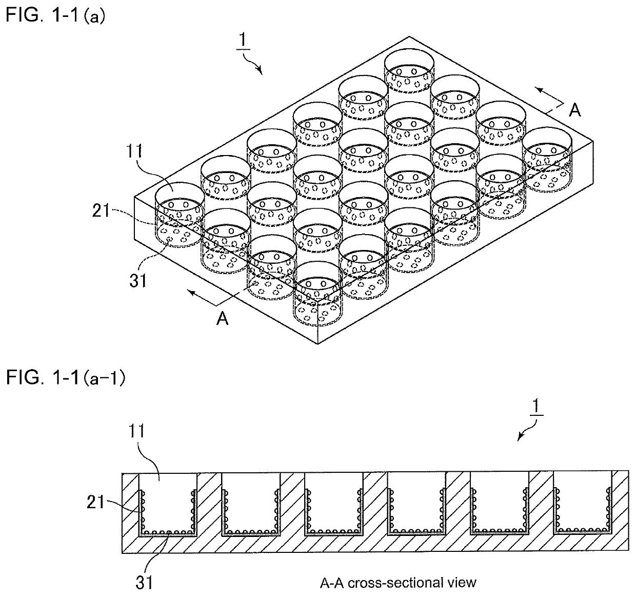
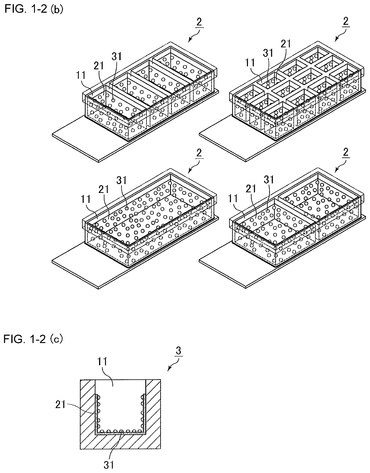
The medical analysis device described in this patent has a special layer on its inner surface that helps to capture specific cells like cancer cells and stem cells from blood or biological fluid. This layer is made of a special compound that reduces the sticking of other proteins and cells, allowing for the selective capture of cancer cells and stem cells.

Problems solved by technology

Unfortunately, however, since the number of circulating tumor cells is very small (several to hundreds of cells / 1 mL of blood), such cancer cells are difficult to capture.
This technique, which involves an antigen-antibody reaction (capture by EpCAM antibody), can only capture cancer cells expressing EpCAM, and the types of cancer cells that can be captured are limited.

Method used


Image

  • Medical analysis device and cell analysis method
  • Medical analysis device and cell analysis method

Examples


example 1

[0085]A 0.25% by mass solution of “SIM6492.7” (Gelest Inc., 2-[methoxy(polyethyleneoxy)propyl]trimethoxysilane, CH3O-(CH2CH2O)6-9-(CH2)3Si(OCH3)3) in water / methanol (50% by mass / 50% by mass) was prepared.

[0086]The solution in an amount of 100 μL was injected into a chamber slide (slide glass part: soda-lime glass, chamber size: 20 mm×20 mm) and allowed to stand at room temperature (17 hours), followed by drying in vacuum at 50° C. for six hours and then washing with water. Thus, an analysis device in which a hydrophilic silane compound layer was formed was prepared.

example 2

[0087]An analysis device was prepared as in Example 1, except that the “SIM6492.7” (Gelest, Inc.) was changed to “SIM6492.72” (Gelest Inc., 2-[methoxy(polyethyleneoxy)-propyl]trimethoxysilane, CH3O-(CH2CH2O)9-12-(CH2)3Si (OCH3)3).

example 3

[0088]An analysis device was prepared as in Example 1, except that the “SIM6492.7” (Gelest, Inc.) was changed to “SIM6492.73” (Gelest Inc., 2-[methoxy(polyethyleneoxy)-propyl]trimethoxysilane, CH3O—(CH2CH2O)21-24-(CH2)3Si(OCH3)3).


PUM


Abstract

The present invention provides a medical analysis device and a cell analysis method, which can capture specific cells such as many types of cancer cells including cancer cells not expressing EpCAM and stem cells. Provided is a medical analysis device including a well portion, the well portion having a hydrophilic silane compound layer formed at least partly on the inner surface thereof.

Description

[0001]The present invention relates to a medical analysis device and a cell analysis method, which can capture specific cells present in blood or biological fluid, such as cancer cells and stem cells present in blood or biological fluid.BACKGROUND ART[0002]Cancer cells, when formed, are known to appear in due course in blood or biological fluid. Such cancer cells in blood are called “circulating tumor cells (CTCs)”. Thus, it has been expected that the circulating tumor cells may be analyzed, e.g., to evaluate the cancer-treating effect, predict prognosis life expectancy, predict the effects of anticancer drugs before administration, or examine treatment methods based on genetic analysis of cancer cells (see, Patent Literature 1).[0003]Unfortunately, however, since the number of circulating tumor cells is very small (several to hundreds of cells / 1 mL of blood), such cancer cells are difficult to capture.[0004]For example, the CellSearch System is known as a technique for capturing ci...

Claims


Application Information

Patent Timeline
Patent Type & Authority Applications(United States)
IPC IPC(8): B01L3/00
CPCB01L2300/161B01L2300/0829B01L3/502776B01L3/502715B01L3/5085B01L2300/163B01L3/5021G01N33/5005
Inventor MINAGAWA, YASUHISATANAKA, MASARUEMURA, HARUKASUTO, KAZUKI
Owner SUMITOMO RUBBER IND LTD
Learn More