Method of estimation physiological parameters using medical image data

a physiological parameter and image data technology, applied in the field of physiological parameter estimation based on medical image data, can solve the problems of consuming substantial time and human resources, fundamentally affecting simulation accuracy, and many manual steps to enable simulations, so as to improve patient outcomes, stable and uniform description of blood-tissue interaction, and accurate physiological parameters

Pending Publication Date: 2021-01-14
KONINKLJIJKE PHILIPS NV
View PDF6 Cites 2 Cited by
  • Summary
  • Abstract
  • Description
  • Claims
  • Application Information

AI Technical Summary

Benefits of technology

[0027]This avoids the problem of an inaccurate or degenerating mesh (caused by movement of tissue, as discussed above), since data points are each treated as individual physical particles with defined mass and density, rather than this physical information being embedded within a general mesh structure. In the latter case, degeneration of the mesh leads to breakdown of the physical model. Meshless frameworks are robust to this, making them better suited to scenarios in which large deformations of constraining structural features are common. Hence this makes them particularly suited for internal cardiac simulations, where deformations of the encompassing anatomical structure occurs with every heart cycle.
[0081]In this case, pressure compensation, making use for example of an expanded Poisson equation for pressure, may help to reduce fluctuations in the pressure in the simulation, and improve stability.

Problems solved by technology

A drawback of known simulation methods is that often several (manual) steps are necessary to enable simulations to be performed on the anatomical data, for example image registration, and segmentation of the images.
Furthermore, in many cases, expertise of a specialist is required to prepare, perform, and post-process such simulations, which consumes substantial time and human resources.
This strong requirement on integrity of the structure information is problematic since even a very detailed 3D image segmentation may contain geometrical imperfections, which will fundamentally undermine the simulation accuracy.
Derived parameters are therefore not reliable.
This often involves skills of a professional engineer, and may take a long time depending on how severe imperfections in the segmentations are.
A further difficulty in known simulation or modeling techniques, in particular in the case of cardiovascular applications, is the typical (relative) low frame rate (frame-per second rate) of registered image sequences, i.e. the low temporal resolution of the image sequence.
Due to this lower frame-per-second (FPS) rate, synchronizing the tissue motion, e.g. left ventricle contraction upon opening and closing of the aortic and mitral valves can be difficult.
Finally, and as mentioned above, a further problem is the frequent, severe deformation of the tissue structure in certain anatomical areas.
This naturally results in a repeated and relative large change in the domain topology, for instance during valve opening.
This is a non-trivial procedure and can be complex and time consuming to perform.
While these techniques assist in resolving some of the above mentioned problems, they will still require finer meshing in the regions of fluid jet development, and also require domains substantially larger than the original geometrical morphology.
Imperfections in the segmentation lead to breakdown in the simulation technique.
In view of this instability of the simulation technique, the clinical workflow may often require adapting, which is inconvenient and inefficient for the clinician.
Significant deformations in the biological tissue structure (e.g. opening of heart valves, or motion of the aorta for instance) are difficult to account for as they alter the topology of the tissue structure.

Method used


Image

  • Method of estimation physiological parameters using medical image data
  • Method of estimation physiological parameters using medical image data

Examples


Embodiment Construction

[0106]The various embodiments will be described with reference to the Figures.

[0107]It should be understood that the detailed description and specific examples, while indicating exemplary embodiments of the apparatus, systems and methods, are intended for purposes of illustration only and are not intended to limit the scope of the the embodiments. These and other features, aspects, and advantages of the apparatus, systems and methods described herein will become better understood from the following description, appended claims, and accompanying drawings. It should be understood that the Figures are merely schematic and are not drawn to scale. It should also be understood that the same reference numerals are used throughout the Figures to indicate the same or similar parts.

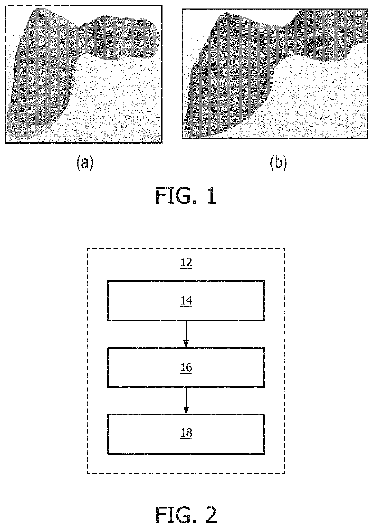
[0108]The various embodiments may provide a method of estimating one or more physiological parameters, in particular of a cardiac region, based on medical imaging data. An input time series of three-dimensional med...


PUM


Abstract

A method (12) of estimating one or more physiological parameters, in particular of a cardiac region, based on medical imaging data. An input time series of three-dimensional medical images is received (14), representative of a cardiac region of a patient. A meshless simulation frame-work is applied (16) for simulating blood flow through at least a portion of the anatomical area captured by the images. The simulation framework comprises in particular simulating force interactions between individual elements of a simulated fluid (representing blood) and individual patches of a tissue structure of the imaged cardiac region. One or more physiological parameters (18) are derived based on the simulated blood flow.

Description

RELATED APPLICATIONS[0001]This application claims the benefit of and priority to both U.S. Provisional Application No. 62 / 643,218, filed Mar. 15, 2018 and U.S. Provisional Application No. 62 / 746,633, filed Oct. 17, 2018, which are incorporated by reference herein.FIELD OF THE INVENTION[0002]This invention relates to estimating physiological parameters based on medical image data, in particular based on application of a simulation framework to medical image data.BACKGROUND OF THE INVENTION[0003]In the field of medical diagnostics and, in particular, cardiac diagnostics, there is a growing demand for automatic quantification of (e.g. hemodynamic) parameters from medical images. Biophysical models and simulation techniques or frameworks can provide a useful tool to assist in this. Not only can modeling and simulation help in automating and streamlining parameter measurement, in many cases, parameters not directly measurable from a medical image can be derived based on the models. There...

Claims


Application Information

Patent Timeline
Patent Type & Authority Applications(United States)
IPC IPC(8): G16H30/40G16H50/20G06F30/28G06T17/20
CPCG16H30/40G06T17/205G06F30/28G16H50/20G16H20/40G16H40/63G16H50/50
Inventor MAESSEN, RALPH THEODORUS HUBERTUSSHULEPOV, SERGEIBARAGONA, MARCO
Owner KONINKLJIJKE PHILIPS NV
Why Eureka
Try Eureka