Alpha-aminoadipate for treatment of vision loss and restoring sight

a technology of alpha-aminoadipate and vision loss, applied in the field of ophthalmology, can solve the problems of inability to address lost or degenerated cells, no treatment that stimulates the retina to regenerate new cells endogenously, and challenges and technical hurdles, and achieves the effect of increasing or restoring the activity/function of the photoreceptor and restoring vision

Active Publication Date: 2021-03-04
THE SCHEPENS EYE RES INST
View PDF0 Cites 0 Cited by
  • Summary
  • Abstract
  • Description
  • Claims
  • Application Information

AI Technical Summary

Benefits of technology

[0004]The present disclosure is based on the surprising discovery that alpha-aminoadipate (α-AA) stimulates proliferation and trans-differentiation of retinal cells to retinal progenitor cells, creates a retinal environment permissive for photoreceptor differentiation by progenitor cells, ultimately resulting in the increased or restored photoreceptor activity / function and restoration of vision.
[0005]Accordingly, compositions for preserving, restoring, or enhancing vision of a subject comprising alpha-aminoadipate or a functional analog thereof are described herein. Methods for preserving, restoring, or enhancing vision of a subject, comprising administering to an injured or diseased eye of said subject a composition comprising alpha-aminoadipate or a functional analog thereof are also described herein. The less invasive chemical- or drug-based strategy of mobilizing endogenous stem cells to repair neural damage presents many advantages over the transplantation approach including decreased risk of immune rejection, neuron integration, and tumor formation.
[0006]As α-AA treatment creates an environment permissive for cell integration and photoreceptor generation, also disclosed herein are compositions for enhancing the efficacy of cell replacement or transplantation in an injured or diseased eye of a subject to the eye of the subject prior to transplantation of exogenous cells. The replacement or transplanted cells are retinal cells, for example, retinal stem cells, retinal progenitor cells, or retinal stem cell-derived neural cells. The cells may be autologous or genetically engineered. As used herein, “enhancing efficacy” relates to increasing engraftment or integration of exogenous retinal cells, increasing photoreceptor activity, or increasing photoreceptor cells. For example, enhancement comprises at least 10%, 20%, 50% or 2-fold, 5-fold, 10-fold, or more of the number or activity of engrafted cells compared to the level in the absence of alpha-aminoadipate. The composition increases the mobility and migration of the endogenous retinal cells. The composition is administered prior to, concurrently with, or after cell replacement or transplantation. Methods for enhancing efficacy of retinal cell replacement in an injured or diseased eye of a subject, comprising administering a composition comprising alpha-aminoadipate or functional analog thereof to the eye of the subject prior to transplantation of exogenous retinal cells are also provided herein.
[0009]As used herein, preserving, restoring, or enhancing vision of a subject relates to increasing photoreceptor activity, increasing photoreceptor regeneration, increasing the number of retinal progenitor cells, increasing the number of retinal neurons, replacing lost or damaged retinal neurons or retinal cells, increasing light retinal response, increasing light perception / detection, increasing visual acuity, or increasing visual contrast. For example, increased number of retinal cells or photoreceptor activity comprises at least 10%, 20%, 50% or 2-fold, 5-fold, 10-fold, or more of the number or activity of engrafted cells compared to the level in the absence of alpha-aminoadipate. For example, the composition increases proliferation, migration, de-differentiation, or trans-differentiation of Muller cells, ciliary epithelial cells, retinal pigment epithelial cells, bone marrow-derived stem cells, mesenchymal stem cells, or iPS cells and iPS-derived retinal progenitor cells. As used herein, “de-differentiation” relates to the process in which a cell of a specific cell type (e.g., a Muller cell) loses at least one cell type-specific (e.g., Muller cell marker). As used herein, “trans-differentiation” relates to the process in which a cell of a specific cell type (e.g., a Muller cell) gains at least one marker of a different cell type (e.g., a marker of a retinal progenitor cell or a photoreceptor cell). Examples of markers for Muller cells include GFAP, CRALBP, and vimentin. Examples of markers for retinal progenitor cells include Nestin and Chx10. Examples of markers for retinal photoreceptor cells include recoverin and rhodopsin.
[0010]Methods for measuring or assessing increased photoreceptor activity, increased photoreceptor regeneration, increased number of retinal progenitor cells, increased the number of retinal neurons, replacement of lost or damaged retinal neurons or retinal cells, increased light retinal response, increased light perception / detection, or increased visual acuity are known in the art. For example, electroretinopathy, patch-clamp recording, spectral domain optical coherence tomography (SD-OCT), or visual evoked potential can be used. Electroretinography (ERG) analysis is a method known in the art for assessing photoreceptor function and neural responses. Advantages of this technique include non-invasiveness, and objective evaluation of retinal function on a layer-by-layer basis. In brief, the flash ERG is assessed in a dark adapted eye. The initial a-wave (initial negative deflection) is primarily derived from photoreceptors where the second half of the a-wave is a combination of photoreceptors, bipolar, amacrine, and Muller cells. The b-wave (positive deflection) originates in retinal cells that are post-synaptic to the photoreceptors and are used as a readout for photoreceptor function. Spectral domain optical coherence tomography (SD-OCT) is also used for detailed and non-invasive evaluation of the retinal architecture in vivo. SD-OCT accurately reflects retinal morphological changes that occur during retinal disease progression, including retinal detachment.

Problems solved by technology

The burden of mortality and morbidity that vision loss and blindness impose on society makes it a pressing public health and medical problem.
For example, anti-VEGF therapy for age-related macular degeneration (AMD) targets abnormal ocular vascularization and eye drops that lower intraocular pressure for treating glaucoma, but such therapies fail to address the lost or degenerated cells.
To date, there are no treatments that stimulate the retina to regenerate new cells endogenously.
Retinal cell transplantation as a therapeutic strategy to replace lost cells is emerging as a promising approach, however challenges and technical hurdles still remain.

Method used


Image

  • Alpha-aminoadipate for treatment of vision loss and restoring sight
  • Alpha-aminoadipate for treatment of vision loss and restoring sight
  • Alpha-aminoadipate for treatment of vision loss and restoring sight

Examples


example 1

Disruption of Retinal Glial Structure by α-AA

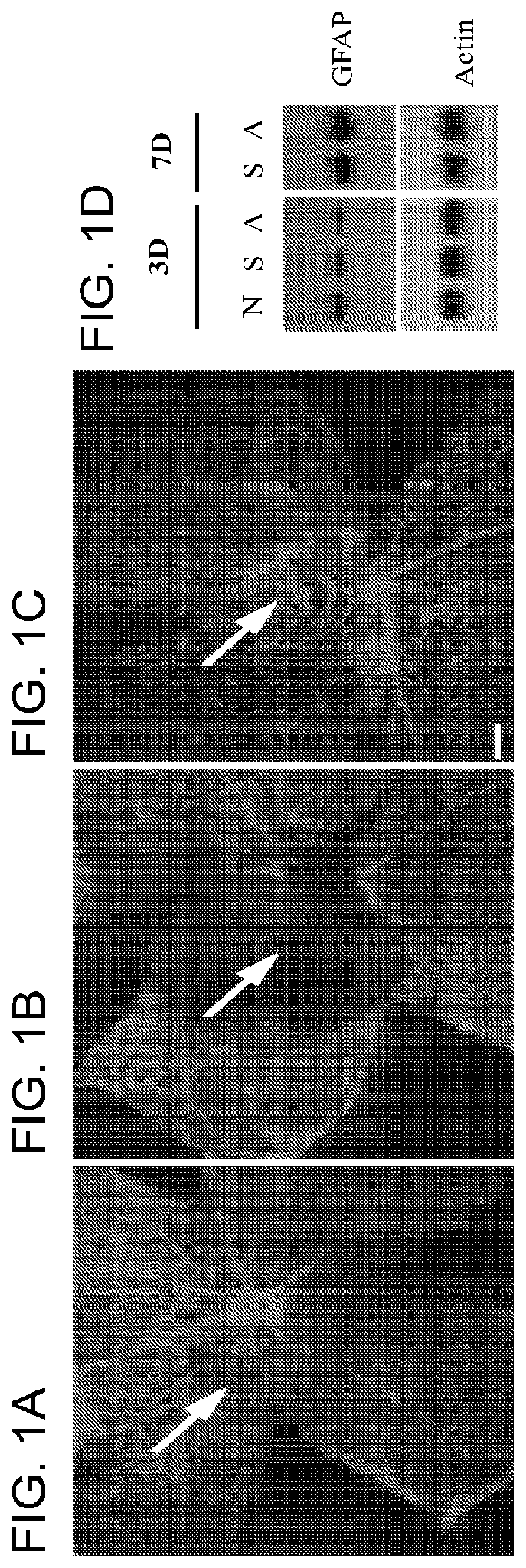
[0099]Retinal astrocytes and Müller glia are central to the homeostatic and metabolic support of retinal neurons; whereas, after injury these cells develop glial scar that forms critical barriers to neuron regeneration and integration by retinal progenitor cells. A solution of α-AA (1 μg / μl) was injected into the subretinal space of adult wild-type mice. Retinal glial morphology was examined by immunolabeling for glial marker GFAP and assessed the level of GFAP expression by Western blot at day 1-7 post injection. Expression of GFAP is widely detected in the normal mouse retinas (FIG. 1a). A single administration of α-AA effectively eliminated GFAP expression around the injection site at 1-3 days post injection (FIG. 1b), indicating the disruption of the glial barrier structure which may lead to the permissive environment for neuron regeneration and integration by retinal progenitor cells. By day 7, however, GFAP immunostaining reappeared...

example 2

afted Cell Integration into α-AA-treated Host Retinas

[0101]If transient disruption of retinal glia with α-AA promotes morphological integration of grafted retinal progenitors in vivo was next investigated. To distinguish implanted cells from the host environment, retinal progenitor cells were isolated as previously described using P0 mouse pups expressing an enhanced green fluorescent protein (EGFP) transgene driven by a chicken β-actin promoter and cytomegalovirus enhancer. At day 2 following saline or α-AA subretinal injection, retinal cells from P0 EGFP mice were injected into the subretinal space of adult wild-type hosts. Three weeks post transplantation, most donor-derived progenitor cells remained at the injection site and failed to migrate or incorporate into the host retina of the untreated or saline-injected wild-type mice (FIG. 3). In contrast, in α-AA-treated mice, numerous grafted cells had migrated away from the injection site and spread widely into the host retinas. Th...

example 3

iation of Transplanted Retinal Progenitors in α-AA-Treated Host Retinas

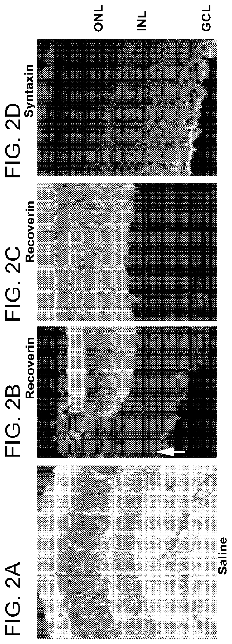
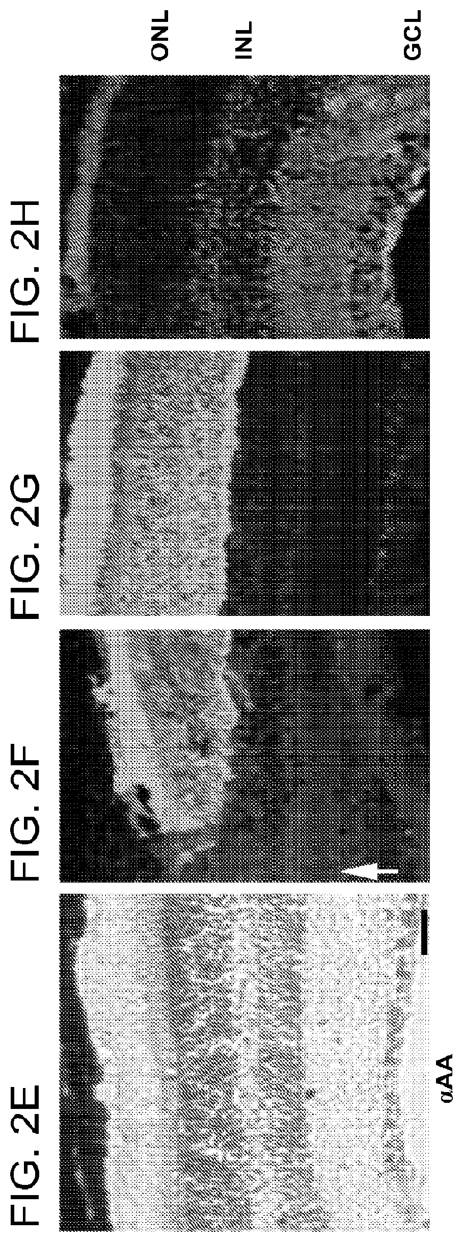
[0102]To determine whether α-AA-treated retina presents a supportive environment that allows appropriate differentiation of retinal progenitors, the behavior of transplanted cells was studied. The fate of transplanted cells was tracked using immunohistochemistry. At day 1 post transplantation, most EGFP+ cells clustered around the injection site and expressed a progenitor cell marker nestin (FIG. 4a-c). By 21 days post transplantation, grafted cells had integrated into the host, and became positive for mature glial cell marker, GFAP (FIG. 4d-g), or neuronal markers, Brn-3b (retinal ganglion cells; FIG. 4h-k) and PCKα (bipolar cells). Many of these cells were localized to the appropriate retinal layers where their corresponding host cells resided. Subretinal injection of α-AA resulted in the majority of grafted cells integrating into the outer nuclear layer (ONL) and expressed the mature photoreceptor cell markers...


PUM

PropertyMeasurementUnit
timeaaaaaaaaaa
pHaaaaaaaaaa
intraocular pressureaaaaaaaaaa

Abstract

The invention provides compositions and methods for preserving, restoring, or enhancing vision of a subject by administering compositions to an injured or diseased eye.

Description

CROSS REFERENCE TO RELATED APPLICATIONS[0001]The present application is a continuation application of U.S. patent application Ser. No. 15 / 029,507, filed on Apr. 14, 2016, now U.S. Pat. No. 10,780,070 issued on Sep. 22, 2020, which is a U.S. national phase application of PCT / US2014 / 061381, filed on Oct. 20, 2014, which claims priority to U.S. Provisional Application No. 61 / 892,822 filed Oct. 18, 2013 and incorporates the disclosure of each of the applications herein by reference in its entirety.FIELD OF THE INVENTION[0002]This invention relates generally to the field of ophthalmology.BACKGROUND OF THE INVENTION[0003]The burden of mortality and morbidity that vision loss and blindness impose on society makes it a pressing public health and medical problem. Current therapeutic strategies for treating loss of vision caused by retinal cell damage almost all have been directed to controlling the cause of the illness or neuron death rather than reversing retinal neuron damage or replacing ...

Claims


Application Information

Patent Timeline
Patent Type & Authority Applications(United States)
IPC IPC(8): A61K31/198A61K9/00A61K35/30A61K31/42
CPCA61K31/198A61K9/0019A61K9/0048A61K31/42A61K35/30A61K45/06
Inventor CHEN, DONG FENGCHO, KIN-SANG
Owner THE SCHEPENS EYE RES INST
Why Eureka
Try Eureka