Method of inhibiting the proliferation of t and/or b lymphocytes via dimethylarginine derivatives and lymphocytes proliferation inhibitor

a technology of dimethylarginine derivatives and inhibitors, which is applied in the field of immunotherapy, can solve the problems of complicated mechanisms of autoimmune diseases and the method of life-long treatment, and achieve the effect of effectively treating arthritis and effectively treating diseases

Active Publication Date: 2021-07-01
GUANGZHOU MEDICAL UNIV
View PDF0 Cites 0 Cited by
  • Summary
  • Abstract
  • Description
  • Claims
  • Application Information

AI Technical Summary

Benefits of technology

[0008]In view of the above-mentioned technical problems in the prior art, the present invention has discovered that dimethylarginine derivatives are effective inhibitors of in vitro T cells and/or B cell

Problems solved by technology

The existing treatment methods for autoimmune diseases are mostly used for specific types of diseases, such as autoimmune liver disease, rheumatoid arthritis and a

Method used


Image

  • Method of inhibiting the proliferation of t and/or b lymphocytes via dimethylarginine derivatives and lymphocytes proliferation inhibitor
  • Method of inhibiting the proliferation of t and/or b lymphocytes via dimethylarginine derivatives and lymphocytes proliferation inhibitor
  • Method of inhibiting the proliferation of t and/or b lymphocytes via dimethylarginine derivatives and lymphocytes proliferation inhibitor

Examples


embodiment 1

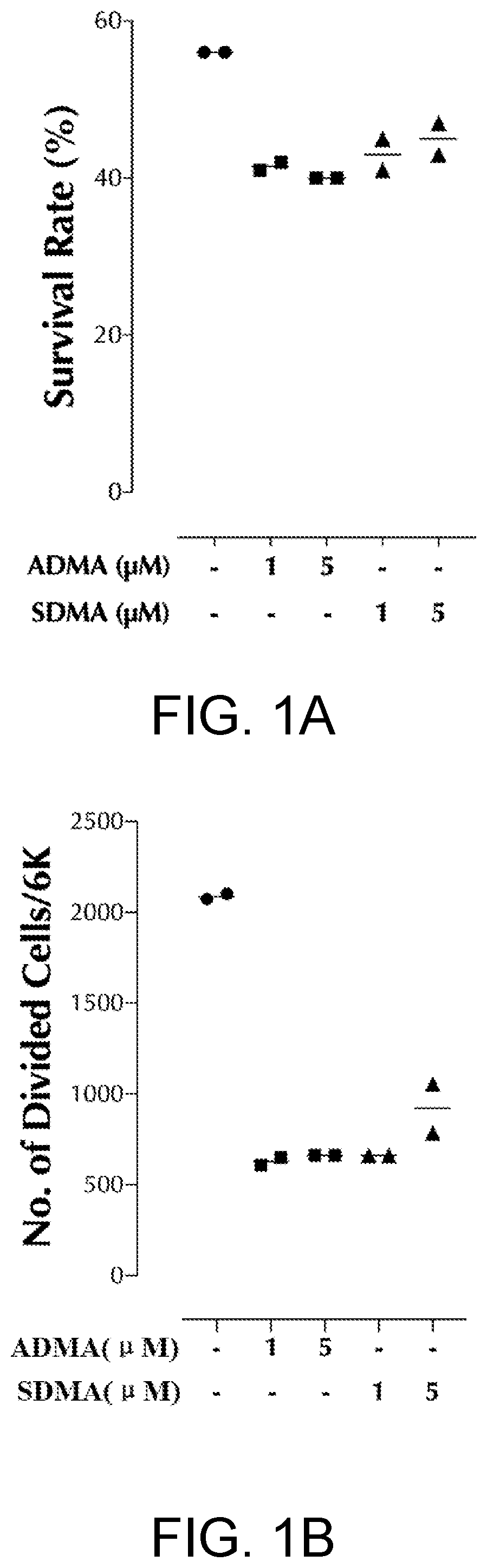
ADMA and SDMA Inhibit Mouse T Cell Proliferation

[0060]Step 1. Mouse T Cells Isolation

[0061]Total splenic T cells were isolated by StemCell isolation kit: From female 6-8 weeks old C57BL mice, spleens were harvested, mechanically disrupted, and incubated with a biotinylated antibody cocktail consisting of antibodies to B220, CD11b, CD11c, CD16 / 32, CD19, CD25, CD105, NK1.1, and TCRγδ (StemCell). After a wash step, cells were incubated with streptavidin-coated magnetic nanoparticles (StemCell). CD3+ cells were then isolated by applying a magnetic field and collecting the unbound cells.

[0062]Step 2. Inhibition of ADMA and SDMA on Mouse T Cells Proliferation

[0063]The total T cells obtained in step 1 were labeled with the proliferation indicator dye CellTrace Violet according to the manufacturer's protocol, and then seeded in 96-well round bottom cell culture plates at 2×10⁵ per well, next the cells were stimulated with anti-CD3 and anti-CD28 antibody coated magnetic beads (Gibco Dynabead...

embodiment 2

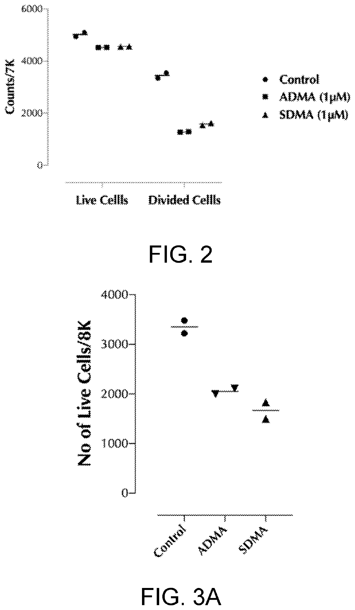
ADMA and SDMA Inhibit Human T Cells Proliferation

[0066]Human T cell isolation and in vitro proliferation experiments: the use of blood from healthy adult blood donors was approved by the Ethics Committee of the Medical Faculty of Guangzhou Medical University, Guangzhou, China. Peripheral blood mononuclear cells (PBMCs) were isolated using Sepmate PBMC isolation tubes following the manufacturer's instructions (StemCell). CD3+ human T cells were positively isolated by magnetic cell sorting (Miltenyi Biotec, Bergisch-Gladbach, Germany). Purified T cells were labeled with the proliferation dye CellTrace Violet according to the manufacturer's protocol. Then the cells (1×10⁵ cells per well in flat bottom 96 well plate) were stimulated in vitro with immobilized anti-CD3 (5 μg / ml) and anti-CD28 (2 μg / ml) antibody (Biolegend, San Diego, USA), and expanded in RPMI1640 medium up to 5 days. Dilution of CellTrace Violet, indicating proliferation, was then evaluated by flow cytometry.

[0067]The ex...

embodiment 3

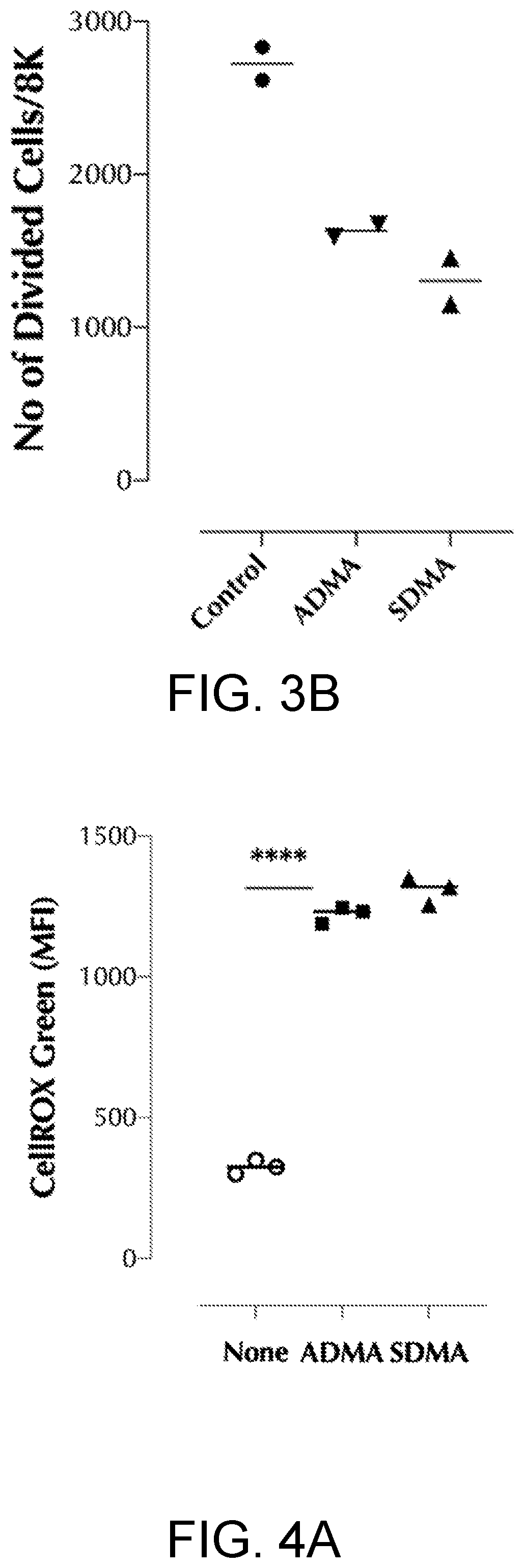
ADMA and SDMA Inhibit Mouse B Cells Proliferation

[0068]Step 1. Isolation of Total Splenocytes

[0069]Spleens were harvested, mechanically disrupted, and resuspend in RPMI 1640 complete medium (10% FBS).

[0070]Step 2. B Cells Proliferation Assay

[0071]Total splenocytes were then labeled with the proliferation dye CellTrace Violet according to the manufacturer's protocol. Then splenocytes (1×10⁵ cells per well in flat bottom 96 well plate) were stimulated with recombinant human IL-4 (1 Ong / me and α-mouse IgM (10 μg / ml), and expanded in RPMI1640 medium up to 5 days. Dilution of CellTrace Violet was then evaluated by flow cytometry. (Note; splenocytes contain T cells and monocytes, which will die off during the assay period.)

[0072]Experimental group: ADMA or SDMA (5 μM) was added (ADMA and SDMA dissolved in sterile endotoxin-free water, the storage solution concentration is 0.5 mM). Control group: the same volume of sterile endotoxin-free water without ADMA or SDMA was added.

[0073]The exper...


PUM

PropertyMeasurementUnit
Molar densityaaaaaaaaaa
Concentrationaaaaaaaaaa

Abstract

The present invention belongs to the technical field of immunotherapy. A method of using dimethylarginine derivatives to inhibit the proliferation of lymphocyte. ADMA, SDMA, and their derivative DMGV can inhibit T cells and/or B cells proliferation in vitro by inducing mitochondrial ROS. ADMA and SDMA can induce mitochondrial ROS generation by the action of alanine-glyoxylate aminotransferase 2 (AGXT2), which converts both ADMA and SDMA into dimethylguanidino valeric acid (DMGV) in the mitochondria. The dimethylarginine dimethylamino hydrolase (DDAH) inhibitor PD404182 also inhibits T cell proliferation by allowing more ADMA and SDMA to be converted to DMGV. As a clinical relevance, DMGV effectively treated arthritis in mice which was caused by activated T cells.

Description

CROSS-REFERENCE TO RELATED APPLICATION[0001]This application claims the priority from Chinese Patent Application serial no. 201911366970.2, filed on Dec. 26, 2019. The entirety of the above-mentioned patent application is hereby incorporated by reference herein and made a part of this specification.FIELD OF THE INVENTION[0002]The present invention relates to the field of immunotherapy, in particular to a method of inhibiting the proliferation of T and / or B lymphocytes via dimethylarginine derivatives and lymphocytes proliferation inhibitor.BACKGROUND OF THE INVENTION[0003]Immunity is human's physiologic function. The human body relies on the function to differentiate “self” and “non-self” components in order to eliminate non-self antigens that enter the human body, or the damaged cells and tumor cells produced by the human body so that homeostasis, the state of resisting or preventing the infection of microorganisms, parasites or other undesirable organisms, is maintained.[0004]The ...

Claims


Application Information

Patent Timeline
IPC IPC(8): A61K31/198A61P35/04
CPCA61K31/198A61P35/04A61K45/00A61P37/08A61P35/00A61P37/02A61P29/00A61P19/02A61P17/06A61P11/06A61P3/10
Inventor LEE, JONGDAEYANG, FENGYUANSHEN, LIYA
Owner GUANGZHOU MEDICAL UNIV
Why Eureka
Try Eureka