Methods and system for autonomous volumetric dental image segmentation

a volumetric dental image and segmentation technology, applied in the field of volumetric dental image segmentation processing in xray computed tomography, can solve the problems of difficult segmentation processing, significant constraint, and prove challenging, and achieve the challenge of error-free segmentation processing

a volumetric dental image and segmentation technology, applied in the field of volumetric dental image segmentation processing in xray computed tomography, can solve the problems of difficult segmentation processing, significant constraint, and prove challenging, and achieve the challenge of error-free segmentation processing

US20220012888A1Pending Publication Date: 2022-01-13CARESTREAM DENTAL LLC

Image

  • Methods and system for autonomous volumetric dental image segmentation
  • Methods and system for autonomous volumetric dental image segmentation
  • Methods and system for autonomous volumetric dental image segmentation

Examples


Embodiment Construction

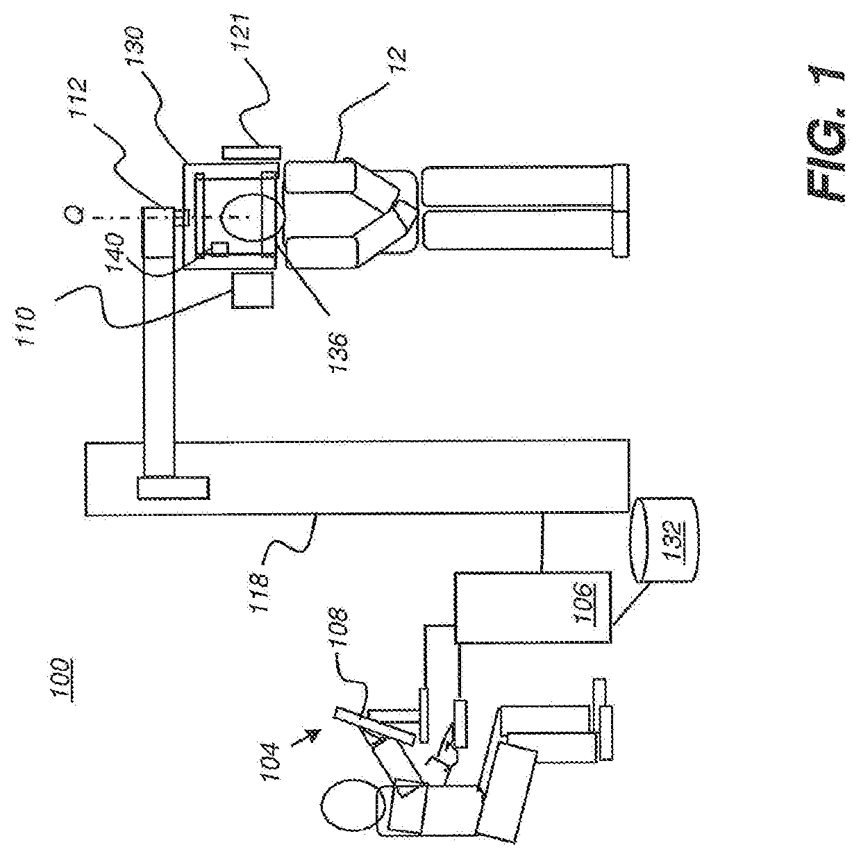
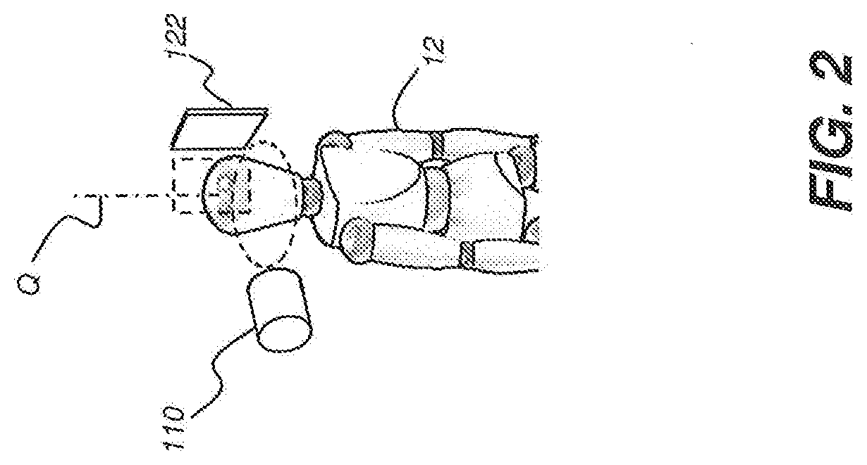
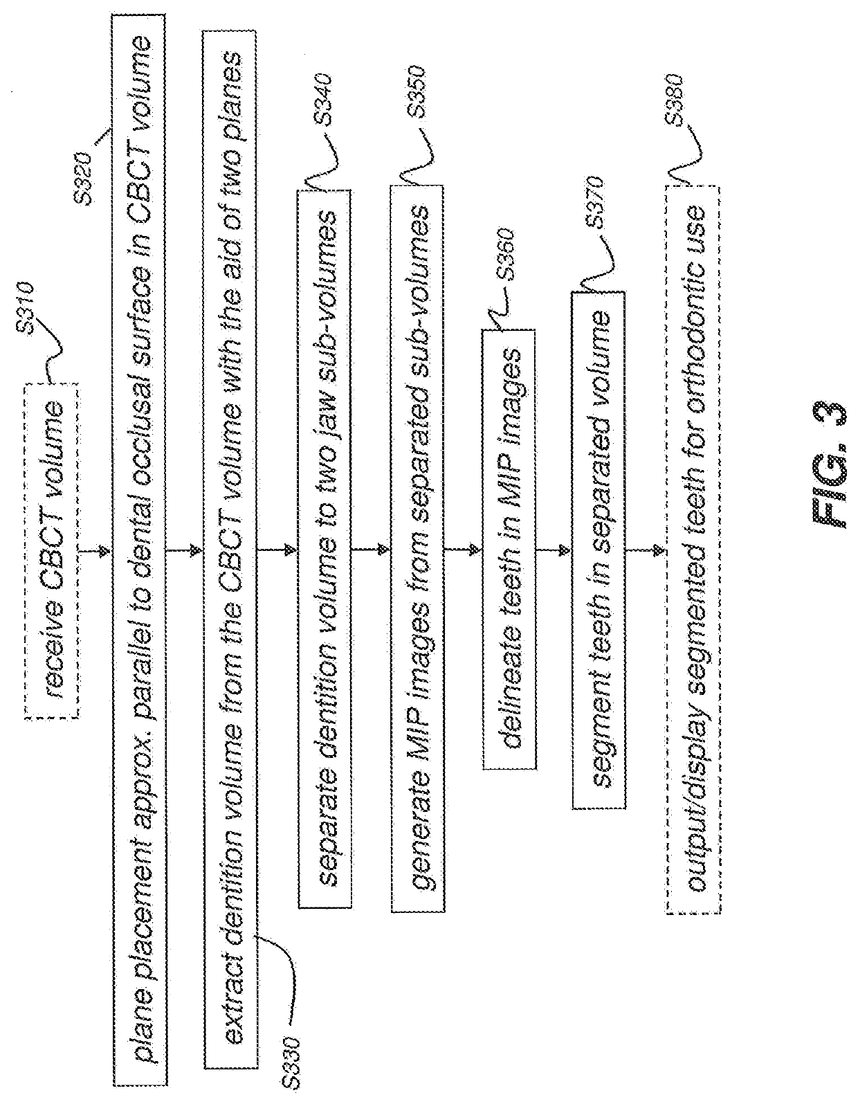
[0034]In the following detailed description of example embodiments of the present invention, reference is made to the drawings in which the same reference numerals are assigned to identical elements or steps in successive figures. It should be noted that these figures are provided to illustrate overall functions and relationships according to embodiments of the present invention and are not provided with intent to represent actual size or scale.

[0035]Where they are used in the context of the present disclosure, the terms “first”, “second”, and so on, do not necessarily denote any ordinal, sequential, or priority relation, but are simply used to more clearly distinguish one step, element, or set of elements from another, unless specified otherwise.

[0036]As used herein, the term “energizable” relates to a device or set of components that perform an indicated function upon receiving power and, optionally, upon receiving an enabling signal.

[0037]In the context of the present disclosure,...


PUM


Abstract

The present disclosure describes a system and methods for autonomous segmentation of volumetric dental images, such as those produced by an imaging system, The methods, implemented by the system, acquire a volume image of a patient and extract a volume of interest comprising patient dentition from the acquired volume image. A first plane is extended through maxillary portions of the patients jaw and a second plane through mandibular portions of the patients jaw. A maxillary sub-volume is generated from the volume of interest according to the first plane and a mandibular sub-volume from the volume of interest according to the second plane. Maximum intensity projection images are formed for each sub-volume and teeth are delineated from these images. Teeth are segmented within each sub-volume according to the tooth delineation for their respective sub-volume.

Description

FIELD OF THE INVENTION[0001]The present invention relates generally to volume image processing in x-ray computed tomography and, in particular, to image segmentation of a three-dimensional (“3D”) volume from digital Cone Beam Computed Tomography (“CBCT”).BACKGROUND[0002]Imaging and image processing for computer-aided diagnosis and improved patient care are areas of interest to dental practitioners. Among areas of particular interest for computer-aided diagnosis, treatment assessment, and surgery is image segmentation, particularly, for tooth regions.[0003]A three-dimensional or volume x-ray image can be of significant value for diagnosis and treatment of teeth and supporting structures. A volume x-ray image for this purpose is formed by combining image data from two or more individual two-dimensional (“2D”) projection images, obtained within a short time of each other and with a well-defined angular and positional geometry between each projection image and the subject tooth and betw...

Claims


Application Information

Patent Timeline
13 Jan 2022
Publication
US20220012888A1
IPC
G06T7/11; G06T7/12; G06T7/00; G06T5/30; A61B6/14; A61C7/00; G16H30/40; G16H50/20; G16H30/20
CPC
G06T7/11; G06T7/12; G06T7/0012; G06T5/30; A61B6/14; A61C7/002; G06T2207/20161; G16H50/20
Inventors
CHEN, SHOUPU; YE, WEI