
Method for Restoring Immune Tolerance In Vivo

a technology of immune tolerance and recombinant proteins, applied in the field of restoring immune tolerance in vivo, can solve the problems of long-term use of immuno-suppressing medications, affecting the immune system, and causing tissue scarification, so as to achieve the effect of restoring immune tolerance and restoring immune toleran

Pending Publication Date: 2022-04-07
AMARNA HLDG
View PDF0 Cites 0 Cited by
  • Summary
  • Abstract
  • Description
  • Claims
  • Application Information

AI Technical Summary

Benefits of technology

The present invention provides a method for restoring immune tolerance in vivo by using a recombinant gene encoding auto-antigens or antigenic parts thereof under the control of a polyomaviral early and late promoter. This is achieved by introducing a polyomavirus DNA incapable of encoding a functional small T antigen into a cell line permissive to the wild-type polyomavirus and culturing them to produce recombinant polyomaviral vector particles. The invention also provides a composition comprising recombinant polyomaviral vector particles that are safe for use in medical treatments as they are incapable of expressing a functional small T antigen. The invention also relates to a method for producing large quantities of polyomaviral vector particles without a single wild-type virus being present. The cell lines used in this invention are genetically modified to express a functional polyomaviral large T antigen and do not express a functional polyomaviral small T antigen.

Problems solved by technology

In addition, chronic inflammation results in amyloid plaque formation and tissue scarification due to collagen deposition.
Current treatments for patients with a degenerative or dystrophic disease non-specifically suppress inflammation and immunity, and thus only alleviate the symptoms and delay the progression to disabling stages.
Furthermore, long term use of immuno-suppressing medication coincides with often severe adverse side effects and enhances the risk of developing autoimmune processes in other tissues.
However, due to the fact that these proteins or peptides are not efficiently delivered to the proper antigen-presenting cells, that they lack the proper accompanying signals for reverting the immune response and / or that they are rapidly degraded in the body, such approaches have been adopted with limited success.
This DNA vaccination approach however, only works in mice.
However, recent clinical gene therapy studies conducted demonstrated that the organ-specific and host-derived promoters used to control transcription of the therapeutic genes are not potent enough to yield therapeutic levels of self-proteins in the treated patients (A. C. Nathwani et al., New England Journal of Medicine 365:2357-2365, 2011).
Although viral vector-based tolerization approaches to treat degenerative diseases using organ-specific and host-derived promoters driving expression of auto-antigens look promising, it has become obvious from the above described prior art that there is an unmet need for treatments that restore immune tolerance to the auto-antigens.
A major disadvantage of the currently used viral gene delivery vectors is the fact that they cannot be produced in sufficient amounts to treat significant numbers of patients.
As a consequence, these vectors can only be administered a single time to a patient, whereas the expression levels of the introduced therapeutic gene rapidly decline.
So far the successes with replicon-based expression systems have been limited.
Replicon systems based on RNA viruses in general produce recombinant proteins for only a short period of time, whereas those derived from DNA viruses in general do not replicate well in the commercially used cell lines.
There is an important disadvantage however to the use of such cell lines.
The emergence of the replication competent wildtype virus particles and the presence of the T antigen oncoproteins in such conventional packaging cell lines have made the use of SV40 vectors for medical purposes impractical.
Since the T antigen oncoproteins are present in HEK293T cells and there is a risk that replication competent SV40 viruses emerge, the use of this cell line for the production of SV40 vectors for medical purposes is undesired and impractical.
Since HEK293TT as a derivative of HEK293T accumulates the small and large T antigen oncoproteins and poorly supports SV40 replication, the use of this cell line to produce recombinant SV40 vectors for medical purposes is also undesired and impractical.
However, the packaging cell lines described herein still accumulate significant amounts of the small and large T antigen oncoproteins.
The packaging cell lines described in WO 08 / 000779 do not provide a solution to the disadvantages of the packaging cell lines of the prior art described herein above.
The level of replication in the CHOP cell lines was not sufficient to make this system attractive for commercial application, possibly due to a splicing bias in favour of the small T antigen or middle T antigen mRNA in CHO cells.

Method used


Image

  • Method for Restoring Immune Tolerance In Vivo
  • Method for Restoring Immune Tolerance In Vivo

Examples


example 1

Construction of the SV40 Derived Gene Delivery Vector

[0136]Six oligonucleotides were designed:

WdV101:(SEQ ID No. 2)CCGCTCGAGTTGCGGCCGCTGTGCCTTCTAGTTGCCAGCCATC

(containing a Xhol and a Notl restriction site) and

[0137]WdV102: GGTACCATAGAGCCCACCGCATCCCCAGCATGCC (SEQ ID No. 3) (containing a Kpnl restriction site) and

[0138]WdV103: GGCCGCTTTATTAATTAAGCCCTGCAGGTTGTTTAAACTTGGCGC GCCTTAT (SEQ ID. No. 4) (contains from 5′ to 3′ subsequently a Notl sticky restriction site, a Padl, SbfI, Pmel and an AscI intact restriction site and a ClaI sticky restriction site) and

[0139]WdV104: CGATAAGGCGCGCCAAGTTTAAACAACCTGCAGGGCTTAATTAAT AAAGC (SEQ ID No. 5) (contains from 3′ to 5′ subsequently a Notl sticky restriction site, a PadI, SbfI, PmeI and an AscI intact restriction site and a ClaI sticky restriction site) and

WdV105:(SEQ ID NO. 6)CGGGATCCAGACATGATAAGATACATTG

(containing a BamHI restriction site) and

WdV106:(SEQ ID No. 13)ATAGTTTAGCGGCCGCAACTTGTTTATTGCAGCTTATAATGG

(containing a Notl restriction site).

[0...

example 3

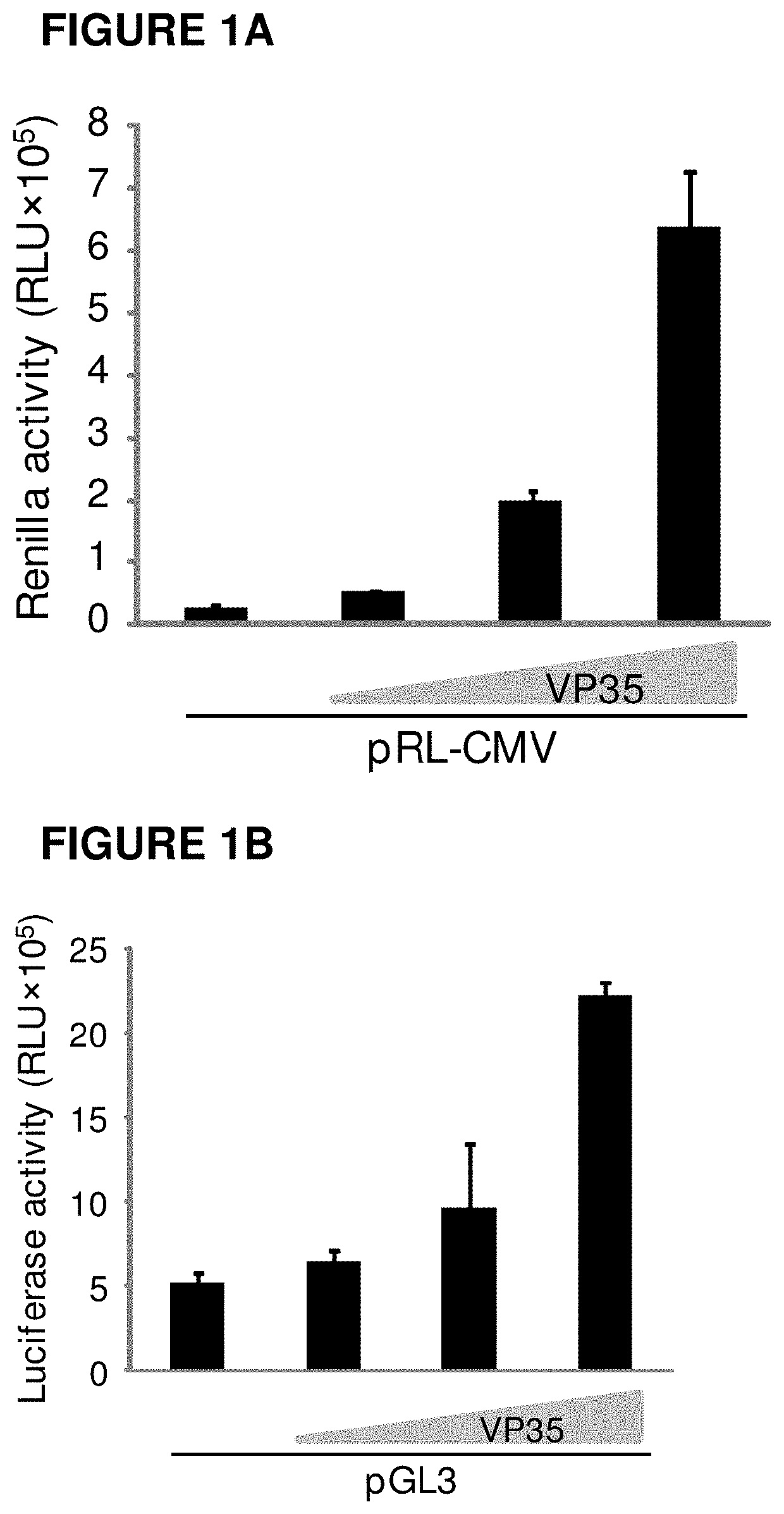
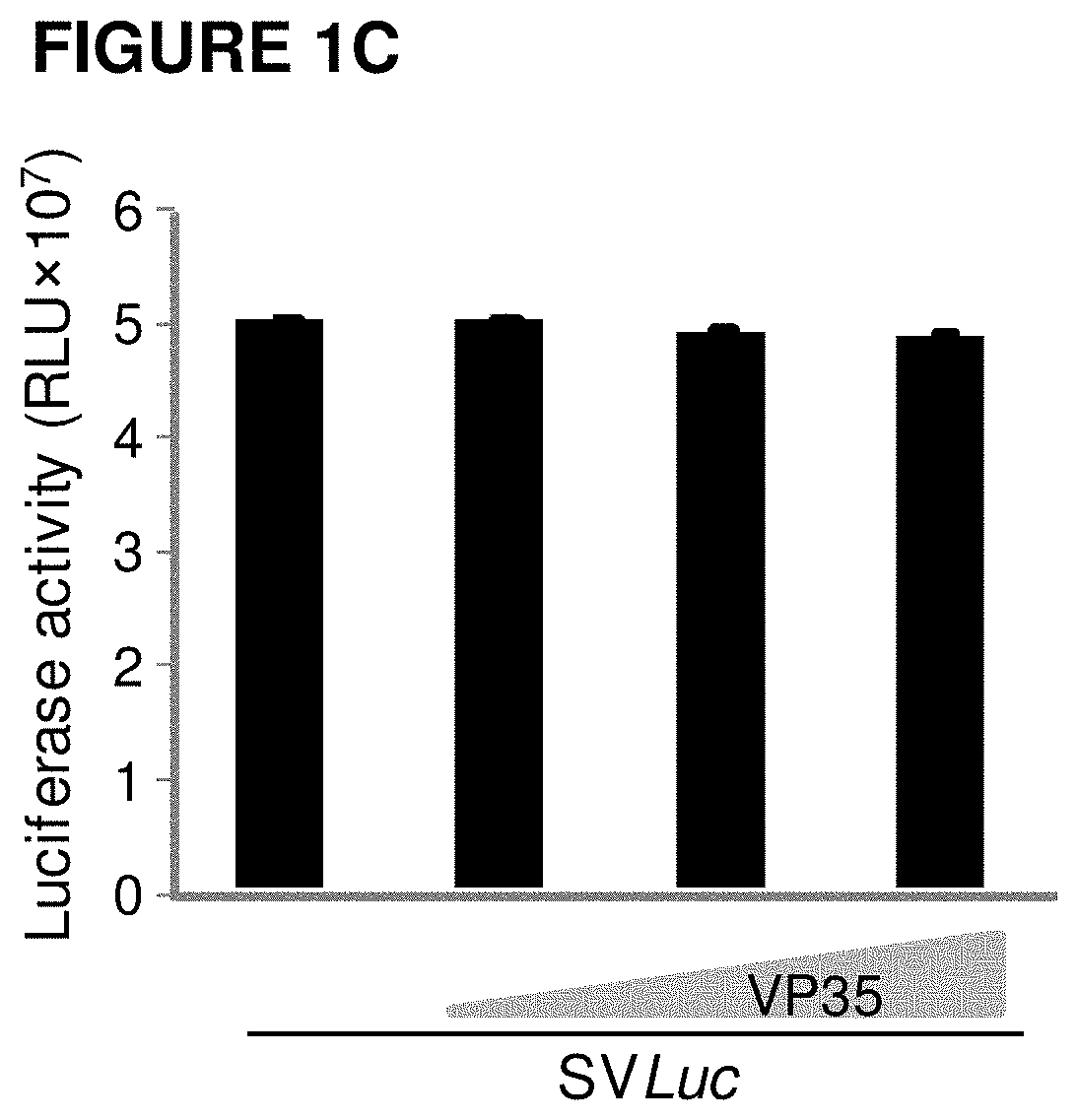
Molecular Cloning of an SV40 Encoding Luciferase Expression Vector (SVLuc)

[0152]The expression plasmid pGL3 (Promega) was used as template for cloning of the firefly luciferase (Photinus pyralis) into pSVac destination vector by PCR. Two oligonucleotides were designed:

[0153]WdV-070: GGGGACAAGTTTGTACAAAAAAGCAGGCTATGGAAGAC GCCAAAAACATAAAGAAAGGC (SEQ ID NO: 10) and

[0154]WdV-071: GGGGACCACTTTGTACAAGAAAGCTGGGTTTACACGGCGA TCTTTCCGCCCTTC (SEQ ID NO: 11) containing, respectively, AttB1 and AttB2 recombination sequences.

[0155]Purified pGL3 plasmid DNA was subjected to PCR using oligonucleotides WdV070 and WdV071. The purified PCR product was subsequently recombined by a BP reaction in pDONR221 (Invitrogen) to create a luciferase entry clone, pAM007. This entry clone was used for a GATEWAY® LR reaction to recombine the firefly luciferase coding sequence into pSVac-dest (pAM006), resulting in the pSVLuc vector plasmid (pAM008).

example 2

Molecular Cloning of a SV40 Luciferase Expression Vector and the Production of Recombinant SV40 Luciferase Vector Particles

[0156]The expression plasmid pGL3 (Promega) was used as template for cloning of the firefly luciferase using PCR. Two oligonucleotides were designed WdV389: 5′-TTGGCGCGCCATGGAAGACGCCAAAAACATAAAGAAAGGC-3′ (SEQ ID NO: 14) and WdV407: 5′-CCCTTAATTAATTACACGGCGATCTTTCCGCCCTTC-3′ (SEQ ID NO: 15) containing respectively restriction sites Ascl and PacI. The PCR amplified luciferase fragment was subsequently Ascl and PacI digested and ligated into pAM005, resulting in pAM006.

[0157]Two oligonucleotides were designed WdV437 5′ GGGATCCAGACATGATAAGATACATTG 3′ (SEQ ID NO: 16) and WdV442: ATAGTTTAGCGGCCGCAATGAATGCAATTGTTGTTGTTAACTTG (SEQ ID NO: 17) containing respectively BamHI and Notl restriction site. The pSL-PL vector was used as template for cloning of the large T antigen trailer sequence using PCR. The resulting PCR fragment was digested with BamHI and Notl and cloned in...


PUM

PropertyMeasurementUnit
Compositionaaaaaaaaaa

Abstract

The present invention provides a method for restoring immune tolerance in vivo. The invention relates to the use of a recombinant gene encoding auto-antigens or parts thereof for restoring immune tolerance to the auto-antigens in vivo, under transcriptional control of polyomaviral early and late promoters. In a preferred embodiment, the invention relates to the use of recombinant polyomaviral gene delivery vector particles, such as simian virus 40 (SV40) viral vector particles encoding one or multiple auto-antigens or parts thereof under transcriptional control of the SV40 early and late promoter, for restoring immune tolerance to the auto-antigens in vivo. The invention also relates to compositions comprising recombinant genes or polyomaviral vectors and uses thereof as treatment for degenerative or dystrophic diseases.

Description

CROSS-REFERENCE TO RELATED APPLICATIONS[0001]This application is a continuation-in-part U.S. application Ser. No. 15 / 038,433, Filed on May 20, 2016, published as US 2016 / 0287685 on Oct. 6, 2016, which is a national phase entry of International Patent Application PCT / EP2014 / 075346, filed Nov. 22, 2014, designating the United States of America and published in English as International Patent Publication WO 2015 / 075213 Al on May 28, 2015, which claims the benefit under Article 8 of the Patent Cooperation Treaty to European Patent Application Serial No. 14153144.2, filed Jan. 29, 2014, and to European Patent Application Serial No. 13194126.2, filed Nov. 22, 2013.TECHNICAL FIELD[0002]The present invention relates to improved methods for the production of viral particles, viral vectors, viral vector particles and recombinant proteins. In particular, the invention relates to improved methods for the production of recombinant polyomaviral vector particles and polyomaviral vector producer ce...

Claims


Application Information

Patent Timeline
IPC IPC(8): A61K39/00C07K14/005A61P37/02
CPCA61K39/0008A61K2039/5254A61P37/02C07K14/005C12N2710/22043C12N2710/22052C12N15/86C12N2710/22022A61K2039/55566Y02A50/30
Inventor DE HAAN, PETRUS THEODORUS
Owner AMARNA HLDG
Learn More