

Locally enhanced raman spectroscopy with an atomic force microscope
a raman spectroscopy and microscope technology, applied in the field ofatomic force microscopes, can solve the problems of complicated interfaces or limited chemical information, and achieve the effect of enhancing raman scattering and raman signal
- Summary
- Abstract
- Description
- Claims
- Application Information
AI Technical Summary
Benefits of technology
Problems solved by technology
Method used
Image

Examples
Embodiment Construction
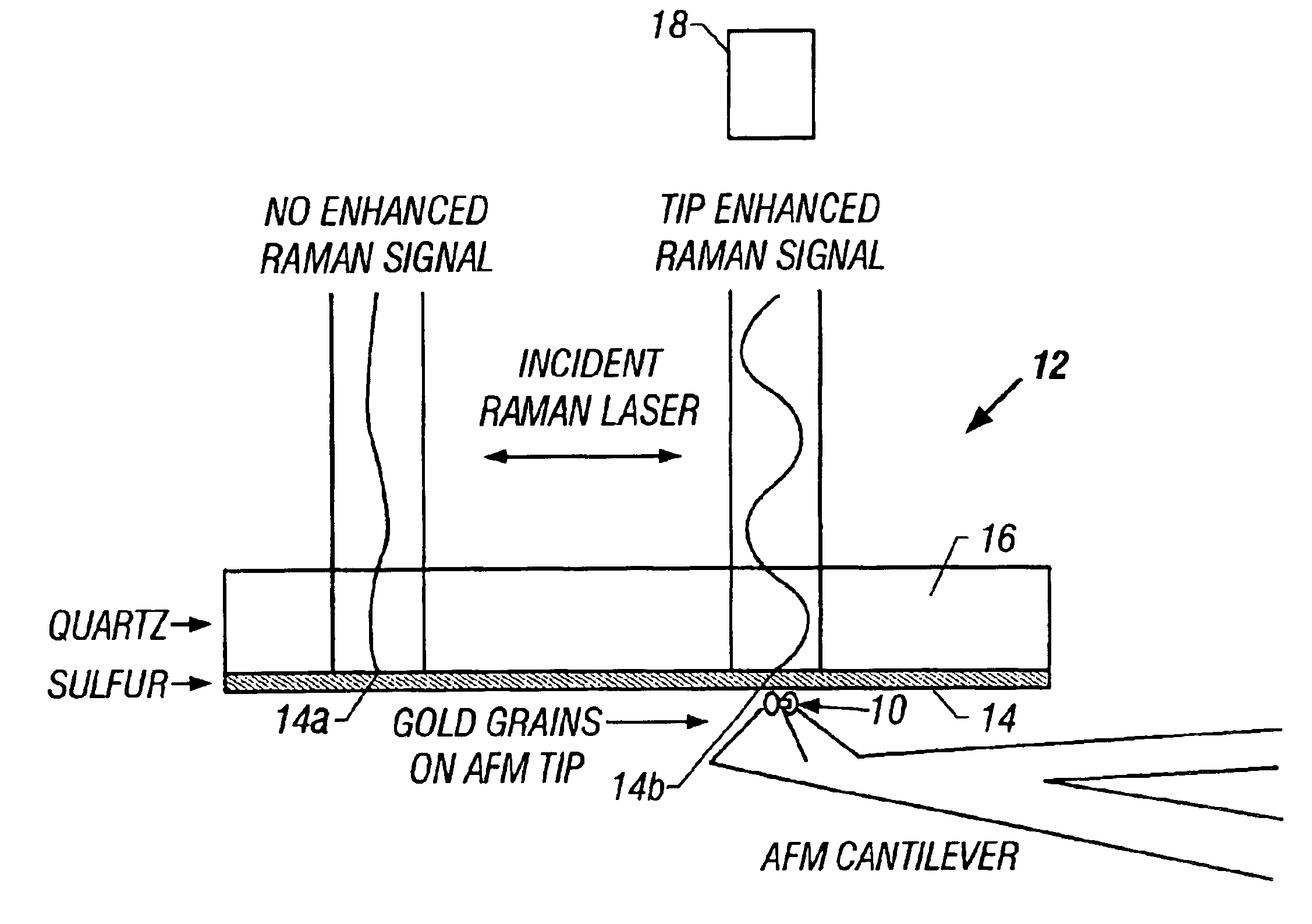
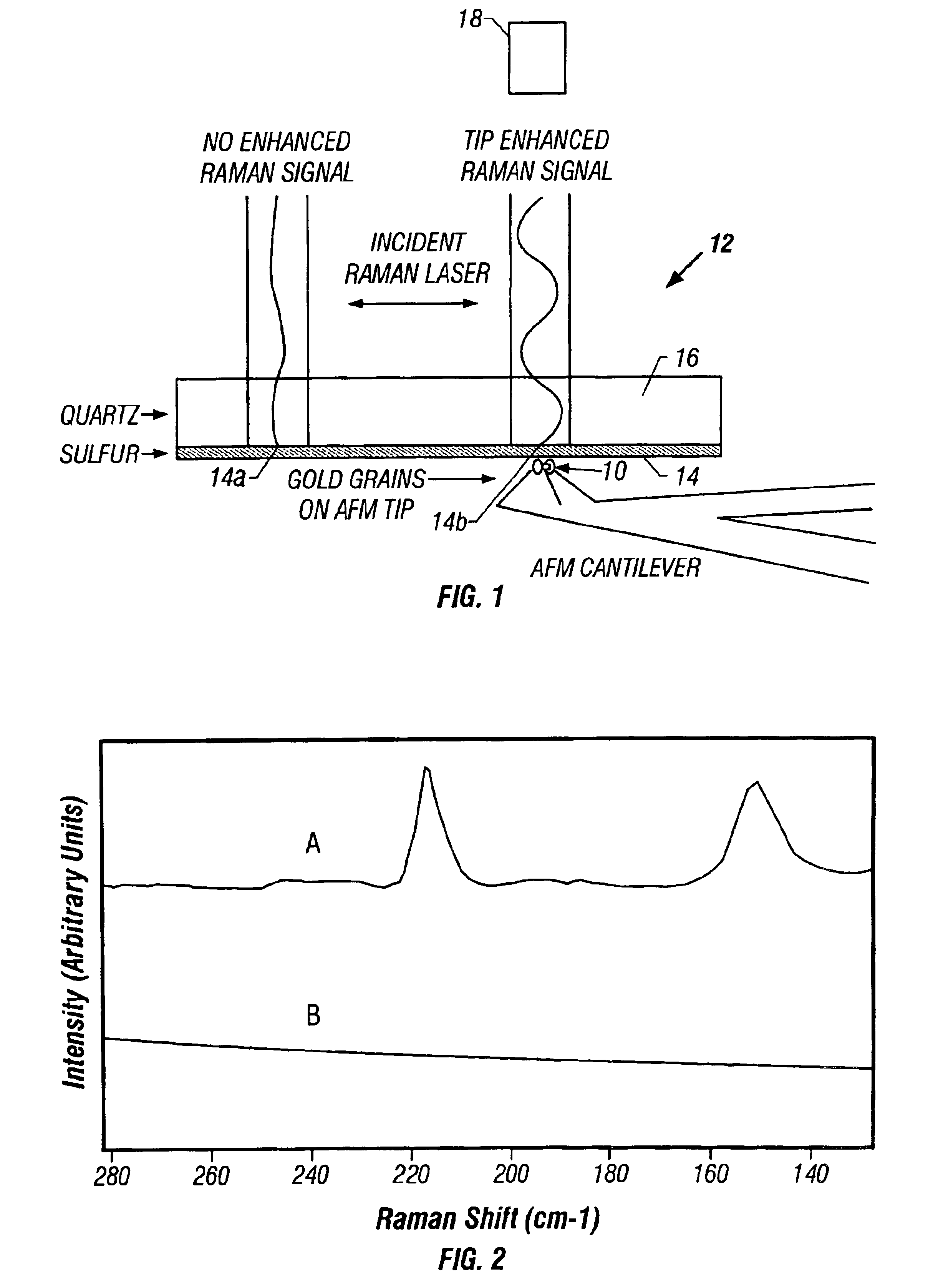
An atomic force microscope (AFM) tip is used to selectively produce surface enhanced Raman scattering (SERS) for localized Raman spectroscopy. Spectra of thin films, normally undetectable with a Raman microprobe spectrometer alone, are readily acquired with the use of a suitably gold-coated AFM tip according to the invention. Alternatively, an AFM tip is used to remove sample layers at the nanometer scale and subsequently serves as a SERS substrate for ultra-trace analysis. The combination of an AFM with a Raman spectrometer thus provides increased sensitivity, selectivity and spatial resolution over a conventional Raman microprobe. An AFM guiding the SERS effect has the potential for even targeted single molecule spectroscopy.
When a conventional AFM tip 10 depicted in FIG. 1, which tip 10 is suitably coated with gold, can provide spatially selective enhancement of a Raman signal from a sample using a surface enhanced Raman scattering (hereinafter SERS) effect. The SERS effect explo...
PUM
 Abstract
Description
Claims
Application Information