
Digital flat panel x-ray receptor positioning in diagnostic radiology

a digital flat panel and radiology technology, applied in the field of radiology, can solve the problems of high initial cost of digital detector, and difficulty in using in procedures where patients are involved, so as to achieve the effect of safe and convenient movemen

Inactive Publication Date: 2005-02-08
HOLOGIC INC
View PDF32 Cites 101 Cited by
  • Summary
  • Abstract
  • Description
  • Claims
  • Application Information

AI Technical Summary

Benefits of technology

An exemplary and non-limiting embodiment comprises a digital, flat panel, two-dimensional x-ray detector system that is not mechanically coupled to x-ray source motion and can safely and conveniently move to any one of a wide variety of positions for standard or other x-ray protocols and can securely maintain the selected position to take x-ray images, thus making it possible to use a standard x-ray source in an x-ray room, such as a ceiling-mounted source, with a single digital flat panel detector for x-ray protocols that might otherwise require plural detectors.
For certain x-ray protocols, it can be desirable to couple vertical motion of the detector to vertical motion of a patient table, and such provisions are included in the disclosed system. To make moving and positioning the detector easier, motion in at least some of the degrees of freedom is regulated with detents that bias the motion to preferred steps and can lock to prevent undesired motion. The motion in one or more degrees of freedom can be motorized. Further, the motion in some or all of the degrees of freedom can be computer-controlled. A collision avoidance system can be provided to help prevent pinch-points and collisions for the motions in one or more of the degrees of freedom. Encoders coupled with moving parts can provide digital information regarding motion and position, and the information can be used by a programmed computer to control the motions in various ways. For example, the information can be used in pinch-point and collision avoidance and / or in computer-controlling motions that position the detector at selected positions and orientations.

Problems solved by technology

On the other hand, digital flat panel detectors currently have a higher capital cost than film cassettes, and are more fragile.
They often incorporate lead shielding to protect radiation-sensitive electronics, and can be heavy.
The high initial cost of the digital detector can hinder outfitting of an x-ray room with multiple detectors pre-mounted in a variety of positions, such as a vertically-mounted unit for chest, and a horizontal unit under a bed.
The fragility, weight, and initial cost of the units make them difficult to use in procedures where the patient cradles the detector.
The unique characteristics of digital flat panel detectors can make conventional film cassette holders impractical for use with flat panel detectors.
A number of other proposals have been made for positioning x-ray film cassettes but the different physical characteristics and requirements of flat panel detectors systems do not allow for direct application of film cassette positioning proposals.

Method used


Image

  • Digital flat panel x-ray receptor positioning in diagnostic radiology
  • Digital flat panel x-ray receptor positioning in diagnostic radiology
  • Digital flat panel x-ray receptor positioning in diagnostic radiology

Examples


Embodiment Construction

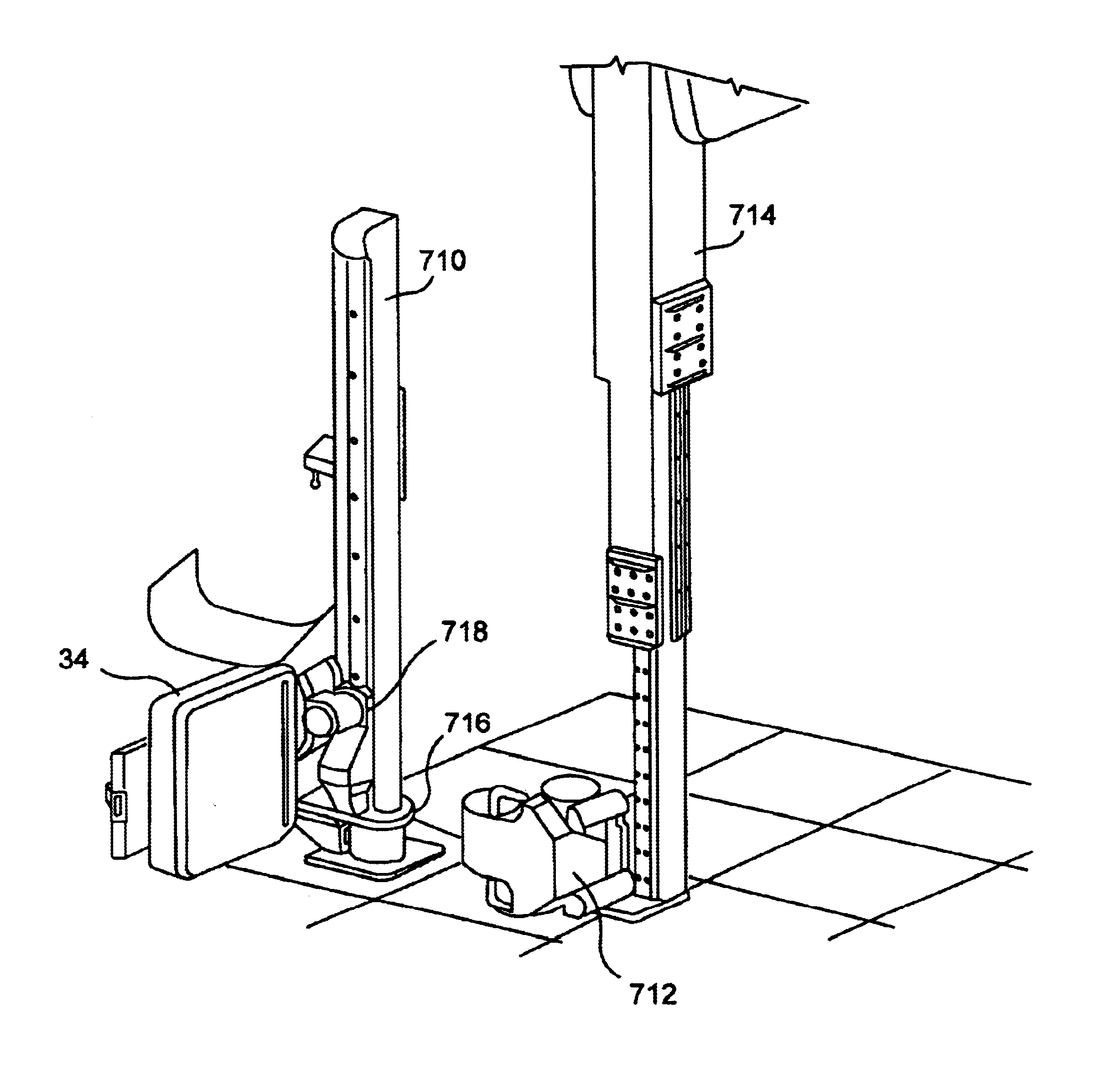
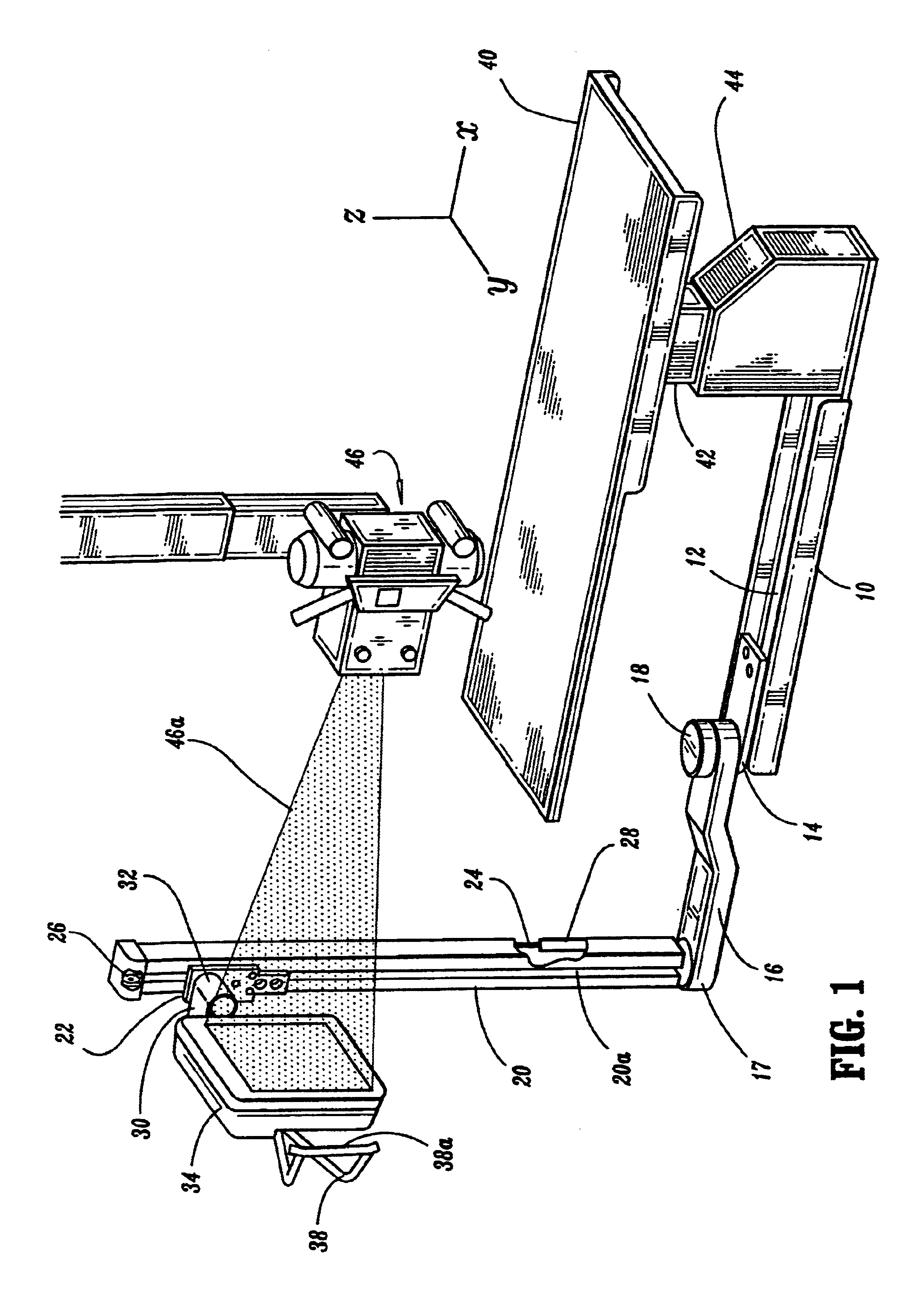
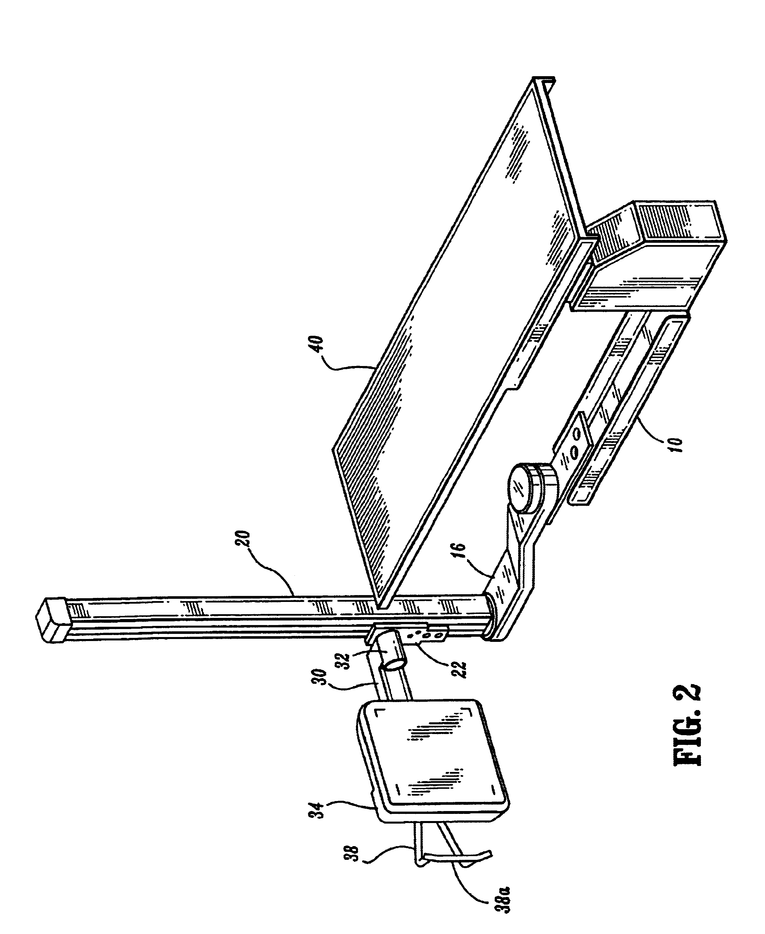
Referring to FIG. 1, a main support 10 can be secured to the floor of an x-ray room (or to a movable platform, not shown), and has a track 12 on which a lower slide 14 rides for movement along an x-axis. Slide 14 supports the proximal or near end of a generally horizontal lower arm 16 through a bearing at 18 allowing rotation of arm 16 about an upwardly extending axis, e.g., a z-axis. The distal or far end of lower arm 16 in turn supports an upwardly extending, e.g., vertical, column 20, mounted for rotation about an upwardly extending, e.g, vertical, axis though a bearing at 17. Column 20 has a slot 20a along its length. An upper slide 22 engages slot 20a to ride along the length of column 20 and is supported by a cable or chain system 24 that reverse direction over pulleys 26 at the top and bottom of column 20 (only the top pulley is illustrated) and connect to counterweights 28 riding inside column 20. Upper slide 22 in turn supports an upper arm 30 through a bearing arrangement ...


PUM


Abstract

A digital, flat panel, two-dimensional x-ray detector moves reliably, safely and conveniently to a variety of positions for different x-ray protocols for a standing, sitting or recumbent patient. The system makes it practical to use the same detector for a number or protocols that otherwise may require different equipment, and takes advantage of desirable characteristics of flat panel digital detectors while alleviating the effects of less desirable characteristics such as high cost, weight and fragility of such detectors.

Description

FIELDThis patent specification is in the field of radiography and pertains more specifically to the field of x-ray equipment using a digital flat panel detector.BACKGROUNDMedical diagnostic x-ray equipment has long used x-ray film contained inside a lightproof cassette, with the cassette at one side of the patient and an x-ray source at the opposite side. During exposure, x-rays penetrate the desired body location and the x-ray film records the spatially varying x-ray exposure at the film. Over the years, medical experience has developed and optimized a variety of standard protocols for imaging various parts of the body, which require placing the film cassette in different positions relative to the patient. Chest x-rays, for example, are often performed with the patient standing, chest or back pressed against a vertical film cassette. Imaging of the bones in the hand might be done with the cassette placed horizontally on a surface, and the hand placed on top of the cassette. In anot...

Claims


Application Information

Patent Timeline
IPC IPC(8): A61B6/04A61B6/00A61B6/06A61B6/10
CPCA61B6/0457A61B6/06A61B6/4233A61B6/4464A61B6/4291A61B6/547A61B6/56A61B6/563A61B6/488A61B6/105A61B6/583A61B2560/0271A61B6/0487
Inventor SMITH, ANDREW P.STEINWILSON, KEVIN E.CABRAL, RICHARD E.ROSSI, REMOMILLER, JAMES D.
Owner HOLOGIC INC
Learn More