Method and apparatus for measuring a intracorporal passage image

a technology of intracorporal passage and measurement method, which is applied in the field of medical surgical procedures, can solve the problems of narrowing of the lumen of these blood vessels, leakage around the catheter, and variety of complications, and achieve the effect of accurately measuring, accurately determining, and accurately determining size/dimension

Inactive Publication Date: 2005-02-22
DALRYMPLE ROBERT
View PDF16 Cites 24 Cited by
  • Summary
  • Abstract
  • Description
  • Claims
  • Application Information

AI Technical Summary

Benefits of technology

An advantage of the invention is that it can be used in conjunction with a variety of imaging techniques.
is that it can be used in conjunction with a variety of imaging techniques.
Another advantage of the invention is that it provides for an accurate selection of a medical catheter to be inserted into an intracorporeal passage (that is, a vessel).
Yet another advantage of the invention is that obliquely disposed intracorporeal passages in an image can be more accurately measured.
Yet another advantage of the invention is that it provides compensation for object to image ratio.
The present invention is a medical measuring apparatus and method for using the same for more accurately determining an appropriately sized / dimensioned catheter for a target intracorporeal passage into which the catheter is to be inserted. In short, the present invention contemplates substituting for “eyeballing” of images in connection with measuring intracorporeal passages.

Problems solved by technology

For example, plaques such as fatty substances, cholesterol, calcium and / or fibrin may build in the inner lining of arteries, thereby causing an occlusion that results in narrowing of the lumen of these blood vessels.
If a catheter having too large of a size (often referred to in French scale, 3F=1 mm) is inserted into a blood vessel, a variety of complications may occur.
Complications include leakage around the catheter, encrustation, irritation, and infection.
A serious type of complication can result from damage to the blood vessel and bleeding occasioned thereby.
Extensive bleeding can lead to a host of various complications such as embolisms and aneurisms, etc., which can result in strokes or heart attacks.
Further, sudden blockage of an artery may occur if the employed catheter is too large.
This can also lead to a stroke, for example, and in some cases, utilizing a catheter of an inappropriate size may be fatal to a patient.
These formulas, however, do not typically take into consideration the specifics of the particular patient being subjected to the intracorporeal surgical procedure.
This approach can lead to inaccurate determinations.
First, it is often difficult to include both the markers and the anatomic region of interest on the same image during a procedure.
Secondly, some conventional marker catheters do not have any mechanisms for ensuring that the line extending between the two markers is perpendicular to the image beam.
This causes the image beam to foreshorten the apparent distance between the two markers on the image which can result in an inaccurate determination of the size of the anatomic structure in question.
Thirdly, some conventional marker catheters are designed exclusively for intravascular use and ignore biliary and bronchial applications.
Finally, a medical professional may begin a procedure with an unmarked catheter and may not realize until the procedure is already under way that it is desirable to measure the size of a structure.
In such a case, it can be difficult, time-consuming, and expensive to exchange an unmarked catheter for a marked catheter.
While the foregoing prior art devices and methods have proved useful, they suffer from a number of disadvantages.
For instance, some are complex to use.
Others are costly or have an inadequate measurement range.

Method used


Image

  • Method and apparatus for measuring a intracorporal passage image
  • Method and apparatus for measuring a intracorporal passage image
  • Method and apparatus for measuring a intracorporal passage image

Examples


Embodiment Construction

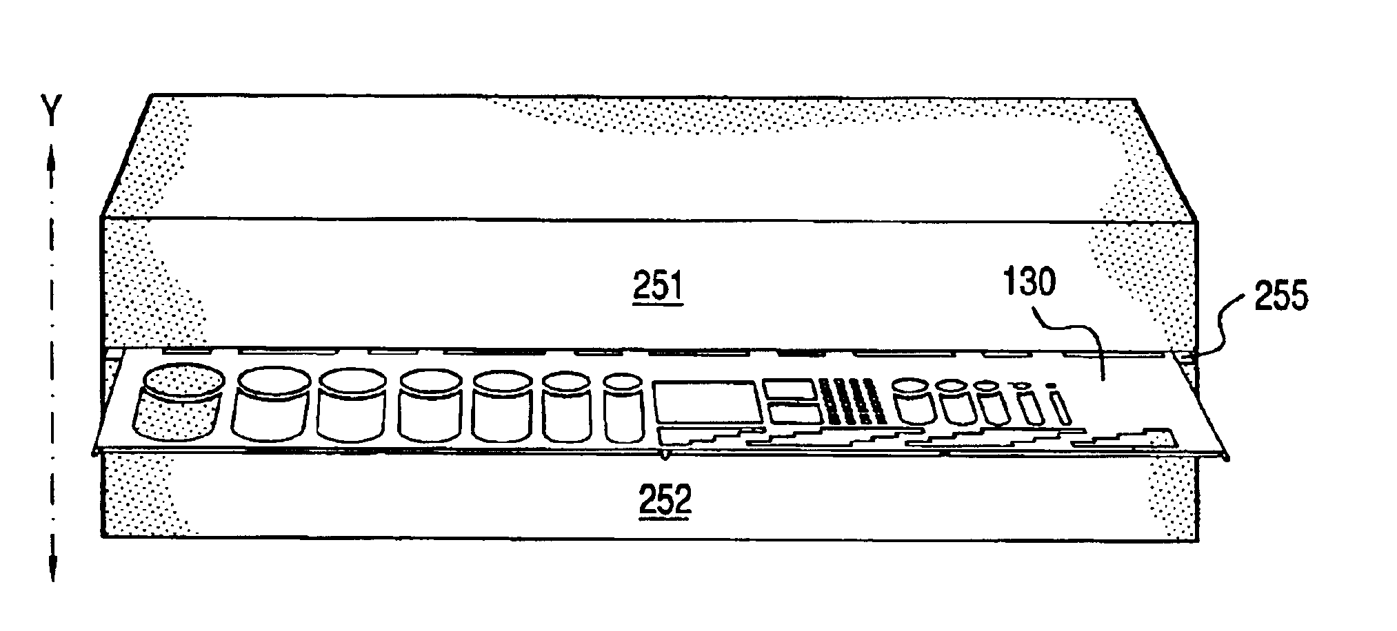
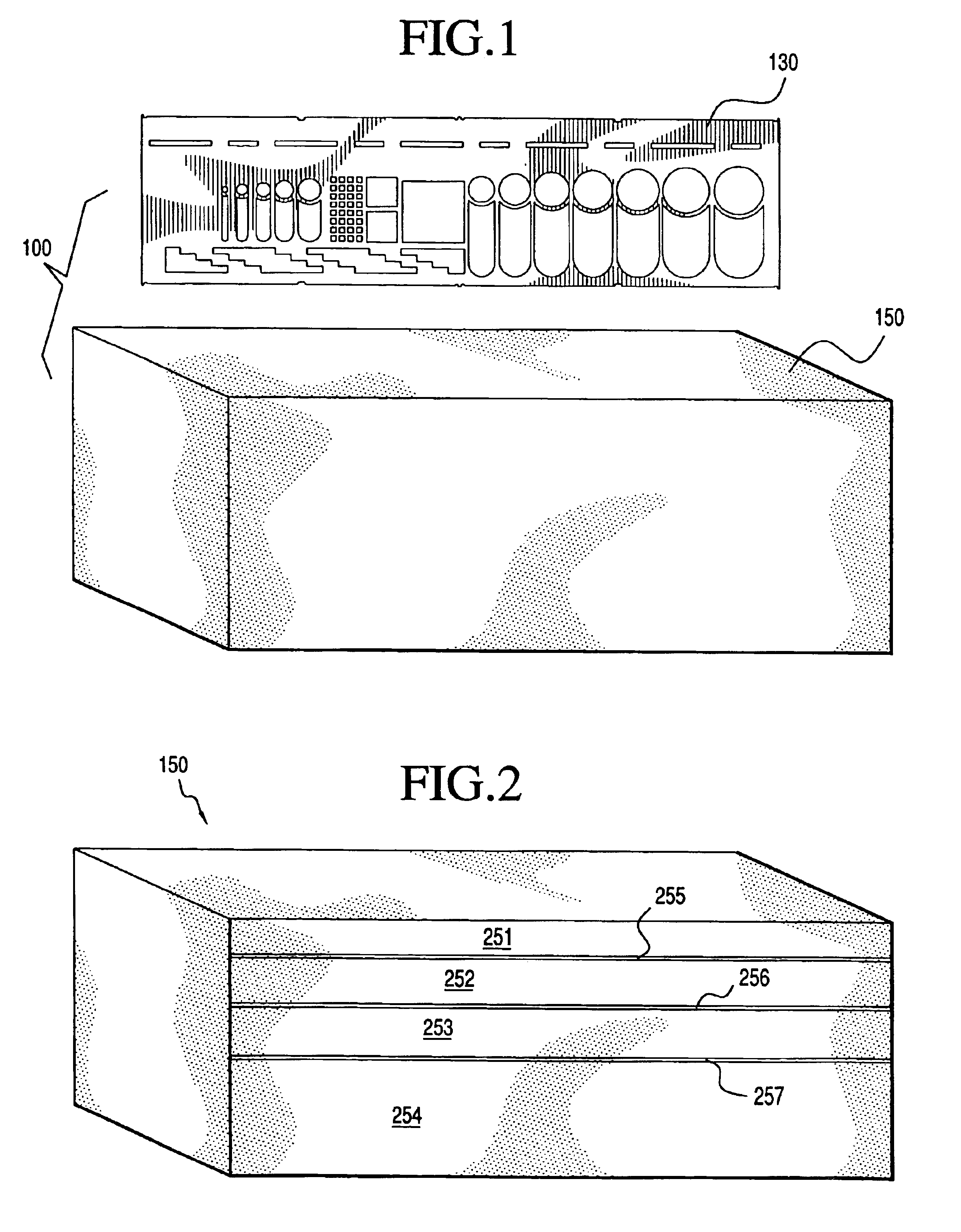
FIG. 1 illustrates the exemplary medical measuring apparatus 100. The apparatus 100 preferably includes a measuring adjunct 130 and an object to image retainer 150 in accordance with the illustrated embodiment of the present invention. In the embodiment depicted in FIG. 1, the object to image retainer 150 is a pad. As will be described below, however, in alternative embodiments, the object to image retainer 150 is not necessarily a pad. It should also be noted that in at least one embodiment of the invention, the object to image retainer 150 may be a mechanically / hydraulically controlled device for adjusting the height of the measuring adjunct 130. In another embodiment, the object to image retainer 150 may not be present. In such an embodiment, the medical measuring apparatus 100 includes only the measuring adjunct 130.

In the embodiment of the invention depicted in FIG. 1, a medical professional operates the medical measuring apparatus 100 preferably by placing the measuring adjunc...


PUM


Abstract

A medical measuring apparatus for measuring a lumen of an intracorporeal passage as it appears in an image wherein the apparatus preferably includes a measuring adjunct and an object to image retainer. The measuring adjunct preferably includes at least one geometric figure outline having a border formed by a portion of the measuring adjunct such that an aperture having a geometric shape is defined for measuring the lumen of the intracorporeal passage. The at least one geometric figure outline preferably includes a corresponding measurement specification (for example, in the metric system). In at least one embodiment, the at least one geometric figure outline is preferably utilized to measure an occlusion associated with the intracorporeal passage (for example, a renal tumor). The object to image retainer is for housing the measuring adjunct and compensates for a magnification ratio relating to the intracorporeal passage and the intracorporeal passage as it appears in the image.

Description

FIELD OF THE INVENTIONThe present invention relates to medical surgical procedures, and more specifically to vascular surgery.BACKGROUND OF THE INVENTIONVascular disease is one of the leading causes of morbidity and mortality in the United States. For example, a common type of vascular disease is known as Atherosclerosis, a vascular disease that occurs in artery segments (for example, in the carotid artery, the renal arteries, and peripheral arteries of the limbs). Atherosclerosis (also known as hardening of the arteries) is a progressive, degenerative arterial disease that leads to occlusion of affected blood vessels, thereby reducing vessel patency, and hence, blood flow through the vessels. For example, plaques such as fatty substances, cholesterol, calcium and / or fibrin may build in the inner lining of arteries, thereby causing an occlusion that results in narrowing of the lumen of these blood vessels.As arteries are internal, special procedures must be employed to locate the oc...

Claims


Application Information

Patent Timeline
Patent Type & Authority Patents(United States)
IPC IPC(8): B43L7/00G01B3/14
CPCG01B3/30A61B5/1076
Inventor DALRYMPLE, ROBERT
Owner DALRYMPLE ROBERT
Why Eureka
Try Eureka