
Dental and orthopedic densitometry modeling system and method

a densitometry and modeling system technology, applied in the field of densitometry modeling system and method, can solve the problems of inability to detect caries in the enamel surface, difficulty in locating using conventional diagnostic equipment and procedures, and inability to detect caries in the beginning, etc., to achieve the effect of convenient monitoring of decalcification and economical operation

Inactive Publication Date: 2005-09-13
OSSEO IMAGING
View PDF46 Cites 2 Cited by
  • Summary
  • Abstract
  • Description
  • Claims
  • Application Information

AI Technical Summary

Benefits of technology

[0016]The principle objects and advantages of the present invention include: providing a dental and orthopedic diagnostic application for densitometry; providing such an application which includes a method for modeling dental and orthopedic structure using densitometry; providing such a method which includes dual-energy, X-ray emission and detection; providing such a method which includes providing a color-coded output model showing dental density; providing such a method which detects incipient caries; providing such a method which is adapted for detecting decalcification beneath the surface of the dental enamel layer; providing such a method which employs scanning X-ray techniques; providing such a method which utilizes commercially available tomography equipment and other equipment as described; providing such a method which detects dental fractures; providing such a method which detects dental apical abscesses; providing such a method which detects dental pathologies at the micron level; providing such a method which facilitates the monitoring of decalcification in dental structures for determining appropriate treatment; providing such a method which is adaptable for monitoring osseointegration; providing such a method which can be practiced with relatively minor changes to existing densitometry equipment; providing such a system and method which have applications in dental and medical diagnosis, treatment, identification, forensics and biometrics; and providing such a system and method which are economical in operation and particularly well adapted for the proposed usage thereof.

Problems solved by technology

Incipient caries, particularly those located within the enamel surface, often go undetected with conventional equipment.
When such caries are finally found, considerable damage to tooth structure may have already occurred.
They are particularly difficult to locate using conventional diagnostic equipment and procedures.
Dental fractures can result from bruxism (teeth grinding), trauma, etc.
The dental structure that has been weakened by various causes, such as decalcification, is particularly susceptible to fractures.
Fracture patterns and configurations can be particularly difficult to locate using conventional X-ray equipment and procedures.
For example, fractures which are generally parallel to the X-ray beam are often undetectable on an X-ray negative.
Pathologies in the dentin and pulp layers are often associated with considerable pain and tooth loss.
Apical abscesses comprise yet another dental condition which can be difficult to diagnose with conventional equipment, particularly in the early stages.
Advanced apical abscesses can cause considerable pain because they involve the neurovascular bundles located in the root canals as well as the osseous tissue around the apex of the root.
Heretofore there has not been available a system or method for applying the technology of densitometry to dental and medical applications such as the detection of caries and decalcification and the monitoring of osseointegration in connection with dental and medical prostheses.

Method used


Image

  • Dental and orthopedic densitometry modeling system and method
  • Dental and orthopedic densitometry modeling system and method
  • Dental and orthopedic densitometry modeling system and method

Examples


first modified embodiment

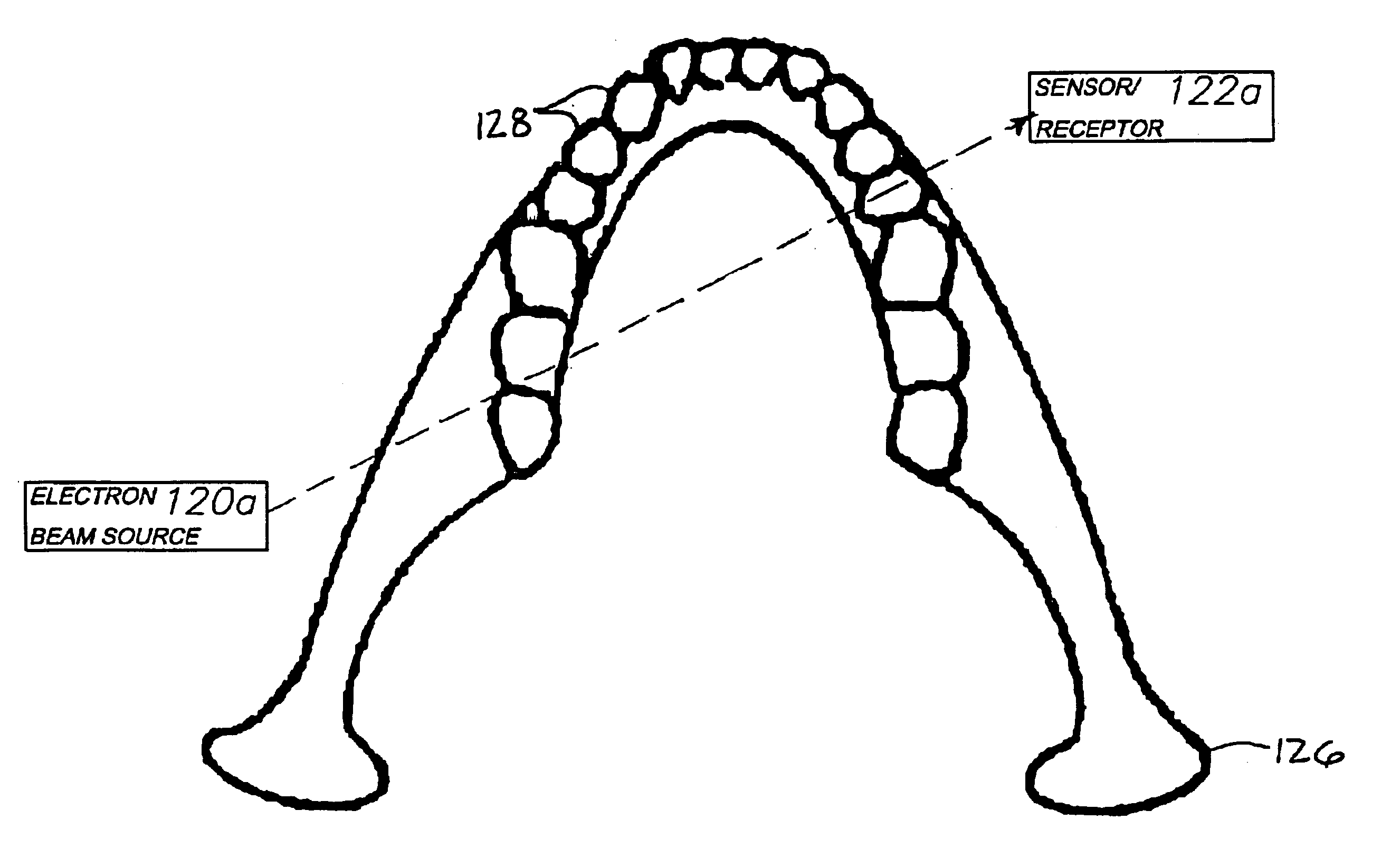
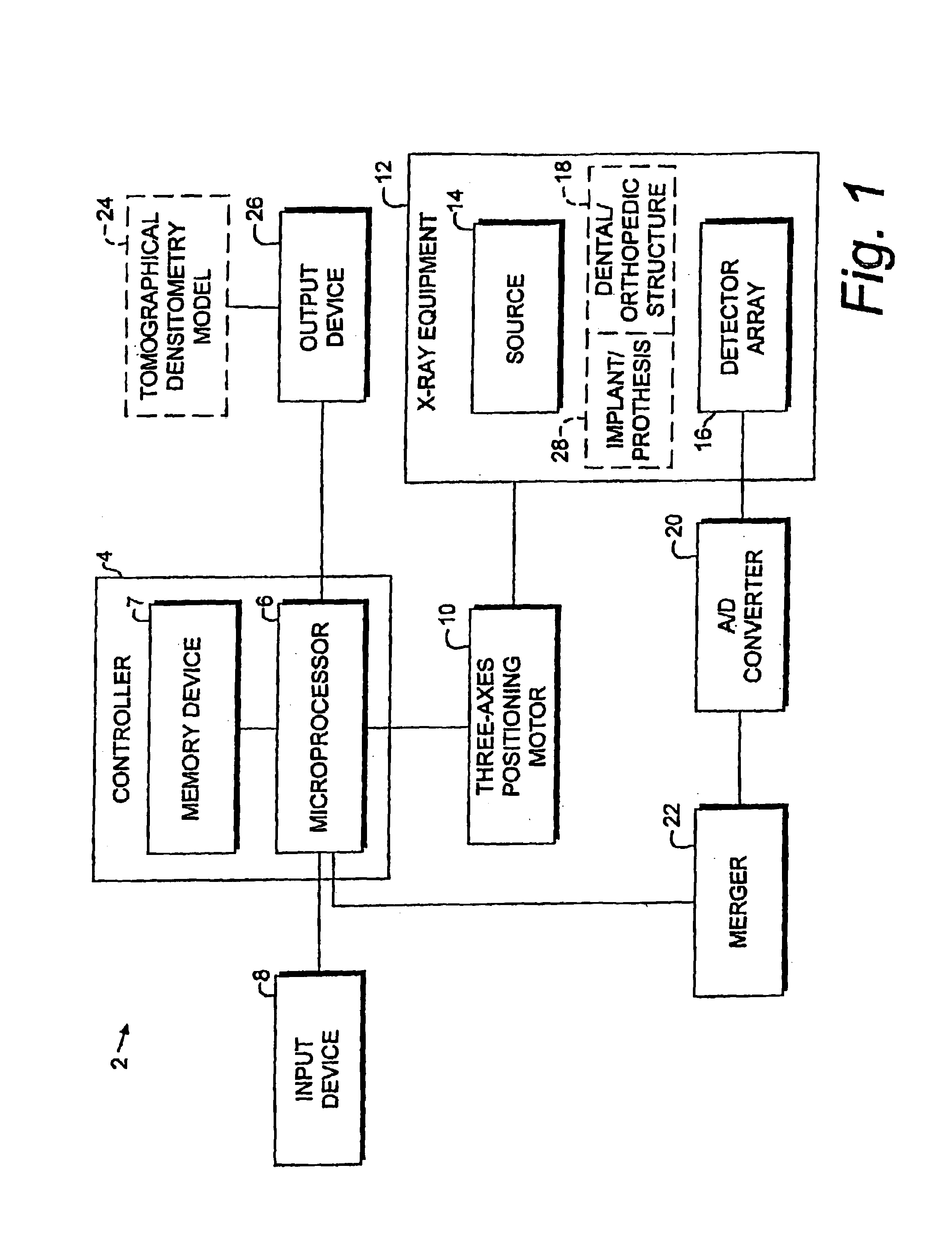
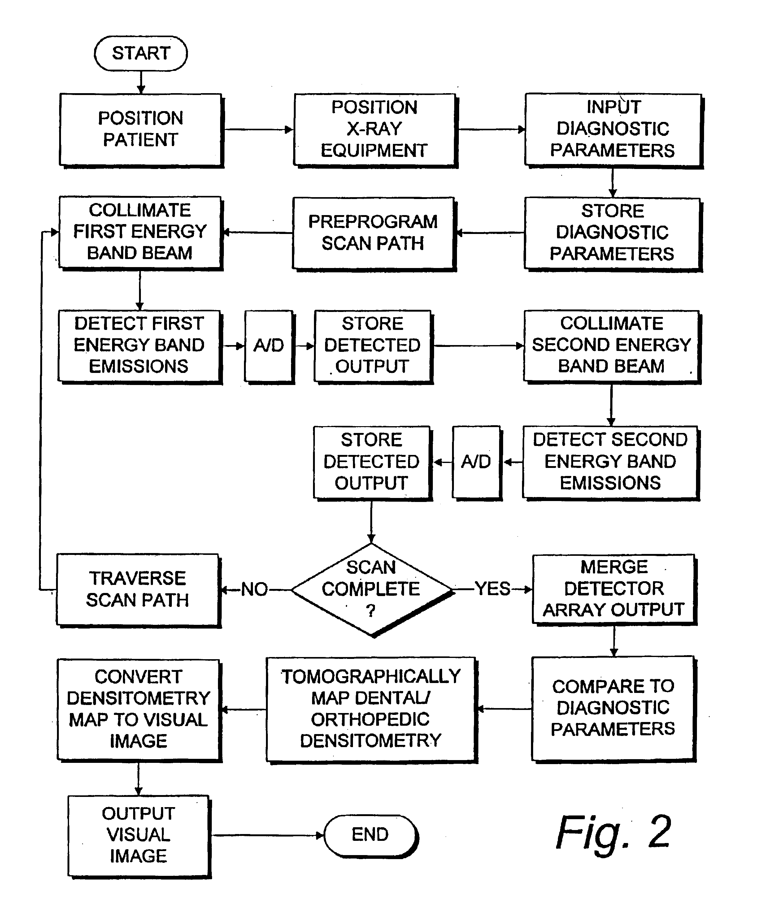
[0038]A densitometry modeling system 102 comprising present invention is shown in FIG. 3 and generally includes a computer 104 with an input 104a and an output 104b. Input and output devices 106 and 108 are connected to the computer input and output 104a,b respectively.

[0039]The computer 102 includes a memory 110, such as a hard drive, a tape drive, an integrated circuit (e.g., RAM) or some other suitable digital memory component, which can be either internal or external to the computer 102. Imaging software 112 is provided for converting the digital data into images, which are adapted for visual inspection by displaying same on a monitor 114 or by printing same on a printer 116 of the output device 108. Such images can also be transmitted by a suitable transmission device 117, such as a fax or modem. The computer 104 also includes comparison software 118, which is adapted for digitally comparing baseline and patient-specific dental and orthopedic densitometry models.

[0040]The input...


PUM


Abstract

A dental or orthopedic densitometry modeling system includes a computer with a digital memory adapted for storing patient densitometry information, an input and an output. A dental or orthopedic input device includes energy source and an energy sensor, both of which can be either external or intraoral to the patient. The sensor transfers densitometry signals to the computer, which creates, stores and compares digital densitometry models. A densitometry modeling method includes the steps of creating a densitometry database consisting of dental or orthopedic information and obtaining current dental or orthopedic densitometry information from a patient. The current information is compared to the database, which can include the patient's previous densitometry models, and an updated patient densitometry model is created.

Description

CROSS-REFERENCE TO RELATED APPLICATIONS[0001]This application is a continuation-in-part of Ser. No. 10 / 134,153, filed Apr. 27, 2002 now abandoned, which is a continuation of Ser. No. 09 / 452,348, filed Dec. 1, 1999, now U.S. Pat. No. 6,381,301 B1, in which priority is claimed and which are incorporated herein by reference.BACKGROUND OF THE INVENTION[0002]1. Field of the Invention[0003]The present invention relates generally to dental and orthopedic diagnosis, forensics, identification, biometrics and treatment, and in particular to a densitometry modeling system and method.[0004]2. Description of the Related Art[0005]The field of dental diagnostics is generally concerned with locating pathologies in the dental structure, i.e. the teeth and the surrounding tissue and bone. Three of the most common pathologies are: 1) caries associated with decay; 2) fractures; and 3) apical abscesses. The system and method of the present invention are primarily, but not exclusively, concerned with det...

Claims


Application Information

Patent Timeline
Patent Type & Authority Patents(United States)
IPC IPC(8): A61C13/00A61B6/14A61B6/00A61B6/03A61B6/51A61C19/00
CPCA61B5/0088A61B6/032A61B6/14A61B6/145A61B6/482A61B6/505A61C13/00A61C13/0003A61C19/00A61B6/512A61B6/51
Inventor MASSIE, RONALD E.
Owner OSSEO IMAGING
Learn More