
Computer-aided diagnosis method for aiding diagnosis of three dimensional digital image data

a computer-aided diagnosis and digital image data technology, applied in the field of computer-aided diagnosis (cadx), can solve the problems of large amount of data for the physician to examine, inability to form a mental model, and inability to diagnose cancer or other diseases

Inactive Publication Date: 2006-01-03
SIEMENS CORP +1
View PDF19 Cites 39 Cited by
  • Summary
  • Abstract
  • Description
  • Claims
  • Application Information

AI Technical Summary

Benefits of technology

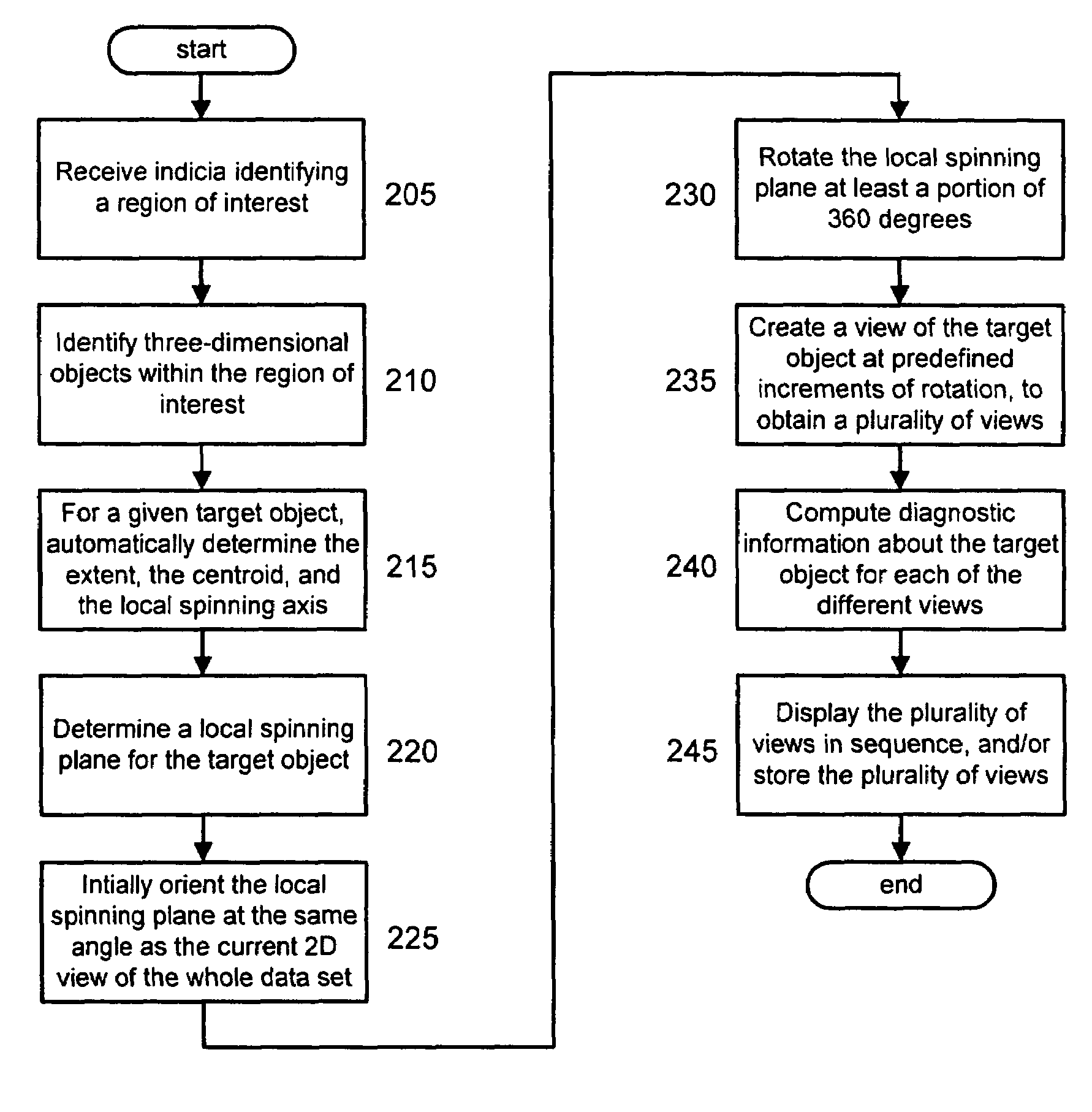
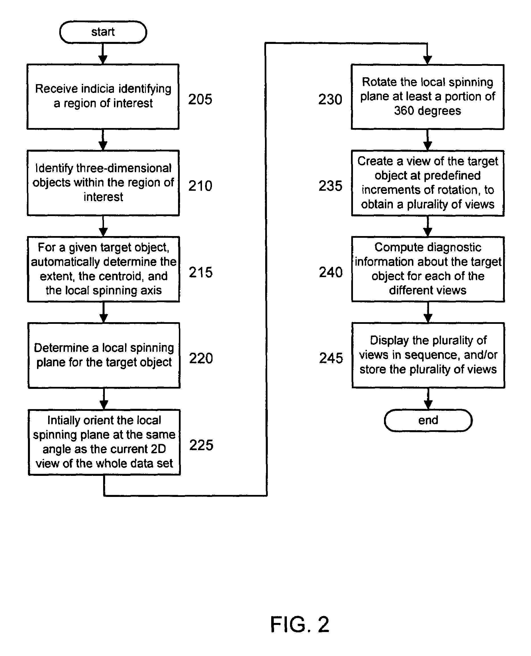
The present invention is a computer-aided diagnosis (CADx) method for assisting diagnosis of three-dimensional digital image data. The method integrates a physician's knowledge into the CADx process seamlessly, performs diagnostic computation accordingly, and then provides a novel presentation of the data that aids the physician in understanding the data. The method includes identifying at least one region of interest in a digital medical image, identifying three-dimensional objects within the region, determining the extent, centroid, and local spinning axis of the object, and creating a view of the object at predefined increments of rotation. The method also includes providing a plurality of views of the object and determining indicia such as the volume, geometrical location, center of mass, intensity values, texture, surface smoothness, regularity measures, and shape measures of the object. The results can be stored in a table for comparison with subsequent scans of the same patient and a confidence value can be assigned to each object. The method can also set thresholds for abnormal objects and identify those that exceed the thresholds. Overall, the invention improves the accuracy and efficiency of CADx for diagnostic purposes.

Problems solved by technology

In current clinical practice, cancer or other diseases may be missed during a physician's un-aided examination of medical image data, in part because of the large volume of data.
This is particularly a problem for screening applications, since there is generally little time to devote to the examination of each patient's data, and the entire range of the data must be examined to make sure it is free from disease.
However, the change from two dimensions to three means that there is a much larger amount of data for the physician to examine.
For complex anatomical structures, the volume rendering may be too cluttered for the physician to form a mental model of the underlying three-dimensional structure of the data, and how it corresponds to normal and abnormal anatomical structure.
Unfortunately, many current CADx systems are not readily accepted by physicians, because their aid is seen as more of a distraction than a help.
Many such systems present the results of the computer's diagnosis to the physician by marks, such as a red circle or arrow on the softcopy, which some physicians feel can create a bias in their interpretation of the data.
Furthermore, many systems are perceived as a “black box”; that is, the physicians feel that they do not have any understanding of how the systems work and how they generate their diagnosis.

Method used


Image

  • Computer-aided diagnosis method for aiding diagnosis of three dimensional digital image data
  • Computer-aided diagnosis method for aiding diagnosis of three dimensional digital image data
  • Computer-aided diagnosis method for aiding diagnosis of three dimensional digital image data

Examples


Embodiment Construction

[0031]The present invention is directed to a computer-aided diagnosis (CADx) method for aiding diagnosis of three-dimensional digital image data. It is to be appreciated that the invention may be used to aid diagnosis of any abnormality in any part of the body which is represented in a digital image.

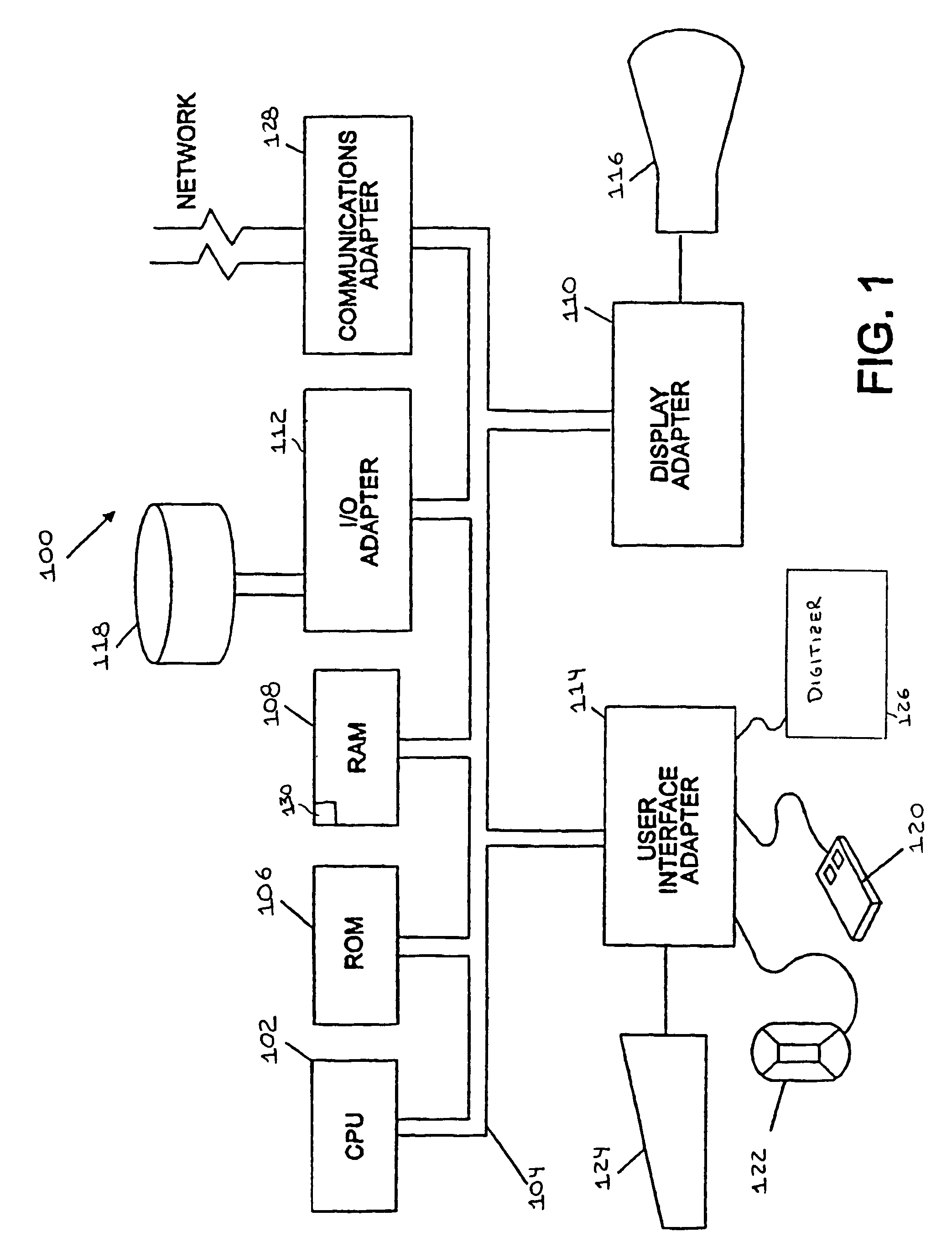
[0032]FIG. 1 is a block diagram of a computer processing system to which the present invention may be applied, according to an embodiment of the present invention. The system 100 includes at least one processor (CPU) 102 operatively coupled to other components via a system bus 104. A read only memory (ROM) 106, a random access memory (RAM) 108, a display adapter 110, an I / O adapter 112, and a user interface adapter 114 are operatively coupled to system bus 104.

[0033]A display device 116 is operatively coupled to system bus 104 by display adapter 110. A disk storage device (e.g., a magnetic or optical disk storage device) 118 is operatively couple to system bus 104 by I / O adapter 112.

[003...


PUM


Abstract

There is provided a computer-assisted diagnosis method for assisting diagnosis of three-dimensional digital image data. The method includes the step of receiving indicia identifying at least one region of interest in a digital medical image. Three-dimensional objects within the at least one region of interest are identified. For a given three-dimensional (3D) object within the at least one region (ROI), an extent, a centroid, and a local spinning axis of the given object are determined. Moreover, a local spinning plane is determined for the given object, the local spinning plane being centered at the centroid and the local spinning axis. The local spinning plane is rotated at least a portion of 360 degrees. The rotating step includes the step of creating a view of the given object at predefined increments of rotation, so as to result in a plurality of views of the given object.

Description

BACKGROUND[0001]1. Technical Field[0002]The present invention relates generally to computer-aided diagnosis (CADx) and, in particular, to a computer-aided diagnosis (CADx) method for aiding diagnosis of three-dimensional digital image data.[0003]2. Background Description[0004]Computer-assisted diagnosis is an important technology in many clinical applications. In current clinical practice, cancer or other diseases may be missed during a physician's un-aided examination of medical image data, in part because of the large volume of data. This is particularly a problem for screening applications, since there is generally little time to devote to the examination of each patient's data, and the entire range of the data must be examined to make sure it is free from disease. Computer analysis that is performed silently in the background can greatly aid physicians in their work.[0005]New technologies that offer three-dimensional (3D) scans of the human body, such as Magnetic Resonance Imagi...

Claims


Application Information

Patent Timeline
Patent Type & Authority Patents(United States)
IPC IPC(8): G06K9/00A61B5/055A61B6/03G06T1/00G06T7/00
CPCA61B6/032A61B6/466G06T7/0012A61B6/469A61B6/467
Inventor NOVAK, CAROLQIAN, JIANZHONGBOBICK, AARON
Owner SIEMENS CORP
Learn More