Therapeutic device and method for treating diseases of cardiac muscle

a technology of therapeutic devices and cardiac muscle, which is applied in the direction of electrotherapy, therapy, heart stimulators, etc., can solve the problems of affecting the performance of the heart, exacerbate the deteriorating condition of the heart, and few therapies address the problem of tissue remodeling, so as to reduce promote healing of diseased or inflamed tissue, and minimize the risk of tissue damage

Inactive Publication Date: 2006-05-02
MEDTRONIC PS MEDICAL
View PDF6 Cites 12 Cited by
  • Summary
  • Abstract
  • Description
  • Claims
  • Application Information

AI Technical Summary

Benefits of technology

[0021]The invention provides many advantages. The present invention provides the heart with a safe therapeutic current to promote healing of diseased or inflamed tissue. The invention mitigates the concerns relating to the application of therapeutic current to the heart, including but not limited to the concerns of uncontrolled or dangerous current level and the possible dangerous asynchronous application of current with respect to the cardiac cycle. The apparatus and method allow for long-term treatment for beneficial remodeling of the heart while minimizing the risk of damaging tissue.

Problems solved by technology

Although therapies have addressed treating specific indications, few therapies address the problem of tissue remodeling.
In those patients, the resulting remodeling adversely affects the heart's performance and exacerbates the deteriorating condition of the heart.
Abnormalities in the matrix's composition and concentration during dilation, hypertrophy and ischemic injury inhibit the function of the heart and may lead to heart failure.
However, drug therapy for inhibiting matrix metalloproteinases may present potentially serious problems.
The systemic inhibition of the matrix metalloproteinases has been found to produce a variety of side effects, such as joint and muscle pain.
The limited blood flow may result in localized tissue death known as an infarction.
The presence of an infarction weakens contraction in that region and therefore degrades the heart's performance.
These surgeries are complicated and dangerous.
However, in practice, direct injection of these angiogenic agents fails to specifically target the ischemic zone.
Further, injection of genetic material within a vector is a more biologically complex process and frequently suffers from a low transfection efficiency.
In addition, the introduction of xenobiotics compounds can be dangerous.
The compound itself may be toxic, virulent and / or allergenic.
In addition, many of the drugs prescribed for CHF patients are primarily for palliative or symptomatic relief These drugs typically do not treat the underlying disease process of CHF and their use frequently results in serious or prohibitive side effects.
Further, the drugs are typically administered systemically and therefore, impact the entire body not just the organ or tissue to be treated.
However, sub-threshold stimuli have not been broadly applied to the heart and adjacent blood vessels to promote healing and tissue remodeling.
However, directly or indirectly applying electrical stimulation to the heart can be dangerous.
There is a risk of inducing a depolarization of the cardiac tissue resulting in an unwanted cardiac contraction.
Further, there is a risk of inducing a life-threatening arrhythmia.
One problem not addressed by prior methods is the inability to accurately assess the field strength generated in the region of the heart.
A particular stimulus level may be safe when applied to one patient, and yet that same stimulus may evoke an unwanted reaction from another patient.
Another problem is the artifact created by an applied stimulus, which may disrupt the sensing and activation of an implanted pacing or defibrillation device.
The disruption could result in stimulation at a high-risk portion of the cardiac cycle, or an undesirable or unnecessary defibrillation shock.

Method used


Image

  • Therapeutic device and method for treating diseases of cardiac muscle
  • Therapeutic device and method for treating diseases of cardiac muscle
  • Therapeutic device and method for treating diseases of cardiac muscle

Examples


Embodiment Construction

[0026]The present invention is described generally as a therapy for promoting remodeling of a patient's heart. Those skilled in the art will recognize the improvements and advantages conferred by the present invention in the treatment of heart disease and heart failure, the specific etiologies including, but not limited to, ischemic cardiomyopathy, idiopathic dilated cardiomyopathy, other cardiomyopathies, myocarditis and atrial fibrillation.

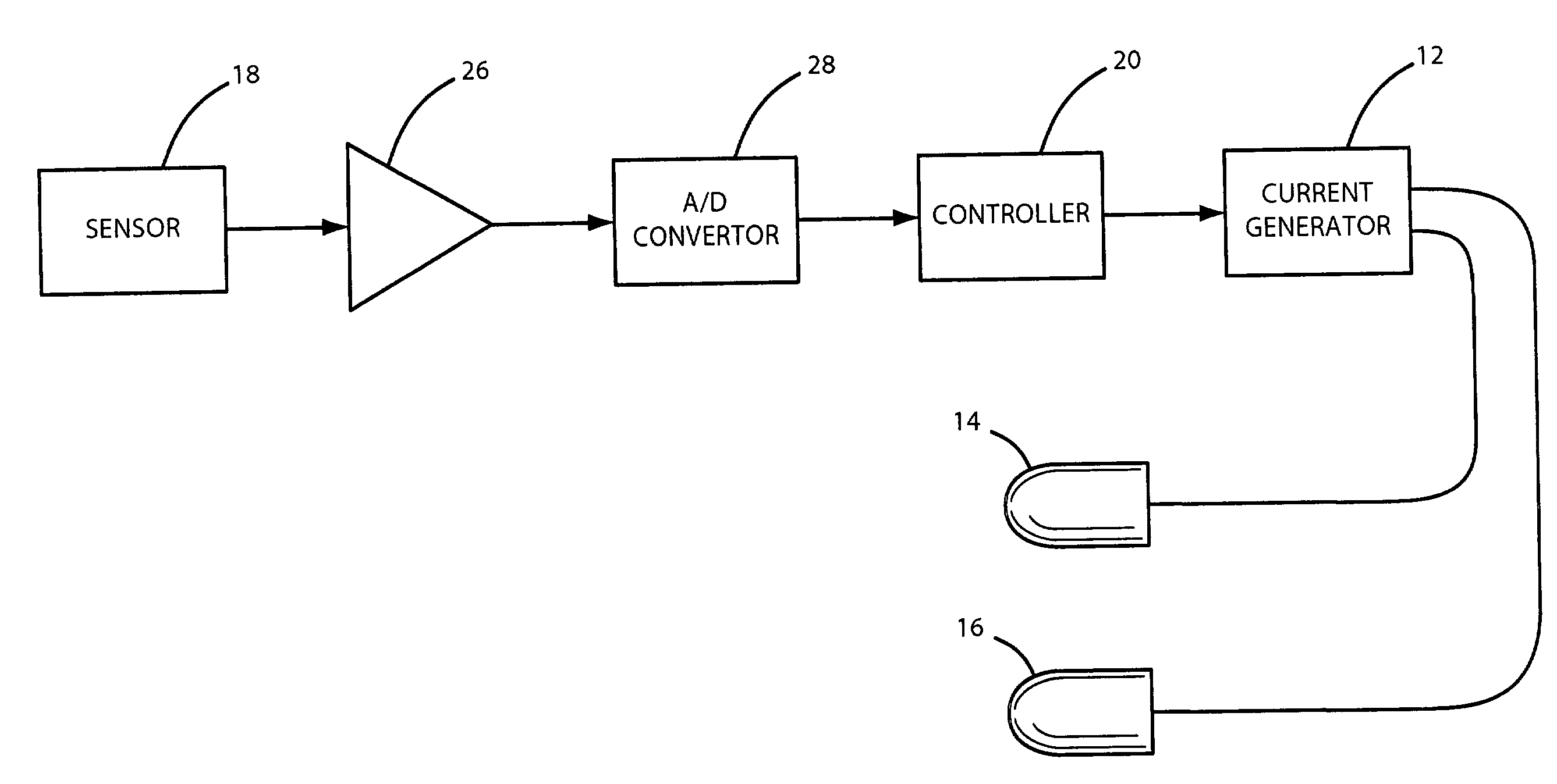
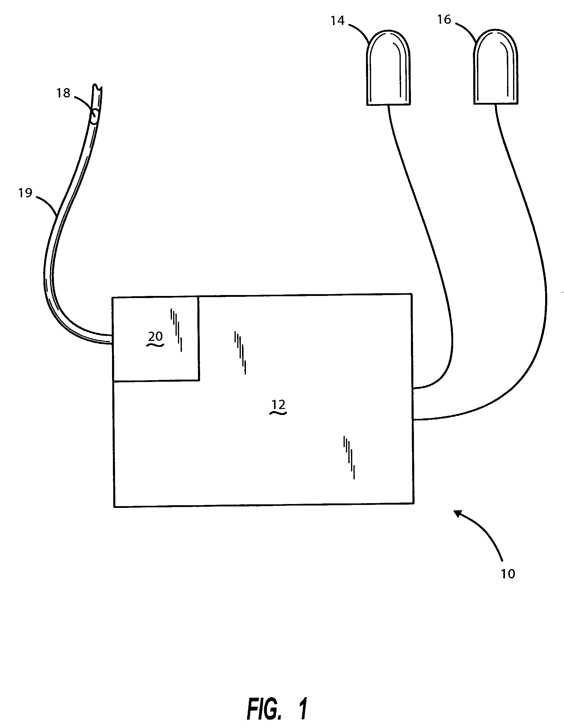
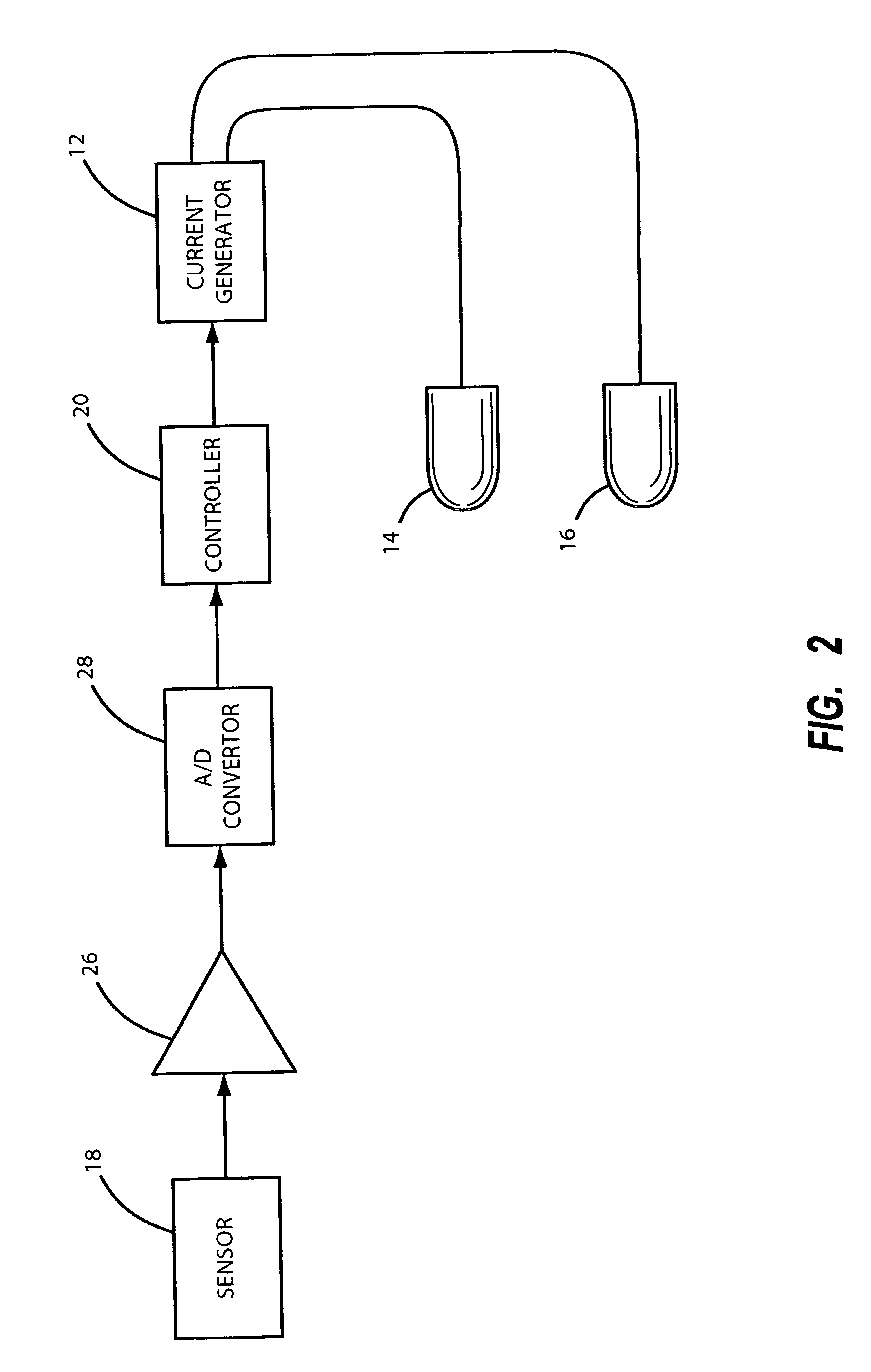
[0027]An apparatus 10 in accordance with the present invention is shown in FIG. 1. Apparatus 10 includes a current generator 12, a first electrode 14, a second electrode 16, a sensor 18, and a controller 20. First drive electrode 14 and second drive electrode 16 are configured to be implanted in a chamber of the heart or within the coronary vessels, or are configured to be secured on or near the heart. Typically, the electrodes are connected to current generator 12 by a conductive wire. Current generator 12 is configured to provide a sub-thresho...


PUM


Abstract

An apparatus and method for conferring a therapeutic current to the heart is provided. The apparatus includes a first electrode, a second electrode, a current generator and a controller. The apparatus may further include a sensor. The sensor is generally configured to measure field strength between the first and second electrode. The sensor may also monitor the cardiac cycle. The method includes applying an electric stimulus to the heart and sensing the electric field generated by the electric stimulus to prevent the level of current from inducing unwanted depolarization.

Description

CROSS-REFERENCE TO RELATED APPLICATIONS[0001]This application is a divisional of continuation-in-part U.S. application Ser. No. 09 / 756,567, filed Jan. 8, 2001, now U.S. Pat No. 6,560,489 which claims priority to a application of U.S. patent application Ser. No. 09 / 638,233, filed Aug. 14, 2000 (now abandoned), which claims priority from U.S. Provisional Application Ser. No. 60 / 143,510, filed Aug. 24, 1999. The above-referenced Patent and Provisional Applications are incorporated by reference in the present application in their entirety.BACKGROUND OF THE INVENTION[0002]1. Field of the Invention[0003]The present invention relates to an apparatus and method for treatment of the heart and, more particularly, to an apparatus and method for providing a therapeutic sub-threshold electrical current to the heart.[0004]2. Discussion of the Related Art[0005]A wide range of therapies is available for the treatment of cardiac tissue damage, heart failure and for treatment of the specific underlyi...

Claims


Application Information

Patent Timeline
Patent Type & Authority Patents(United States)
IPC IPC(8): A61N1/362A61N1/08A61N1/32
CPCA61N1/08A61N1/326A61N1/3627
Inventor HAUCK, JOHN A.
Owner MEDTRONIC PS MEDICAL
Why Eureka
Try Eureka