Method for modulating light penetration depth in tissue and diagnostic applications using same

a technology of light penetration depth and tissue, applied in the field of non-invasive determination of in vivo concentrations of analytes or evaluation of a disease state, can solve the problems of inability to achieve acceptable precision and accuracy, inability to provide non-invasive glucose measurement accuracy, and inability to achieve non-invasive metabolite testing, such as glucose monitoring, to achieve the effect of reducing the effect of repositioning error

Inactive Publication Date: 2006-05-09
ABBOTT DIABETES CARE INC
View PDF79 Cites 405 Cited by
  • Summary
  • Abstract
  • Description
  • Claims
  • Application Information

AI Technical Summary

Benefits of technology

[0042]The method of this invention is preferable to prior art methods for the determination of diabetic status and other disease conditions based on measurement of body analytes in that the method relies on two measurements performed at the same site on the surface of the biological sample, thereby decreasing the effect of repositioning error and the possibility of the optical probe being in contact with different micro-structural regions of the biological sample during different single measurements.

Problems solved by technology

In its advanced stages, diabetes can cause blindness, coma, and ultimately death.
Although a variety of detection techniques have been disclosed in the art, there is still no commercially available device that provides non-invasive glucose measurements with an accuracy that is comparable to the current commercially available invasive devices.
As a result, current approaches to non-invasive metabolite testing, such as glucose monitoring, have not achieved acceptable precision and accuracy.

Method used


Image

  • Method for modulating light penetration depth in tissue and diagnostic applications using same
  • Method for modulating light penetration depth in tissue and diagnostic applications using same
  • Method for modulating light penetration depth in tissue and diagnostic applications using same

Examples


example 1

[0135]FIG. 2 through FIG. 5 illustrate an apparatus suitable for the measurement of optical properties, and hence the concentration of different analytes at various depths in tissue. Co-pending U.S. application Ser. No. 09 / 098,049, filed Nov. 23, 1998, assigned to the assignee of this application, describes in detail many of the components used in the apparatus of this application. The apparatus is capable of introducing light into the skin of human subjects and measuring the light re-emitted therefrom.

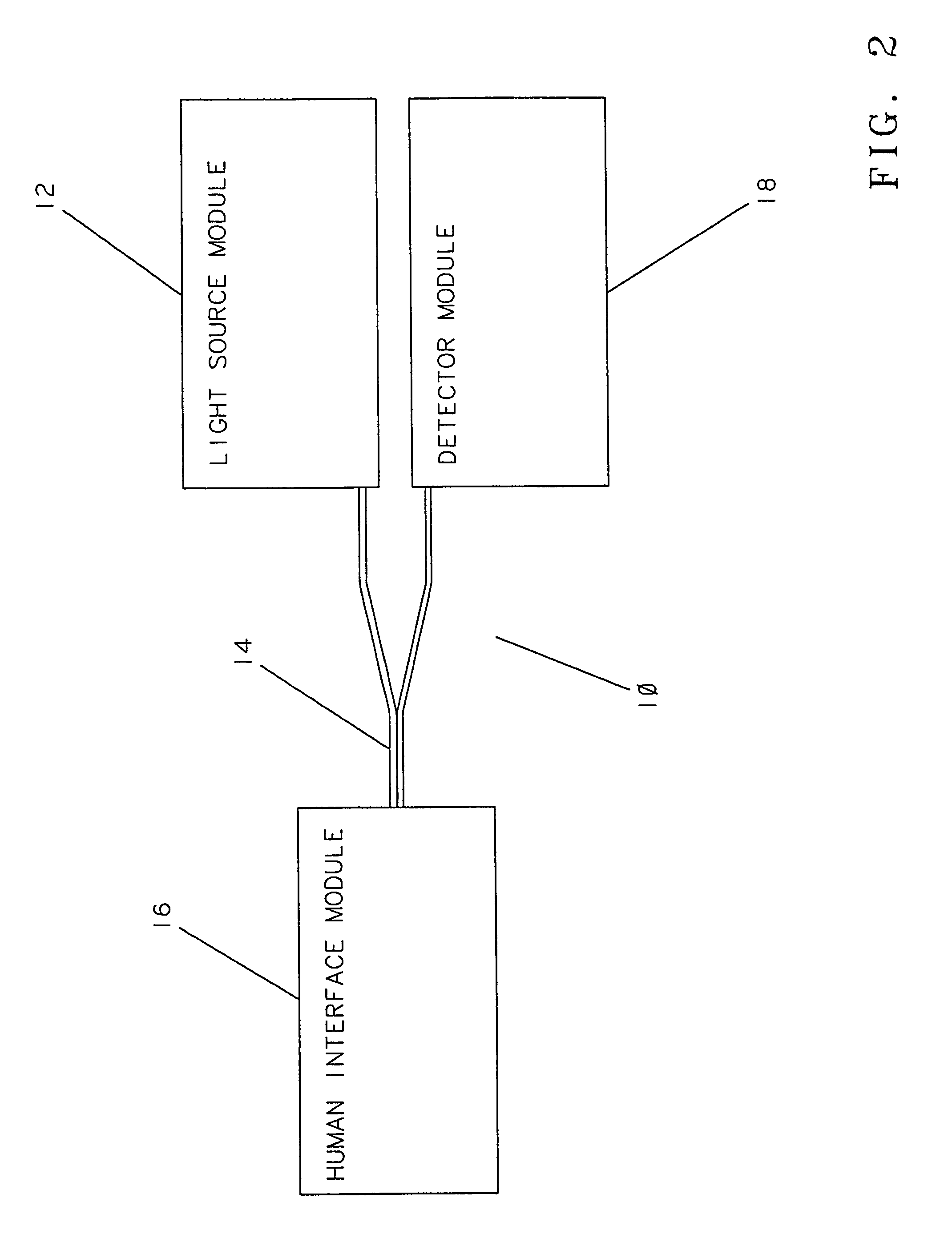
[0136]FIG. 2 is a block diagram illustrating an embodiment of an apparatus 10 of this invention. The apparatus 10 comprises a light source module 12, a bifurcated optical fiber bundle 14, a human interface module 16, and a detector module 18. The light source module 12 includes a source of modulated light (not shown), such as a Gilway L1041 lamp modulated with a Stanford Research Optical Chopper. A prism, a dichroic beam splitter, or the like (not shown) may be used to direct a portio...

example 2

[0146]The apparatus described in Example 1 was used to study the effect of temperature changes on the optical properties of human skin. FIG. 6A shows the change in reflectance as a function of temperature for a Caucasian subject for light having wavelengths of 590 nm, 650 nm, and 800 nm. The temperature was increased from 24° C. to 34° C., then lowered to 22° C. and finally raised to 38° C. The reflectance values are normalized to the value at 24° C. Temperature was maintained at each value for four (4) minutes during which the reflectance was measured; the transition between two temperatures consumed a period of about one minute. The μs′ and μa values were determined by measuring spatially resolved diffuse reflectance in a temperature-controlled area of the human forearm.

[0147]The normalized reflectance curves were analyzed by means of a Monte Carlo simulation and calibration grid generated with a set of absorbing and / or scattering phantoms to generate a calibration grid. The absor...

example 3

[0149]The apparatus described in Example 1 was used to test the change in optical properties of the human forearm for several subjects of different skin color. Nine subjects were tested. Testing was performed at least two hours after the breakfast meal and was completed between 10 A.M. and 12 noon each day. Testing on nine individuals was performed over the period of several days. Two of the subjects were Type II diabetics. Temperature was varied between 38° C. and 22° C. Values of μs′ and μa were calculated by means of the method described in Example 2 of this application.

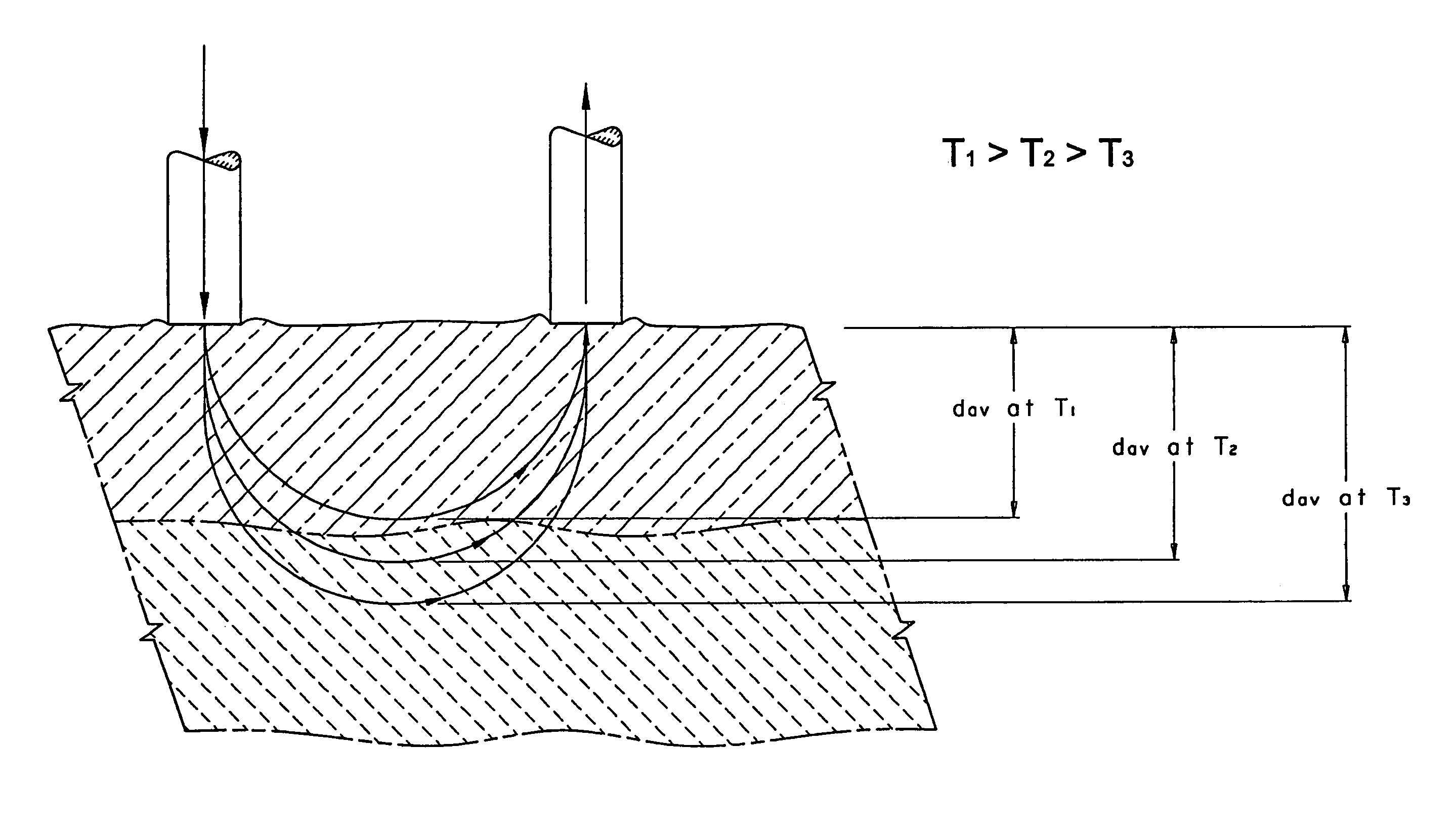
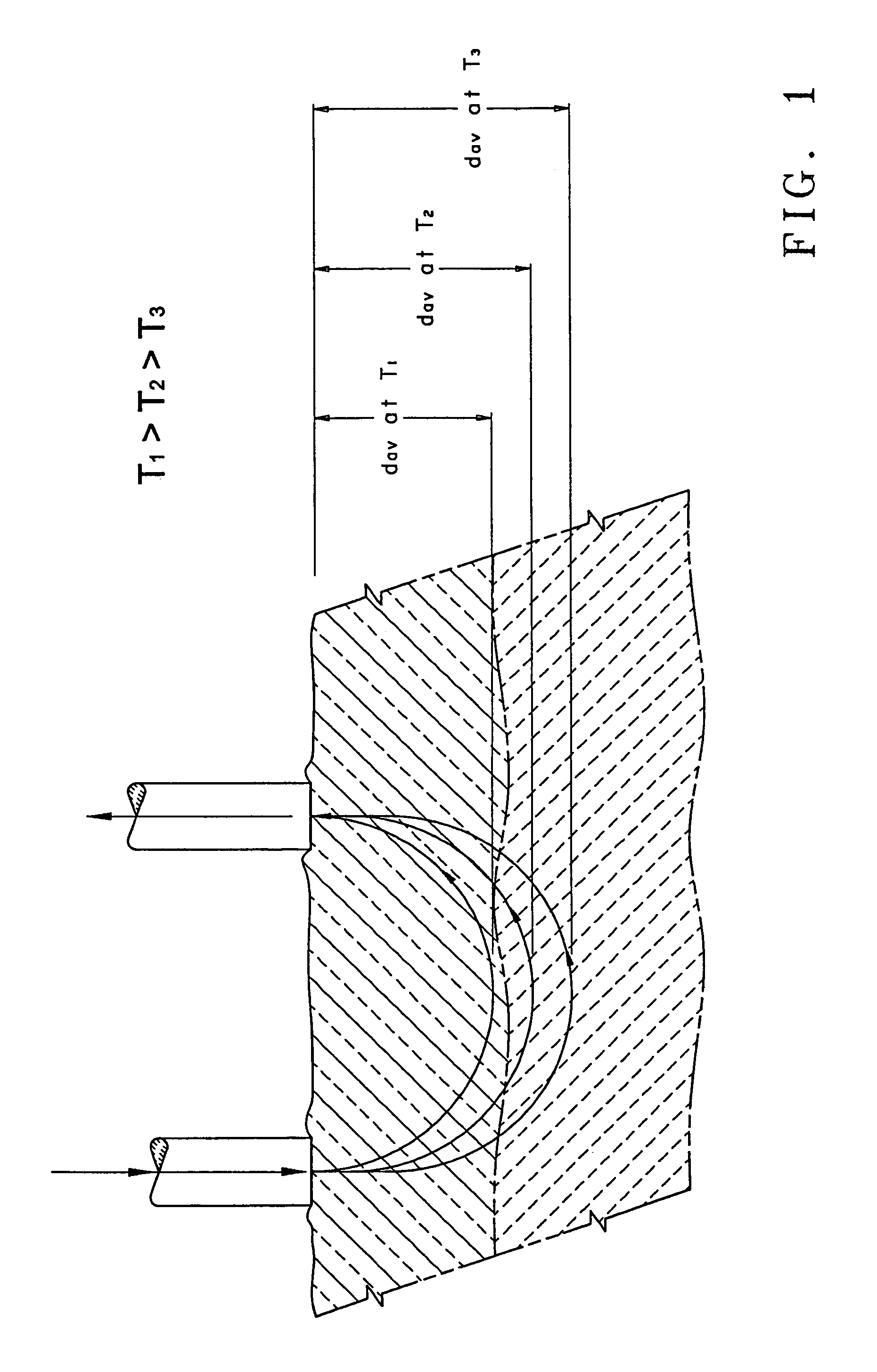
[0150]The value of μeff was calculated using Equation (2). Light penetration depth δ for a given temperature was calculated as δ=1 / μeff. Change in light penetration depth (Δδ) as a result of changing temperature between 38° C. and 22° C. was then calculated. The following three tables summarize the changes in light penetration depth in tissue resulting from modulating temperature between 38° C. and 22° C. Data for...


PUM


Abstract

Devices and methods for non-invasively measuring at least one parameter of a sample, such as the presence of a disease condition, progression of a disease state, presence of an analyte, or concentration of an analyte, in a biological sample, such as, for example, a body part. In these devices and methods, temperature is controlled and is varied between preset boundaries. The methods and devices measure light that is reflected, scattered, absorbed, or emitted by the sample from an average sampling depth, dav, that is confined within a region in the sample wherein temperature is controlled. According to the method of this invention, the sampling depth dav, in human tissue is modified by changing the temperature of the tissue. The sampling depth increases as the temperature is lowered below the body core temperature and decreases when the temperature is raised within or above the body core temperature. Changing the temperature at the measurement site changes the light penetration depth in tissue and hence dav. Change in light penetration in tissue as a function of temperature can be used to estimate the presence of a disease condition, progression of a disease state, presence of an analyte, or concentration of an analyte in a biological sample. According to the method of this invention, an optical measurement is performed on a biological sample at a first temperature. Then, when the optical measurement is repeated at a second temperature, light will penetrate into the biological sample to a depth that is different from the depth to which light penetrates at the first temperature by from about 5% to about 20%.

Description

[0001]This invention is a continuation-in-part of U.S. Ser. No. 09 / 080,470, filed May 18, 1998, now U.S. Pat. No. 6,662,030, and is a continuation-in-part of U.S. Ser. No. 09 / 302,207, filed Apr. 29, 1999, now U.S. Pat. No. 6,241,663.BACKGROUND OF THE INVENTION[0002]1. Field of the Invention[0003]This invention relates to devices and methods for the noninvasive determination of in vivo concentrations of analytes or evaluation of a disease state, and more particularly, the noninvasive determination of in vivo concentrations of analytes or evaluation of a disease state wherein temperature is controlled and varied between preset boundaries.[0004]2. Discussion of the Art[0005]Non-invasive monitoring of concentrations of analytes in the human body by means of optical devices and optical methods is an important tool for clinical diagnosis. “Non-invasive” (alternatively referred to herein as “NI”) monitoring techniques measure in vivo concentrations of analytes in the blood or in the tissue...

Claims


Application Information

Patent Timeline
Patent Type & Authority Patents(United States)
IPC IPC(8): A61B5/00
CPCA61B5/0059A61B5/14532A61B5/1455A61B2562/0242A61B5/1495
Inventor KHALIL, OMAR S.YEH, SHU-JENWU, XIAOMAOKANTOR, STANISLAWHANNA, CHARLES F.JENG, TZYY-WEN
Owner ABBOTT DIABETES CARE INC
Why Eureka
Try Eureka