Method for supporting cell image analysis

a cell image and image technology, applied in the field of gene analysis, can solve the problems of not analyzing individual cells but a texture of an entire image, disadvantage of a person having to visually check images one by, and extremely difficult to process images in large quantities generated in experiments conducted for multiple cdnas

Inactive Publication Date: 2006-05-23
FUJITSU LTD +3
View PDF9 Cites 16 Cited by
  • Summary
  • Abstract
  • Description
  • Claims
  • Application Information

AI Technical Summary

Problems solved by technology

However, the apparatus does not analyze individual cells but a texture of an entire image or a total extension of elongate structures.
These conventional techniques have, as described above, a disadvantage of a need for a person to visually check images one by one in a screening.
With such a method for visually checking an experimental result and extracting individual cell regions manually, it is disadvantageously, extremely difficult to process images in large quantities generated in experiments conducted for multiple cDNAs.
Thus, since the cells in which the cDNA is injected are present only sporadically, it disadvantageously takes lots of time and labor to locate these cells and to manually name each protein localization pattern and each cellular morphogenesis.
Further, because of multiple types of fluorescent patterns and a feeble pattern in a background, such as a noise, it is disadvantageously difficult to discriminate each cell pattern from the background.
Thus, even if the experimenter visually checks the result, it is often difficult to make a clear determination.
As a result, it is disadvantageously impossible to improve experimental and analytical efficiencies.

Method used


Image

  • Method for supporting cell image analysis
  • Method for supporting cell image analysis
  • Method for supporting cell image analysis

Examples


Embodiment Construction

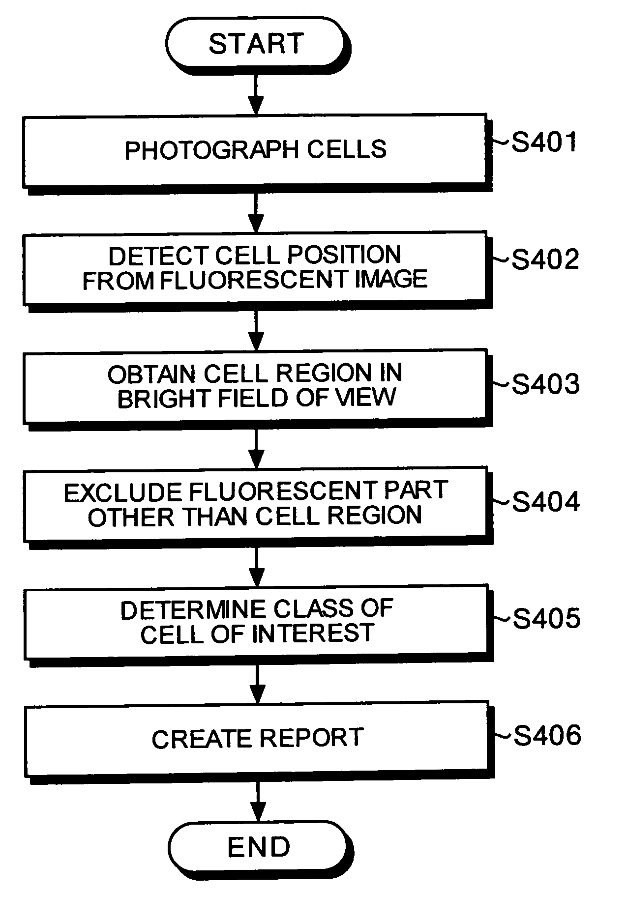
[0037]Exemplary embodiments of an image analysis supporting method, a computer-readable recording medium, and an image analysis supporting device will be explained in detail with reference to the accompanying drawings.

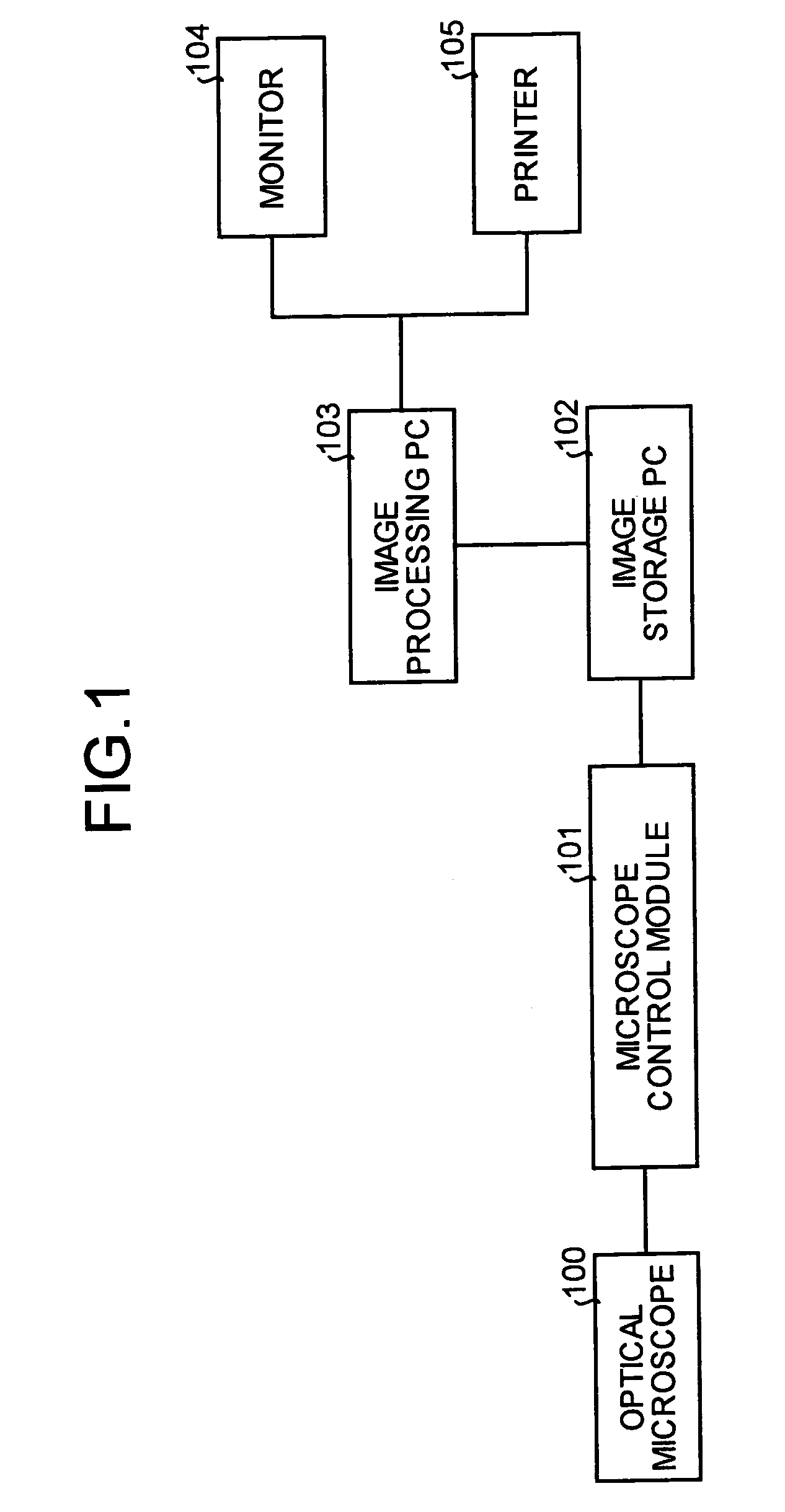
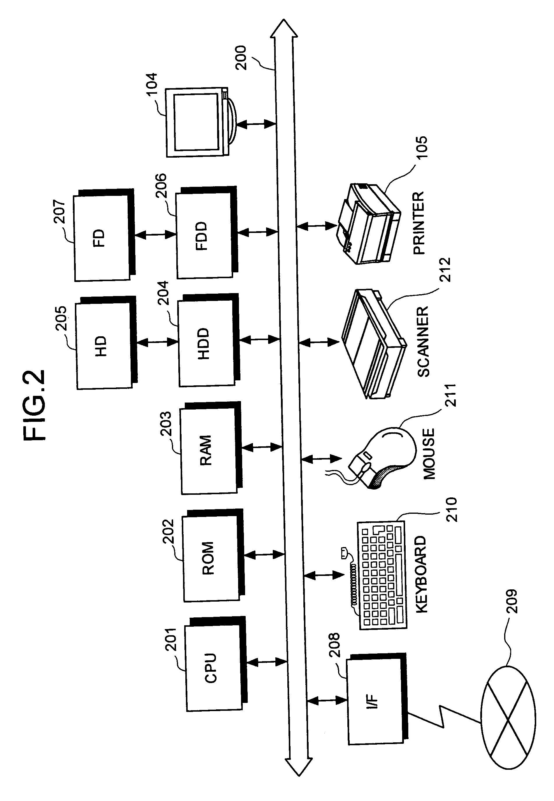
[0038]FIG. 1 is an explanatory view of one example of the hardware configuration of an image processing apparatus according to the embodiment of the present invention.

[0039]A microscope control module 101 controls an optical microscope 100 to photograph a fluorescent image, a bright-field image, and a phase difference image. The microscope control module 101 is set so that a cell, which is successfully injected with a fluorescent marker included in the cDNA, reacts to fluorescence. The optical microscope 100 is set so that the fluorescent image, the bright-field image, and the phase difference image can be photographed in the same field of view. The microscope control module 101 includes xyz stages, a fluorescent (mercury light) shutter, a halogen light shutter, and th...


PUM

PropertyMeasurementUnit
lengthaaaaaaaaaa
fluorescent imageaaaaaaaaaa
phase difference imageaaaaaaaaaa

Abstract

A fluorescent image, and either a bright-field image or a phase difference image of a plurality of cells are acquired by a microscope. A position of each cell is specified in the fluorescent image, and then a cell region of each cell is specified using the bright-field image or the phase difference image. A fluorescent part other than the cell region is thereby excluded. A class of each cell is determined, that is, the cells are classified. Thereafter, based on these results, a report is created.

Description

[0001]This application is a continuing application, filed under 35 U.S.C. §111(a), of International Application PCT / JP02 / 11624, filed Nov. 7, 2002.BACKGROUND OF THE INVENTION[0002]1) Field of the Invention[0003]The present invention relates to a genome analysis on a cell image photographed by a microscope or the like for biotechnological research and development, drug manufacturing, and the like. More specifically, the present invention relates to efficiency improvement in the analysis by automatically clipping images of each cell from the cell image, specifying types of each cell, and displaying the images and the types of each cell as a list.[0004]2) Description of the Related Art[0005]Recently, following an advancement of genome science, a study of identifying a protein localization pattern and observing a morphogenetic change for a cell into which a cDNA is injected, that is, a study of identifying functions of a DNA by performing a quantitative analysis on a morphogenetic chang...

Claims


Application Information

Patent Timeline
Patent Type & Authority Patents(United States)
IPC IPC(8): G06K9/00G01N15/14
CPCG01N15/1475G06K9/00127G01N2015/1488G06V20/69
Inventor MURAO, KOHEISUDA, SHUJIUSAMI, HITOHIDENARUSE, MASATOSHIGOTOH, TOSHIYUKIKAGEI, SEIICHIROUKABUYAMA, NORIKO
Owner FUJITSU LTD
Why Eureka
Try Eureka