Enclosures housing cell-coated supports for treating tumors

a cell-coated support and enclosure technology, applied in the field of enclosures housing cell-coated supports, can solve the problem that systemic administration is very toxic to the human body

Inactive Publication Date: 2006-06-06
RGT UNIV OF MICHIGAN
View PDF23 Cites 8 Cited by
  • Summary
  • Abstract
  • Description
  • Claims
  • Application Information

AI Technical Summary

Benefits of technology

[0031]The present invention is not limited by the nature of the enclosure; however, in one embodiment, the pores are large enough to permit the cells from the beads to exit the enclosure into the wound, while in another embodiment, the pores are too small to permit cells from the beads to exit the enclosure, but large enough to permit cellular factors to exit the enclosure or wound fluid components to enter the enclosure. In certain embodiments, the enclosures are replaced every few days until the wound heals (e.g., once a week).
[0032]In additional embodiments, the enclosure comprises a mesh material, having pores. In certain embodiments, the mesh material comprises polyethylene. In one embodiment, the pores are large enough to permit the cells from the beads to exit the enclosure into the wound, while in another embodiment, the pores are too small to permit cells from the beads to exit the enclosure, but large enough to permit cellular factors (e.g., cytokines) to exit the enclosure or wound fluid components to enter the enclosure.

Problems solved by technology

However, systemic administration is very toxic to the human body.

Method used


Image

  • Enclosures housing cell-coated supports for treating tumors
  • Enclosures housing cell-coated supports for treating tumors

Examples


example 1

[0124]The experiments of this example demonstrate that human culture keratinocytes grown on macroporous microcarriers and contained in a porous enclosure improve healing in surgically created wounds in mice.

A. Experimental Methodology

Preparation of Human Keratinocytes

Isolation and Growing of Human keratinocytes: Human keratinocytes (AATB certified; University of Michigan cultured keratinocyte program) were isolated at The University of Michigan Burn / Trauma Unit from split thickness skin.

[0125]Trypsinization of the split thickness skin was effected as follows. The skin was placed dermis-side down in 150 mm Petri dishes. The pieces were cut into smaller pieces (about 2 cm×about 0.3 cm) and were soaked in a sterile solution of 30 mM HEPES, 10 mM glucose, 3 mM KCl, 130 mM NaCl, 1 mM Na2HPO4 buffer, pH 7.4 containing 50 units of Penicillin and 50 μg Streptomycin (Sigma, P-0906). After soaking for 1–2 hr at 4° C. the buffer was aspirated off, and 0.09% trypsin (Sigma, Type IX) in a Penici...

example 2

[0152]The experiments of this example demonstrate that human culture keratinocytes grown on macroporous microcarriers and contained in a porous enclosure that is then covered with a wound dressing material improve healing in surgically created wounds in mice.

A. Experimental Methodology

[0153]The experiments of this example were performed as described in Example 1, with the following exceptions. The group of mice that received the keratinocyte-coated CYTOLINE 1™ macroporous microcarrier beads (Pharmacia Biotech) (i.e., the beads / bags group) comprised five animals, while the group that received only the bags (i.e., the bags only group) comprised four animals. (They are labelled 2 to 5 because Mouse 1 expired during anesthesia.) In this example the bags from both the beads / bags group and the bags only group were covered with a polyurethane film dressing (TEGADERM, 3M Health Care, St. Paul, Minn.) with a cellophane product.

[0154]More specifically, the wounds were dressed either with huma...

example 3

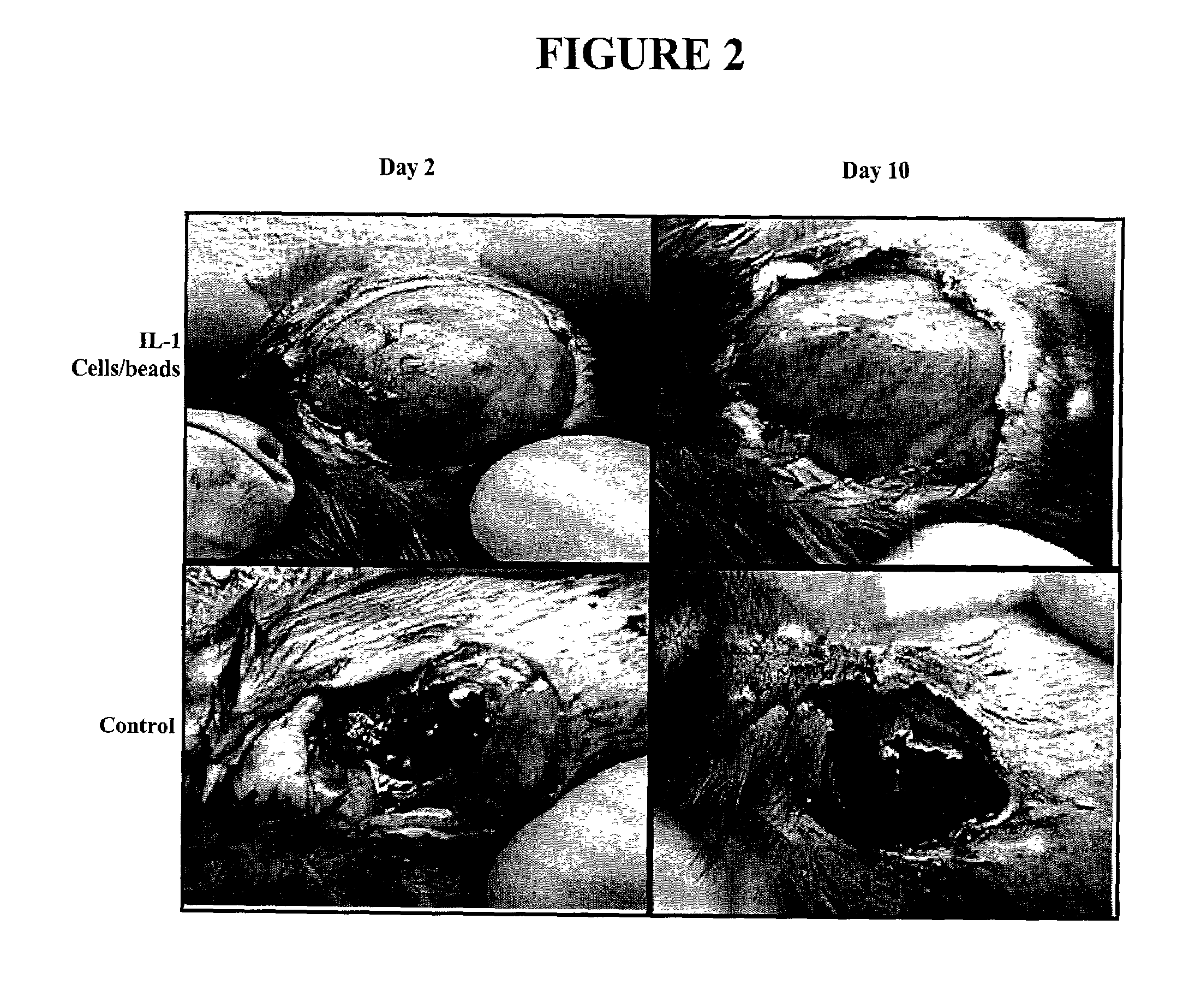
[0164]The experiments of this example demonstrate that a tetr-expressing cell line transfected with hEFG and grown on macroporous microcarriers and contained in a porous enclosure, improves healing in surgically created wounds in mice.

A. Experimental Methodology

Cells

[0165]Osteosarcoma line U20S were grown and maintained in Dulbecco's modified Eagle's medium (DMEM) supplemented with 10% fetal bovine serum. The tetR—expressing cell line, U2CEP4R-11, was cotransfected with pcDNA3, pcmvtetOEGF and EcoRI-linearized pcmvtetOEGF to establish cell lines that expressed tetR and HEGF. Medium containing hygromycin B and G418 was used to select cells resistant to hygromycin B. The HEGF-expressing cell lines were determined by analysis of hEGF expression in the presence or absence of tetracycline. [See, Yao et al., Hum Gene Ther. Feb. 10, 1999;10(3):419–27; and Yao et al., Hum Gene Ther. Sep. 1, 1998;9(13):1939–50].

Cytoline 1™ Bead Wash

[0166]Five grams of CYTOLINE 1 macroporous microcarrier bead...


PUM

PropertyMeasurementUnit
sizeaaaaaaaaaa
sizeaaaaaaaaaa
sizeaaaaaaaaaa

Abstract

The present invention relates to devices, systems and methods for treating tumors. In particular, the present invention relates to enclosures housing cell-coated supports for promoting regression of tumors, such as cancerous tumors, papillomas, and warts. In preferred embodiments, the present invention provides methods of promoting tumor regression employing enclosures secreting therapeutic proteins.

Description

[0001]The present application is a Continuation-in-part of U.S. application Ser. No. 09 / 640,990, filed Aug. 18, 2000, now abandoned which claims priority to U.S. Provisional Application No. 60 / 149,744, filed Aug. 19, 1999, the contents of which are both hereby incorporated by reference.[0002]The present application was funded in part with government support under NIH grant numbers FM 44918 and GM 50401. The government has certain rights in this invention.FIELD OF THE INVENTION[0003]The present invention relates to devices, systems, kits and methods for treating tumors. In particular, the present invention relates to enclosures housing cell-coated supports for promoting regression of tumors, such as cancerous tumors, neoplasms, papillomas, and warts.BACKGROUND OF THE INVENTION[0004]Each year, about one million people in the United States are diagnosed with skin cancer. Skin cancer is the most common type of cancer in the United States. According to recent estimates, 40 to 50 percent ...

Claims


Application Information

Patent Timeline
Patent Type & Authority Patents(United States)
IPC IPC(8): A61K48/00A61K9/70C12N15/00C12N15/63C12N5/00A61K35/12A61K38/00A61K38/18A61K38/20A61L15/40A61L15/44C12N5/071C12N5/0786
CPCA61K38/1808A61K38/2006A61L15/40A61L15/44C12N5/0629C12N5/0645A61K35/12A61L2300/258C12N2531/00A61L2300/426A61L2300/64C12N2510/02A61L2300/416
Inventor REES, RILEYKIM, JIYOUNREMICK, DANIELADAMSON, BELINDA
Owner RGT UNIV OF MICHIGAN
Why Eureka
Try Eureka