
Hybrid molecules and their use for optically detecting changes in cellular microenvironments

a technology of hybrid molecules and microenvironments, which is applied in the direction of viruses, fusion polypeptides, dna/rna fragmentation, etc., can solve the problems of limited application, damage, and thereby altering the cells being recorded, and the detection of changes in the microenvironment involving active cells is a particular challeng

Inactive Publication Date: 2006-08-22
MEMORIAL SLOAN KETTERING CANCER CENT
View PDF1 Cites 6 Cited by
  • Summary
  • Abstract
  • Description
  • Claims
  • Application Information

AI Technical Summary

Benefits of technology

[0021]By providing a means of detecting release of exocytotic vesicles in discrete cell populations this invention provides a means of identifying the contribution of specific proteins or cell processes to exocytosis by measuring such processes in cells which have been altered in some way, for example by the inactivation of ce

Problems solved by technology

Detection of changes in the microenvironments involving active cells presents a particular challenge because many methods involve destruction or damage to the cells being analyzed.
Although significant information has been obtained using electrophysiological techniques, these techniques have limited application for studying complex real time cell physiology and also may damage and thereby alter the cells being recorded.
Also, electrophysiology is limited to detecting events involving a change in electrical potential.
While current electrophysiological methods allow the activity of individual synapses to be recorded, they do not permit populations to be studied.
There is a practical limit to the number of cells that can be impaled simultaneously with intracellular electrodes, and importantly, an invasive method requires an a priori decision on which cells to study, making discovery difficult.
Extracellular field recordings with multiple electrodes avoids some of these problems and thereby allows the collective activities of many cells to be measured, but does not permit activity to be ascribed to individual synapses or neurons (6,7).
However, this method is intrinsically incapable of resolving individual quanta, which can cause only a small decrease in total fluorescence.
Radiochemical labels have high sensitivity but are hazardous and must be used with appropriate caution.
In addition, these labels are not useful for real time localization.
Their usefulness is therefore limited by their accessibility to the appropriate molecular target, nonspecific binding or diffusion to inappropriate locations and availability of appropriate binding pairs.

Method used


Image

  • Hybrid molecules and their use for optically detecting changes in cellular microenvironments
  • Hybrid molecules and their use for optically detecting changes in cellular microenvironments
  • Hybrid molecules and their use for optically detecting changes in cellular microenvironments

Examples


example 1

Synaptolucins

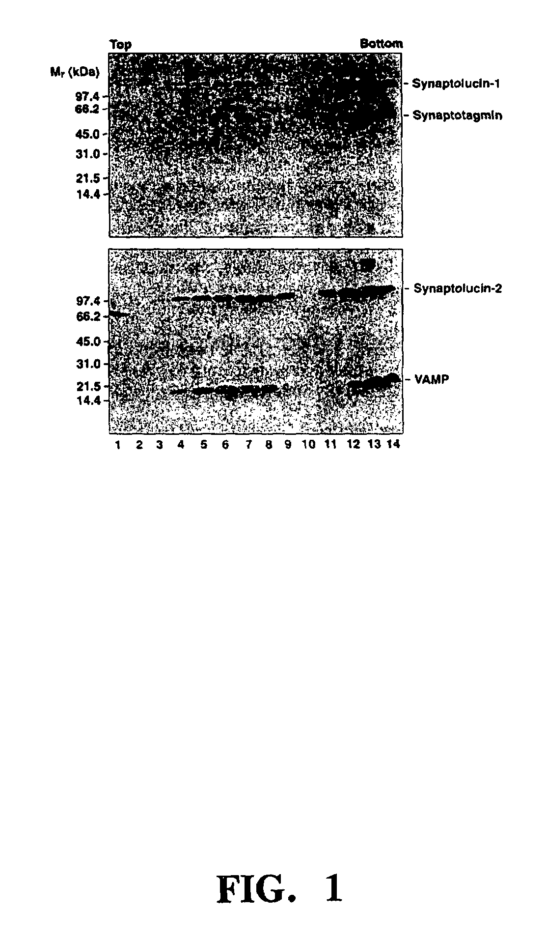
[0162]Synaptolucins. Total Cypridina RNA extracted with TRISOLV (Biotecx: Houston, Tex.) served as the template for the RT-PCR synthesis of a cDNA encoding the luciferase (16). The PCR product was subcloned into the amplicon plasmid pα4“a” (17,18) and its sequence determined with SEQUENASE 2.0 (United States Biochemical; Cleveland, Ohio). To construct synaptolucins-1 and -2, the appropriate portions of the open reading frames for Cypridina luciferase (16) and, respectively, rat synaptotagmin-I (19) and VAMP / synaptobrevin-2 (20,21) were fused via stretches of nucleotides encoding the flexible linker -(Ser-Gly-Gly)4-.

[0163]The amplicon plasmids were transfected into E5 cells (17,22) with the help of LIPOFECTAMINE (Gibco BRL; Bethesda, Md.), and replicated and packaged into virions after infection with 0.1 PFU / cell of the HSV deletion mutant d120 (22). The primary virus stock was passaged on E5 cells until the vector-to-helper ratio exceeded 1:4; the ratio was estimated as...

example 2

SynaptopHluorins

[0180]To generate pH-sensitive GFP mutants, a combination of directed and random strategies was employed. In the folded conformation of GFP, the chromophore is located in the core of the protein, shielded from direct interactions with the environment by a tight beta-barrel structure. The remarkable stability of the fluorescent properties of wild-type protein under environmental perturbations, such as pH changes, is due to the protected position of the chromophore. To allow GFP to function as a pH sensor, a mechanism had therefore to be found by which changes in external proton concentration could be relayed to and affect the chromophore.

[0181]The exemplified approach to converting GFP to a pH sensor involves amino acid substitutions that couple changes in bulk pH to changes in the electrostatic environment of the chromophore. To obtain such substitutions, residues adjacent to 7 key positions were mutated. These key positions are known from X-ray crystallography (Ref....


PUM

PropertyMeasurementUnit
Nanoscale particle sizeaaaaaaaaaa
Lengthaaaaaaaaaa

Abstract

The invention relates to methods and compositions which utilize the emission of light to monitor changes in microenvironments involving cells. The invention is especially useful for monitoring exocytotic activity such as detecting quantal release of synaptic vesicles. Fusion proteins of Cypridina luciferase and synaptotagmin-I or VAMP / synaptobrevin-2 were targeted to synaptic vesicles and, upon exocytosis, formed light-emitting complexes with luciferin present in the extracellular medium. Photon emissions in the presence of a depolarizing stimulus can be observed with these systems. pH-sensitive mutants of green fluorescent protein are also provided, which are useful for visualizing exocytosis and for imaging and measuring the pH of intracellular compartments.

Description

[0001]This application is a divisional of application Ser. No. 09 / 023,946, filed Feb. 13, 1998, now U.S. Pat. No. 6,670,449, which claims priority to U.S. Provisional Applications 60 / 038,179 filed Feb. 13, 1997, and 60 / 036,805, filed Feb. 14, 1997.TECHNICAL FIELD OF THE INVENTION[0002]This invention relates to compositions and methods for monitoring changes in the local environment inside or outside of cells by detecting optical changes in an environment-sensitive reporter molecule. In particular, this invention relates to compositions which may be specifically incorporated into cells, or may be caused to be produced by them, which produce an optically detectable signal in response to a change in the environment in which the optically sensitive reporter molecule is present. This invention is particularly applicable to detecting the release of substances stored in exocytotic vesicles of cells, such as synaptic vesicles, wherein the compositions of the invention are localized in the v...

Claims


Application Information

Patent Timeline
IPC IPC(8): C07H21/04C07K14/00C12Q1/68A01K67/027C07K14/435C07K14/47C07K14/705C12N5/02C12N5/10C12N9/02C12N15/09C12N15/62C12N15/85C12Q1/02
CPCC07K14/43595C07K14/47C07K14/705C12N9/0069C12N15/62C12N15/8509A01K2217/05A01K2267/0393C07K2319/00C12N2799/021C07K2319/055C07K2319/60C07K2319/61C07K2319/03
Inventor MIESENBÖCK, GERODE ANGELIS, DINOROTHMAN, JAMES E.
Owner MEMORIAL SLOAN KETTERING CANCER CENT
Learn More