Method and system of measuring characteristics of an organ

a technology of organs and characteristics, applied in the field of organ characteristics measurement methods and systems, can solve the problems of limiting the application of the technique to routine clinical care, affecting the accuracy of routine clinical use, and requiring a long time to outline the endocardial and epicardial boundaries of the left ventricl

Inactive Publication Date: 2006-12-26
AUCKLAND UNISERVICES LTD
View PDF10 Cites 87 Cited by
  • Summary
  • Abstract
  • Description
  • Claims
  • Application Information

AI Technical Summary

Benefits of technology

[0008]In broad terms in one aspect the invention comprises a method of measuring characteristics of an organ or part thereof of a subject from multiple images of the subject's organ or part thereof, the method comprising the steps of defining the spatial position of at least two of the slices; defining a reference model of the organ or part thereof scaled according to the distance between the slices; defining one or more boundary guide points associated with one or more slices for which the spatial positions have been defined; converting the guide points to three-dimensional coordinates; defining an estimate model by fitting the reference model to the guide points; and calculating the characteristics from the estimate model.
[0009]In broad terms in another aspect, the invention comprises a system for measuring characteristics of an organ or part thereof of a subject from multiple image slices of the subject's organ or part thereof, the system comprising a memory in which is stored the spatial position of at least two of the slices; reference model definition means arranged to define a reference model of the organ or part thereof scaled according to the distance between the slices; boundary guide point definition means arranged to define one or more boundary guide points associated with one or more slices for which the spatial positions are stored in the memory; conversion means arranged to convert the guide points to three-dimensional coordinates; estimate model definition means arranged to define an estimate model by fitting the reference model to the guide points; and calculation means arranged to calculate the characteristics from the estimate model.
[0010]In yet another form the invention comprises a computer program for measuring characteristics of an organ or part thereof of a subject from multiple image slices of the subject's organ or part thereof, the program comprising storage means arranged to store the spatial position of at least two of the slices; reference model definition means arranged to define a reference model of the organ or part thereof scaled according to the distance between the slices; boundary guide point definition means arranged to define one or more boundary guide points associated with one or more slices for which the spatial positions are stored in the memory; conversion means arranged to convert the guide points to three-dimensional coordinates; estimate model definition means arranged to define an estimate model by fitting the reference model to the guide points; and calculation means arranged to calculate the characteristics from the estimate model.

Problems solved by technology

A major limitation of the MRI slice summation method is the prohibitive time required to outline the endocardial and epicardial boundaries of the left ventricle in each slice.
This severely limits application of the use of the technique to routine clinical care.
In the past, many semi-automated image segmentation algorithms have been applied to this problem, but these solutions are frequently not sufficiently robust and accurate for routine clinical use.
In particular the image pixel intensities are insufficient to adequately constrain the segmentation problem, due to the limited temporal and spatial resolution, presence of image artifacts, and lack of contrast between blood and muscle.
The amount of time spent on manual editing and correction of contours obtained from these previous solutions renders automated methods nearly as slow as manual contouring in clinical practice.
Both techniques require the use of edge detection algorithms and in doing so suffer from the disadvantages discussed above.

Method used


Image

  • Method and system of measuring characteristics of an organ
  • Method and system of measuring characteristics of an organ
  • Method and system of measuring characteristics of an organ

Examples


Embodiment Construction

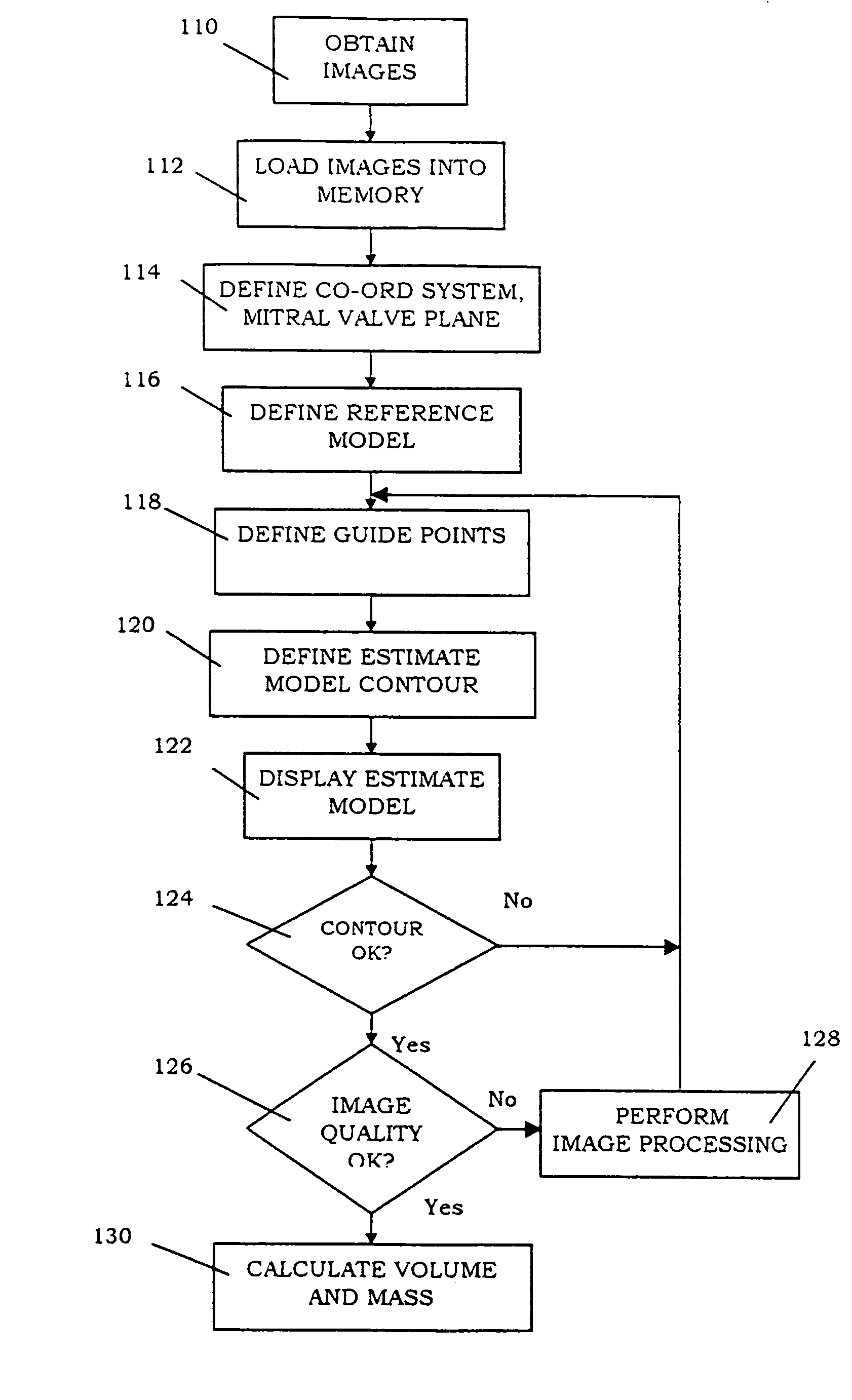
[0021]FIG. 1 sets out a preferred form of method of the invention.

[0022]A number of images are first obtained of the left ventricle of a subject as indicated at. The images could be acquired from an MRI scanner, or may alternatively be acquired by an ultra-fast CT, 3-dimensional ultrasound machine or echocardiography, or other suitable imaging modality. The images could also be obtained from confocal microscopy, electron microscopy or histology. The images are typically 2-dimensional cines or movies of the heart and are taken at standard orientations, for example, short axis and long axis, or at entirely arbitrary positions depending on the nature of the pathology and imaging modality.

[0023]The preferred images are acquired in a number of spatial locations, having a lowest or apical slice, a highest or basal slice, one or more middle slices and one or more long axis slices. The preferred images are acquired in between 2 and preferably 20 spatial locations, and typically 12 spatial l...


PUM


Abstract

The invention provides a method of measuring characteristics of an organ or part thereof from multiple images of the organ or part thereof, the method comprising the steps of defining the spatial position of at least two of the images; defining a reference model of the organ or part thereof scaled according to the distance between reference markers on the images; defining one or more boundary guide points associated with one or more images for which the spatial positions have been defined; converting the guide points to three-dimensional coordinates; defining an estimate model by fitting the reference model to the guide points; and calculating the characteristics from the estimate model. The invention also provides a system and a computer program for measuring characteristics of an organ or part thereof of a subject from multiple images of the subject's organ or part thereof.

Description

FIELD OF THE INVENTION[0001]The invention relates to a method and system of measuring characteristics of an organ or part thereof. The method and system of the invention are particularly suited to measuring cardiac function, volume, and / or mass of a ventricle of the heart of a subject from multiple image slices obtained by magnetic resonance imaging (MRI). The method and system of the invention may also be used to calculate the volume and / or mass and other characteristics of other organs such as a lung, kidney or brain, to measure the position of the wall of a blood vessel for the purposes of analysis of flow, or to calculate the volume and / or mass of a bone, tumour or cyst.BACKGROUND TO INVENTION[0002]Ventricular mass, volumes and wall thickness at end diastole and end systole are essential clinical parameters for diagnosis and management of many cardiac diseases. Magnetic resonance imaging (MRI) may be used to estimate heart wall motion by reconstructing the shape and motion of th...

Claims


Application Information

Patent Timeline
Patent Type & Authority Patents(United States)
IPC IPC(8): G06K9/00G01R33/54A61B5/055A61B5/107A61B6/00A61B6/03A61B8/00G06T1/00G06T3/00G06T5/00G06T7/60
CPCA61B5/055G06T7/0012G06T7/0083G06T7/602G06T7/606A61B5/107G06T2207/10088G06T2207/20101G06T2207/20192G06T2207/30048A61B5/7425G06T7/12G06T7/62G06T7/66
Inventor COWAN, BRETTYOUNG, ALISTAIR
Owner AUCKLAND UNISERVICES LTD
Why Eureka
Try Eureka