
Lenticular net instruments

a technology of lenticular nets and instruments, applied in the field of ocular medical instruments, can solve the problems of prolonged recovery time, cataracts, corneal decompensation and/or uveitis, and the inability to use an additional instrument unnecessarily, so as to achieve enhanced control over proper guidance, greater control over the movement of the net, and avoid the effect of extra cos

Inactive Publication Date: 2007-03-06
SABET SINA J
View PDF9 Cites 11 Cited by
  • Summary
  • Abstract
  • Description
  • Claims
  • Application Information

AI Technical Summary

Benefits of technology

[0013]Another object of the present invention is to prevent nuclear fragments from migrating into the vitreous due to a capsular rupture during cataract surgery.
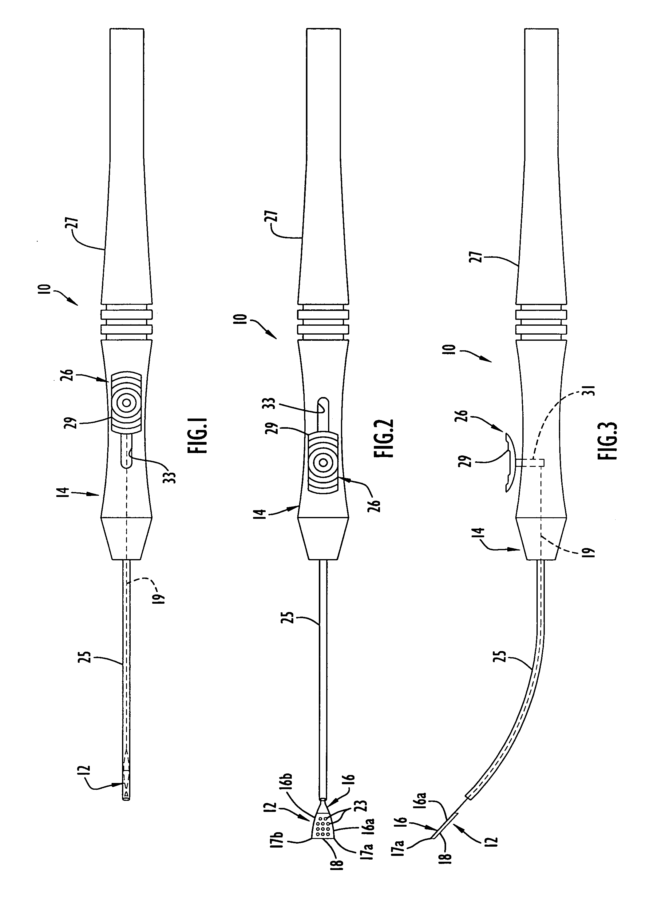
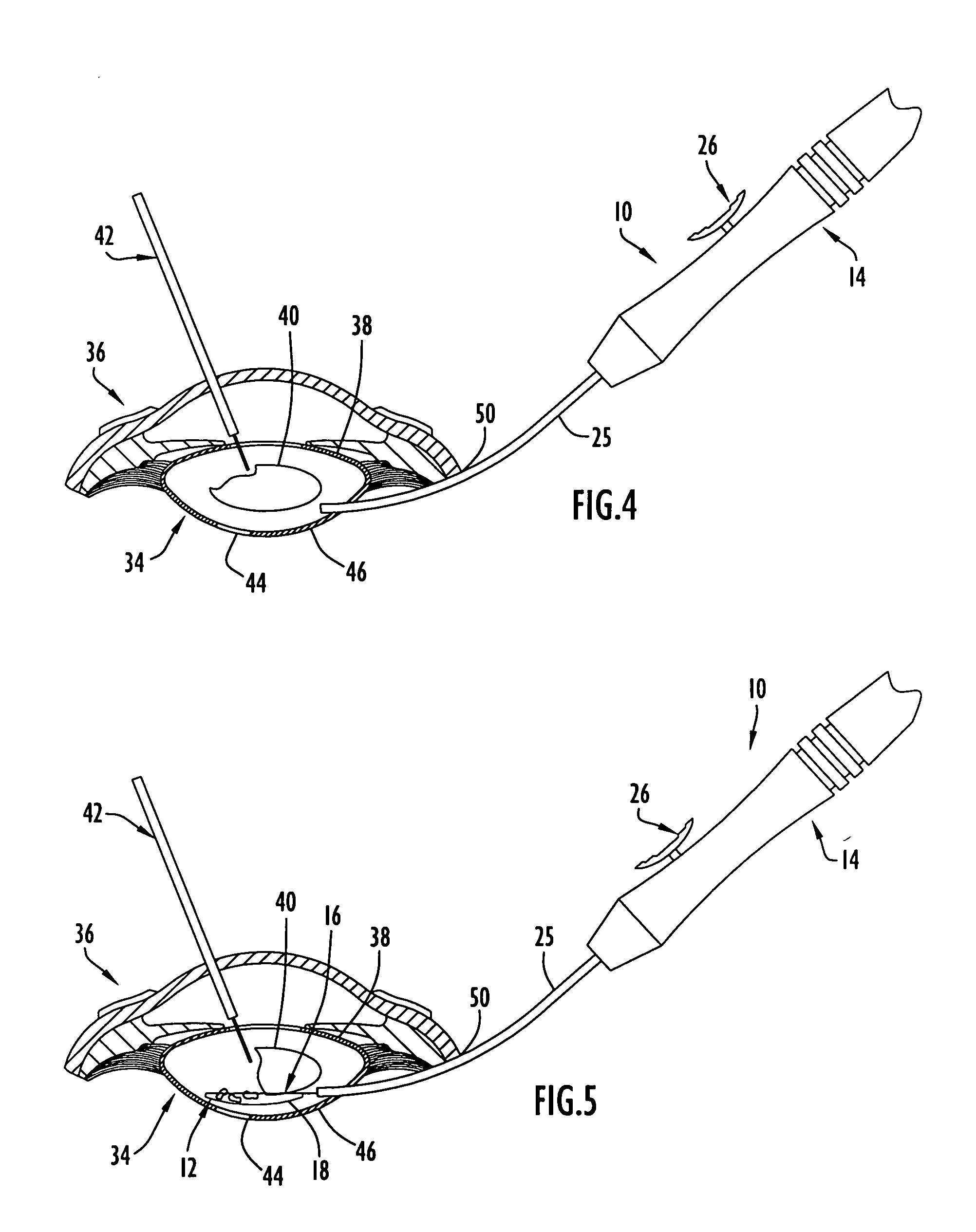
[0021]These and other objects, advantages and benefits are realized with the present invention as generally characterized in a lenticular net instrument for use in extracapsular cataract surgery in which a fragmenting instrument is used to fragment a cataractous nucleus within a lens capsule. The lenticular net instrument comprises a lenticular net, a handle attached to the net and an actuator on the handle for selectively moving the net to a contracted configuration and to an expanded configuration. The net in the contracted configuration is mechanically constrained to assume a narrow, contracted profile permitting insertion of the net in a lens capsule through a relatively small incision in the eye and positioning of the net in the lens capsule at or adjacent a desired deployment site behind a cataractous nucleus which has started to sink behind a ruptured capsule into the vitreous cavity. The net is movable via the actuator from the contracted configuration to the expanded configuration in which the net has a predetermined expanded profile between the cataractous nucleus and the ruptured capsular wall. In the expanded configuration, the predetermined expanded profile of the net covers the rupture and may have a length and width to encompass the cataractous nucleus posteriorly. The predetermined expanded profile may correspond substantially to the anatomical curvature of the posterior capsular wall. The net in the expanded configuration between the cataractous nucleus and the ruptured capsular wall blocks or collects migrating fragments of the cataractous nucleus produced via fragmentation of the cataractous nucleus with a fragmenting instrument so that the fragments are prevented from falling further into the vitreous cavity while fragmentation of the nucleus continues during the cataract surgery. The net has a plurality of holes or openings therein of a size to prevent the passage therethrough of nuclear fragments while permitting flow therethrough to deter undue traction on the vitreous gel. Once fragmentation of the nucleus is completed and the nuclear fragments have been aspirated from the lens capsule, the net is moved from the expanded configuration back to the contracted configuration via the actuator for withdrawal from the eye through the small incision. The net may comprise a frame and a layer of openwork material attached to the frame, with the holes being provided in the openwork material.

Problems solved by technology

During their lifetimes, many people become afflicted with cataracts, i.e. lenticular opacities that interfere with vision.
Intracapsular cataract surgery has numerous disadvantages including the need for undesirably large incisions to accommodate the lens capsule as it is withdrawn from the eye, prolonged recovery times, the risk that the vitreous body, normally contained behind the posterior capsular wall, may move forward into the anterior chamber of the eye, and potential complications including retinal detachment, cystoid macular edema, glaucoma, corneal decompensation and / or uveitis.
Following intracapsular cataract surgery, vision is typically restored in the patient with corrective eye glasses or contact lenses, since the absence of the lens capsule makes it difficult for an intraocular lens implant to be properly supported in the eye.
Although extracapsular cataract surgery is usually preferred over intracapsular cataract surgery, extracapsular cataract surgery presents the risk that the posterior capsular wall may be ruptured inadvertently, such as due to unintended contact with the fragmenting instrument and / or due to the suction used to remove the nuclear fragments.
Rupture of the posterior capsular wall during extracapsular cataract surgery gives rise to many of the same disadvantages associated with removal of the lens capsule in intracapsular cataract surgery.
Particularly, mixing of nuclear material with the vitreous body may manifest as uveitis, resulting in an inflammatory reaction in the eye.
A vitrectomy may be necessary to remove nuclear fragments or pieces from the vitreous body; however, a vitrectomy itself has potential adverse consequences, including retinal complications, for the patient.
Complete removal of the nuclear material from the vitreous is difficult to accomplish; and, if all of the nuclear fragments or pieces are not removed, the patient is at risk for various complications including cystoid macular edema, glaucoma, corneal decompensation and uveitis.
Often, removal of these fragments requires a secondary procedure by a retinal specialist, at considerable cost to the patient and / or third party payer.
Since damage to the posterior capsular wall is not an inevitable occurrence during cataract surgery, the patient may be unnecessarily placed at increased risk from the shield being routinely inserted in the lens capsule.
Accordingly, the shield must be inserted in the eye prior to removal of the cataractous nucleus, and is not suited for insertion in the eye where some of the nucleus has already been removed.
Grasping the shield with an instrument is tedious and time consuming and may require numerous trial and error attempts.
Since the shield is solid, undesired pressure imbalances or differentials in the eye may occur due to there being no air or fluid flow through the shield.

Method used


Image

  • Lenticular net instruments
  • Lenticular net instruments
  • Lenticular net instruments

Examples


Embodiment Construction

[0036]A lenticular net instrument 10 according to the present invention is illustrated in FIGS. 1–3 and includes a lenticular net 12 attached to an elongate handle 14. The lenticular net 12 includes a frame 16 and a layer of openwork material 18 connected to frame 16. Frame 16 comprises first and second arms or frame parts 16a and 16b biased to extend angularly outwardly away from one another in opposite directions from a distal end of an elongate operating member 19 in an expanded configuration for net 12 as shown in FIGS. 2 and 3. In the case of lenticular net instrument 10, the central longitudinal axis of operating member 19 is disposed in a plane, and the arms 16a and 16b extend angularly outwardly on opposite sides of the plane in the expanded configuration. The arms 16a and 16b comprise wires of minimal cross-sectional size extending distally from the distal end of the operating member 19 to distal ends or tips 17a and 17b, respectively. In the expanded configuration for net ...


PUM


Abstract

A lenticular net instrument comprises an elongate handle coupled with a lenticular net movable to contracted and expanded configurations via an actuator of the handle. The net in the contracted configuration has a narrow profile for insertion in and removal from a lens capsule through a small incision in the eye. The net is movable to the expanded configuration within the lens capsule behind a cataractous nucleus. The net has a plurality of openings therein of a size to prevent fragments of the cataractous nucleus produced by fragmentation with a fragmenting instrument from passing therethrough such that the fragments do not pass into the vitreous cavity. A method of cataract surgery involves deploying a net behind a cataractous nucleus upon the occurrence of a rupture in the capsular wall.

Description

CROSS-REFERENCE TO RELATED PATENT APPLICATION[0001]The subject patent application claims priority from prior provisional patent application Ser. No. 60 / 340,480 filed Dec. 18, 2001, the entire disclosure of which is incorporated herein by reference.BACKGROUND OF THE INVENTION[0002]1. Field of the Invention[0003]The present invention relates generally to ocular medical instruments and, more specifically, to safety net instruments for use in cataract surgery.[0004]2. Discussion of the Related Art[0005]During their lifetimes, many people become afflicted with cataracts, i.e. lenticular opacities that interfere with vision. Indeed, cataracts may be considered the most prevalent visually disabling eye disease in the world. The cloudiness or opacity associated with cataract arises in the nucleus of the anatomical lens, which includes a lens capsule also known as the capsular bag, the nucleus within the lens capsule, and gelatinous cortical material between the nucleus and the lens capsule....

Claims


Application Information

Patent Timeline
Patent Type & Authority Patents(United States)
IPC IPC(8): A61F9/00A61F9/007
CPCA61F9/007A61B2017/00287A61F9/00736
Inventor SABET, SINA J.
Owner SABET SINA J
Learn More