
Filter devices for depositing material and density gradients of material from sample suspension

Inactive Publication Date: 2007-05-01
PERCEPTRONIX MEDICAL
View PDF26 Cites 10 Cited by
  • Summary
  • Abstract
  • Description
  • Claims
  • Application Information

AI Technical Summary

Benefits of technology

[0019]Another object of the present invention is to provide a filter from which several deposits can be made in a time-efficient manner.
[0020]Another object of the present invention is provide a filter from which a concentration gradient can be made without the need to prepare multiple dilutions of the sample suspension.
[0021]Another object of the present invention to provide a filter from which a concentration gradient can be captured and deposited in a time-efficient manner.
[0022]Another object of the present invention is to provide a filter which provides the ability to make several deposits from the material collected by the filter in which all of the deposits are representative of the sample suspension from which the material was collected.
[0023]Briefly, and in accordance with the foregoing, the present invention provides a simple and inexpensive apparatus for and method of depositing material from a sample suspension. Accordingly, cells or material may be deposited in near mono-layers for the detection of disease. The apparatus and method of the present invention collects material from a single sample suspension. The collected material is then used to make multiple deposits on a single slide or on multiple slides. A filter assembly of the present invention also provides for the collection and deposit of concentration gradients. Thus, providing a desired range of cell concentrations for cytological examination and ensuring that an appropriate concentration of material is present on the receiving surface for the desired analysis.

Problems solved by technology

Achieving an appropriate concentration and distribution of material for examination or analysis is a limitation of many sample preparation techniques.
This prior art does not teach or derive advantage by capturing material on a filter which extends beyond the dimensions of the receiving surface.
While capturing material on filters, filter dimensions are typically kept to a minimum since filter deformation may cause inconsistencies in flow and thus material capture by the filter.
Under less favorable conditions the filter itself could tear, otherwise fail or its characteristics may be compromised.
Similarly, although filters are often supplied in a support structure, when material distribution is important for analysis, flow impediments in the vicinity of the carrier or support structure are viewed as problematic.
One limitation of using multiple small filters to capture multiple portions of material is that flow rate and other conditions for cell capture must be monitored closely to prevent non-representative samples or inadequate preparations.
Similarly, excess material, sparseness or substantial variations in cellular concentration may impede or otherwise confound diagnosis.
In order to achieve the appropriate concentration of material, however, these techniques are sometimes complicated and require expensive equipment and a substantial amount of time to perform.
Although the concentration of the collected material can be monitored, one difficulty with using multiple filters results not from the design of the device for monitoring the concentration, but from the nature of biological samples.
A related difficulty is that once any material is removed from the sample suspension, the characteristics of the sample have changed and replicates are no longer possible.
Unfortunately, with filter transfer methods, as cells are captured on the membrane, the concentration of constituents in the sample suspension are altered and therefore subsequent preparations from this sample suspension may no longer be representative.
And in some cases, once any material has been removed from the original sample suspension, additional preparations from this may not even be suitable for the intended use.
Similarly, repeated blots from the same area of a filter will not produce representative slides.
Another limitation for many analysis techniques, including cytology, is that to be effective, the concentration of material must fall within a target range.
Unfortunately, in the majority of cases the concentration of cells and constituents, in any given sample, is not known a priori.
While cell counters, sample dilutions, differential pressure and other techniques are commonly employed to monitor or otherwise control the concentration of cells deposited, these require additional equipment, time and expertise.
Even then for a variety of reasons the resulting cell deposition may be inadequate.
In addition, a more restricted set of applications would benefit from a cell or material deposition in the form of a concentration gradient.

Method used


Image

  • Filter devices for depositing material and density gradients of material from sample suspension
  • Filter devices for depositing material and density gradients of material from sample suspension
  • Filter devices for depositing material and density gradients of material from sample suspension

Examples


Embodiment Construction

[0042]While the invention may be susceptible to embodiment in different forms, there is shown in the drawings, and herein will be described in detail, specific embodiments with the understanding that the present disclosure is to be considered an exemplification of the principles of the invention, and is not intended to limit the invention to that as illustrated and described herein.

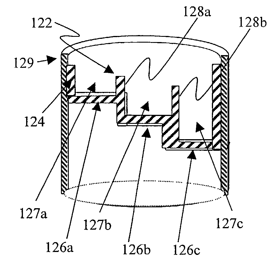
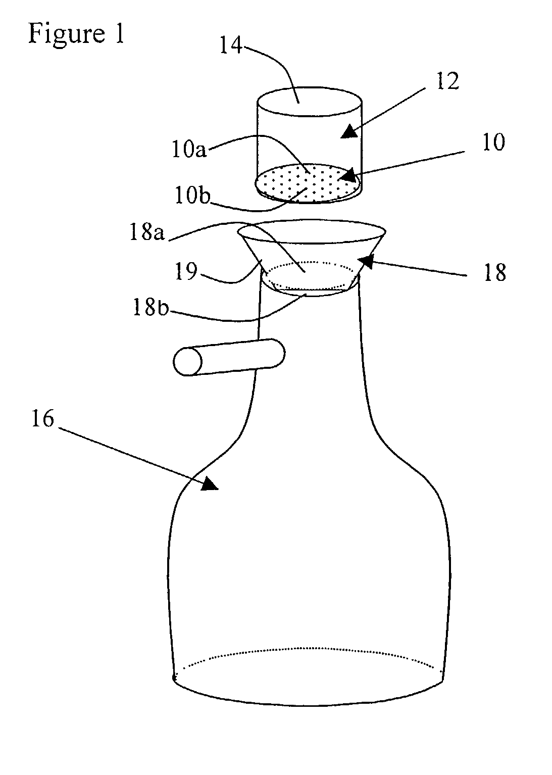
[0043]FIG. 1 shows a generally disk-shaped filter 10 installed at the bottom of a sample chamber 12. The filter 10 has an upper collection surface 10a and a lower surface 10b. The sample chamber 12 is generally cylindrical, providing a top opening 14 for the introduction of a sample suspension. The perimeter of the filter 10 is slightly smaller than the interior dimension of the sample chamber 12. Thus the perimeter of the filter 10 extends to the interior surface of the sample chamber 12. The sample chamber 12 is placed over a vacuum assembly 16 and is preferably fitted thereto through an adapter cone 18...


PUM

PropertyMeasurementUnit
Concentrationaaaaaaaaaa
Densityaaaaaaaaaa
Dimensionaaaaaaaaaa

Abstract

The present invention provides a simple, inexpensive apparatus for and method of capturing biological or other material from sample suspensions on a collection surface and for depositing captured material on a receiving surface, such as a microscope slide, for observation and analysis. Accordingly, cytological material may be deposited in near mono-layers for the detection of disease. In one embodiment of the present invention, density gradients of material are captured and deposited, thus providing a desired range of concentrations for examination. In another embodiment multiple depositions or multiple slides may be made from the same sample suspension. The method and apparatus to exploit this new method are both presented for various applications. The dimensions of material captured and deposited may be controlled and used in conjunction with microscope slides that confine material to specific regions of interest.

Description

BACKGROUND OF THE INVENTION[0001]The present invention relates to the capture of cells or other material from a sample suspension and the deposition of cells or other material onto an appropriate receiving surface, such as a microscope slide for observation.[0002]In the field of cytology, human and machine vision systems perform effectively on near mono-layer depositions of cellular material. Expanded use of machine vision for slide examination, a growing number of special stains and the development of molecular markers have increased the need to prepare multiple representative depositions or multiple slides from the same sample suspension. However, care must be taken in the deposition of cellular material used for cytological examination. In particular, to diagnose disease, slides must be representative of the sample suspension, which ideally is representative of the patient. And if multiple cellular depositions are made on the same or on different slides, then each of these deposi...

Claims


Application Information

Patent Timeline
IPC IPC(8): G01N30/00C12M1/12G01N1/18G01N1/28G01N15/02
CPCG01N1/18G01N1/2813G01N15/0272G01N1/4077Y10T436/25375Y10T436/25Y10T436/255
Inventor FERGUSON, GARY WILLIAMUS-KRASOVEC, MARIJAFLEZAR, MARGARETAZGANEC, MARIOLAVRENCAK, JAKAPALCIC, BRANKO M.
Owner PERCEPTRONIX MEDICAL
Learn More