System for cutting the cornea of an eye

a technology of corneal surgery and cutting device, which is applied in the field of surgical systems for cutting cornea of, can solve the problems of loss of vision, and long recovery time of penetrating keratoplasty, and achieve the effect of reducing the risk of expulsive suprachoroidal hemorrhag

Active Publication Date: 2007-05-29
SHIUEY YICHIEH
View PDF26 Cites 48 Cited by
  • Summary
  • Abstract
  • Description
  • Claims
  • Application Information

AI Technical Summary

Benefits of technology

[0031]One advantage of the present system is that it is able to create a pocket of uniform depth within the cornea. Another advantage of the present system is that it is able to create a cut into the cornea wherein the cut has an external opening that is smaller than the internal dimensions of the pocket. Accordingly, the present invention may be used to transplant a portion of the inner layer of the cornea with the advantage that such transplantation may occur in a relatively closed system protected from atmospheric pressure. This advantageously reduces the risk of expulsive suprachoroidal hemorrhage. Additionally, having the external opening be smaller than the internal dimensions of the pocket will also make the eye much more resistant to trauma than would be the case in penetrating keratoplasty.

Problems solved by technology

Disorders of the cornea, which adversely affect its shape or clarity, can cause loss of vision.
However, there are distinct disadvantages of penetrating keratoplasty.
For example, penetrating keratoplasty has a long recovery time and typically takes between 6 to 12 months to achieve good vision.
Moreover, because the donor corneal tissue is sutured manually, even in the hands of an experienced corneal surgeon, irregularities in the shape of the cornea frequently occur and can produce decreased vision because of induced astigmatism.
The donated corneal tissue can also be rejected by the recipient's immune system with resulting loss of transparency of the donated cornea.
Penetrating keratoplasty also has the potential for a devastating complication called expulsive suprachoroidal hemorrhage.
The terrible result is that the retina, vitreous, and crystalline lens may be expulsed from the opening in the cornea resulting in blindness.
Endophthalmitis (i.e. infection of the inside of the eye) is another serious complication that can occur and can also cause blindness if treatment is unsuccessful.
Finally, after penetrating keratoplasty, the eye is very sensitive to injury, since the junction of the transplanted cornea and the recipient cornea can be easily disrupted with even mild trauma.
In Fuchs' endothelial dystrophy, the endothelium is damaged and is unable to adequately pump fluid out of the cornea, which results in swelling and opacification of the cornea.
Unfortunately, corneal tissue removal and replacement by the free hand method is extremely difficult to perform.
Under the best of circumstances, it usually results in irregular astigmatism that is caused by irregularities in the thickness of the corneal tissue removed as well as in the thickness of the transplanted tissue.
Unfortunately, a problem that can occur with this technique is that the transplanted donor disk may be dislodged with relatively minor trauma, even after prolonged periods of time.
This can occur because the cap of corneal tissue is only held in place by the relatively weak healing between the layers of donor and recipient tissue and there is no support against lateral or vertical pressure.
Unfortunately, the main disadvantage of such free hand technique is that it is very difficult to manually create a pocket in the corneal tissues, wherein the pocket is of uniform depth.
Unfortunately, the inability to create a uniform pocket will necessitate the abandonment of posterior lamellar keratoplasty and will require conversion to traditional penetrating keratoplasty.
A disadvantage of this technique is that, like penetrating keratoplasty, the inside of the eye is exposed to atmospheric pressure and therefore there is also a risk of suprachoroidal hemorrhage with this technique.
Another disadvantage is that post-operatively the eye is still fairly vulnerable to injury.
For example, even minor trauma could result in flap dislocation or rupture of the transplant-recipient junction.
Two disadvantages of this technique are the high cost of lasers and potential difficulty for the laser to create incisions in corneas that are scarred or opacified.
Ametropia, the incorrect focusing of light rays onto the retina, is the most common cause of decreased vision in humans.
In this procedure, there is considerable variability in the size and shape of the laser treatment.
However, with current corneal surgical devices the size and shape of the flap that covers the laser treatment is unfortunately rather limited.
The disadvantage of either a manual technique or a non-motorized technique is that the uniformity of the pocket is largely dependent on the surgeon's skill and experience and therefore there can be a high degree of variability.

Method used


Image

  • System for cutting the cornea of an eye
  • System for cutting the cornea of an eye
  • System for cutting the cornea of an eye

Examples


Embodiment Construction

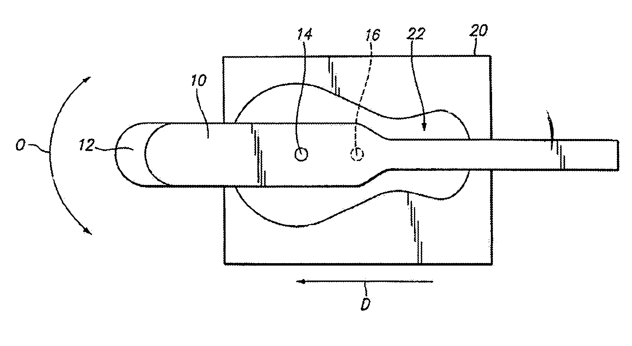
[0051]In preferred aspects, the present invention provides a corneal surgery system that can be used to cut a live or donor cornea to form a pocket, flap or cap by separating the layers of the cornea. Specifically, the present invention provides a system for automatically creating a pocket of uniform depth, which can be of various shapes and sizes, between the layers of a live or donor cornea. The present invention may also be used to create a flap or cap of corneal tissue in a live or donor cornea.

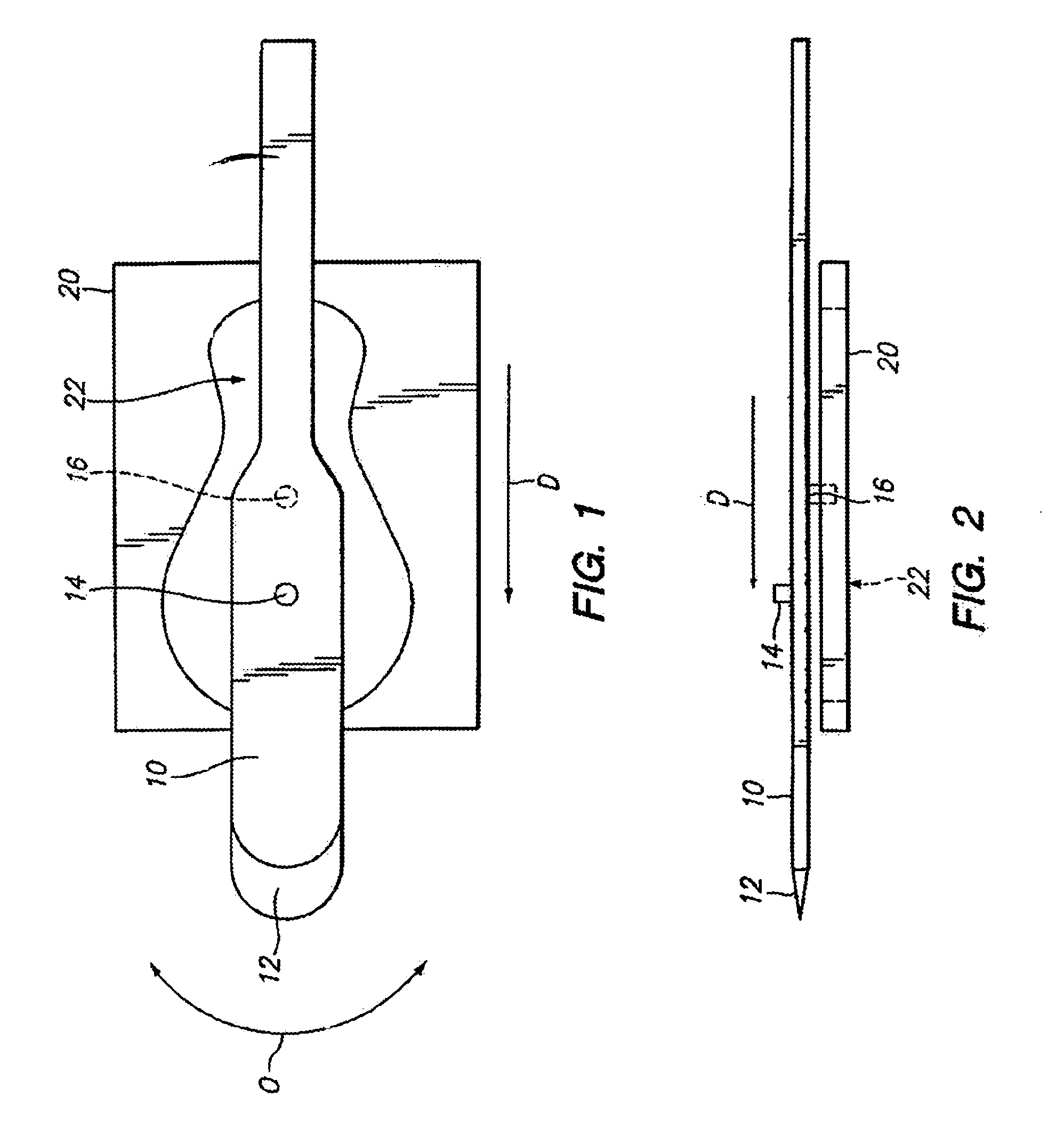
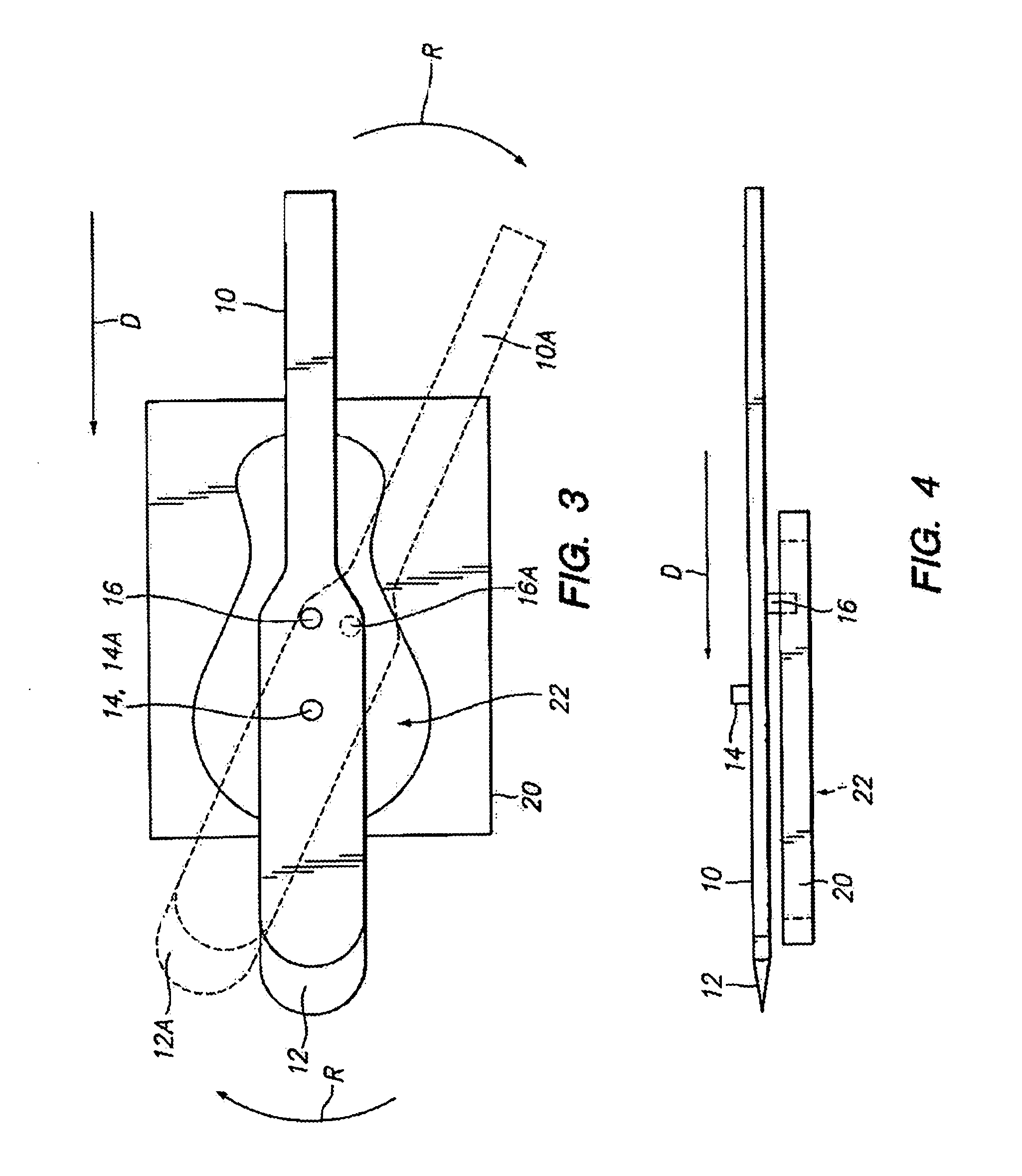
[0052]In accordance with the present invention, a system for cutting a cornea is provided. The system comprises a cutting blade that is moved back and forth in an arcuate path while simultaneously being advanced to cut through a cornea. As will be explained, the degree of angular movement of the cutting blade is limited by contacts between a moveable member (to which the cutting blade is attached) and a cutting guide.

(b) Components of the Present Invention

[0053]Operation of the present in...


PUM


Abstract

A system for cutting the cornea of an eye includes a moveable member with a cutting blade at one end, a pivot element disposed thereon, a cutting guide restraint disposed thereon, a mechanism for oscillating the moveable member around the pivot element, and a cutting guide configured to engage the cutting guide restraint on the moveable member to thereby limit the degree of angular movement of the cutting blade as the moveable member oscillates about the pivot element. A pocket can be cut in the cornea by advancing and oscillating the moveable member.

Description

TECHNICAL FIELD[0001]The present invention relates to surgical systems for cutting the cornea of a patient's eye.BACKGROUND OF THE INVENTION(a) The Cornea, Its Diseases and Current Treatments:[0002]The cornea is the clear cover of the eye and is also the main focusing lens in the eye. Disorders of the cornea, which adversely affect its shape or clarity, can cause loss of vision. Such disorders include Fuchs' endothelial dystrophy, pseudophakic bullous keratopathy, keratoconus, and herpes virus infection. When these conditions are severe the most common treatment is a full thickness corneal transplant which is also known as penetrating keratoplasty.[0003]Penetrating keratoplasty is the removal of a full-thickness disk of diseased corneal tissue followed by the replacement of the diseased full-thickness disk of tissue by a full thickness disk of donated healthy corneal tissue. Currently, the diseased tissue is removed by the use of a non-automated or automated corneal trephine combine...

Claims


Application Information

Patent Timeline
Patent Type & Authority Patents(United States)
IPC IPC(8): A61F9/00A61F9/013
CPCA61F9/0133A61F2/142A61F9/013
Inventor SHIUEY, YICHIEH
Owner SHIUEY YICHIEH
Why Eureka
Try Eureka