[0008]Such a set-up and such a method enable the required measurements to be performed in one process. This is substantially more pleasant for the person undergoing such a measurement. Moreover, this set-up also ensures that a detection of the optical properties of
layers beneath the cornea occurs with a required local calibration, because such a local calibration is enabled by the detection of the surface
topography of the cornea. It is thus no longer necessary that the person rigidly stares into the focusing light during the measurement of the optical properties of layers disposed beneath the cornea, which is physiological not possible. Rather, smaller deviations can be measured accordingly by the detected surface
topography. Thus, the apparatus and the method of the present invention allow for the first time determination of the optical properties of the entire eye with adequately high local precision.
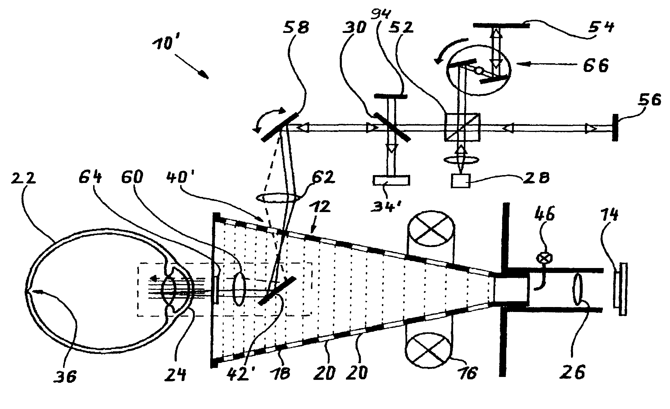
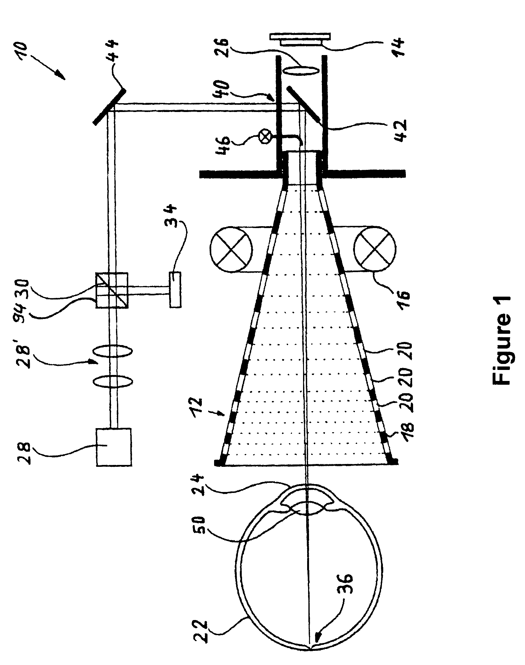
[0013]The measurements will be particularly precise and easy to perform when a Placido Topometer is used for projecting the pattern onto the surface of the cornea. In this process, the
laser beam can be guided on their path towards the eye and back through the beam of the Placido Topometer for detecting the optical properties of layers disposed beneath the cornea. For this purpose suitable deflection means such as tilted mirrors or deviating prisms are used.
[0014]For determining the optical properties of layers disposed beneath the cornea it is possible to introduce a known beam profile or a known wave front of a
laser source in the zone of the pupillary opening and to direct the same onto the cornea and the lower sections of the eye. By determining the profile form of a wave front that is reflected by the eye and by comparing such profile with the wave front that is sent into the eye, it is possible to detect the optical properties. A Hartmann-Shack
detector is particularly suitable for this purpose. Such an arrangement will yield a particularly precise picture of the optical properties of the layers of the eye that are disposed beneath the cornea.
[0015]Alternatively or cumulatively, it is also possible to provide
optical coherence tomography (OCT) in order to determine the optical properties of the layers disposed beneath the cornea. Such a coherence
tomography has proven its worth and reliably supplies tomographs from the entire eye and also contains information on the
layer thickness of the individual portions of the eye that are relevant for the
refraction (biometry). In particular, the method and apparatus of the present invention can be used for a substantial improvement of
optical coherence tomography because movement artifacts can be respectively corrected by the continuous simultaneous detection of the surface topography of the cornea. Mathematical calculations based on these surface topographies can be used to compensate for corneal movements during
data acquisition, and can thus also be used to compensate the movement errors for the OCT measurement.
[0016]The
corneal topography in the central corneal area (e.g., the central portion of the
cornea surface that is equal to or less than ½ of the area of the total
cornea surface) can also be determined in particular in such a way that in the central Placido-ring-free area of the cornea, a short-coherent measurement
system, preferably a
laser measurement
system, is mirrored in and is aimed at the cornea and the lower sections of the eye coaxially to an
optical axis extending through the
pupil and the
retina. In addition, the OCT offers the
advantage of measuring the morphology and other optical features of the different layers inside the eye. For example, these data can be used to measure and analyze the
corneal wound healing process in the stromal tissue after
refractive surgery.
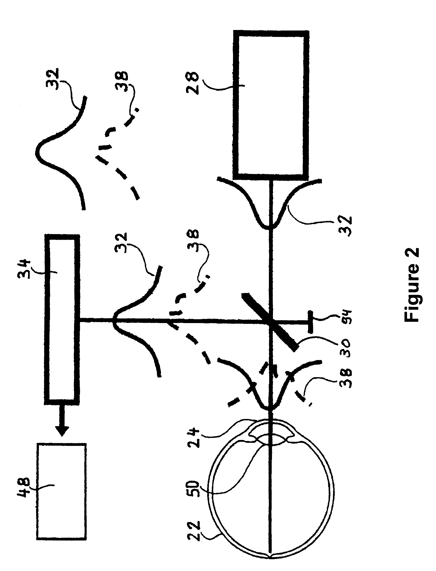
[0020]OCT measurements of more than one point (centrally and paracentrally) allow the measurement of the lens in situ. Such measurements can be achieved by splitting the OCT (either statically using prisms, or dynamically using a scanning device) and subsequently assessing the differences between each beam's run-time.
Beam splitting in one of the described manners allows
triangulation measurements of the lens position, which is essential morphometric information.

