Method and apparatus for tracking an internal target region without an implanted fiducial

a target region and internal technology, applied in the field of tracking the location can solve the problems that the target region in the abdomen or the chest cannot be treated with this method alone, the x-ray camera system cannot be used to determine the position of the target region, and the difficulty of aiming

Inactive Publication Date: 2007-08-21
ACCURAY
View PDF7 Cites 193 Cited by
  • Summary
  • Abstract
  • Description
  • Claims
  • Application Information

AI Technical Summary

Benefits of technology

[0009]The invention may be used in conjunction with one or more real-time sensors to track the position of the target region on a real-time basis. In order to achieve real time tracking, the signal from the real-time sensor is correlated with the position of the target region. The correlation model is produced by simultaneously taking an x-ray and reading the signal from the real-time sensor, then using the x-ray to identify the best-matching three-dimensional image that shows the target position. Once the correlation is established, the position of the target region can be tracked real time during treatment by reading the signal from the real-time sensor almost continuously.

Problems solved by technology

However, this x-ray camera system cannot be used to determine the position of a target region if the position of the target region relative to a bony landmark changes because the target, e.g., a tumor, is generally not visible in x-ray images.
For example, a target region in a patient's abdomen or chest cannot be treated with this method alone.
While accurate aiming of the beam is not difficult when the tumor is in a body part that does not move, such as the brain, aiming becomes a challenge when the tumor is in or near a body part that moves, such as the lungs.
As the change in the position of the tumor does not necessarily correlate with the change in the position of an external surface of the patient, a tumor cannot be easily tracked based on external measurements alone.
For example, placing external sensors on a patient's chest and tracking the movement of the sensors does not provide accurate information about the position of the tumor inside the chest cavity because a certain soft tissue structure may move in one direction while bones move in another direction.
Nor can the tumor be located with x-ray systems, because in most cases, neither the target region nor the surrounding soft tissues are visible in the x-ray images.
The two-dimensional nature of x-ray images compromises the accuracy with which the radiation is applied to the target region.
Moreover, even if x-ray provided accurate enough data, real-time tracking of target region cannot be performed with x-ray imaging alone because of the excess radiation that the patient would be exposed to.
Unfortunately, use of internal fiducials in the above manner has its disadvantages.
First, the x-ray imaging process is too slow and too invasive to track the respiration motion in real-time.
Second, the implanting of the fiducials is an invasive and expensive procedure because the procedure usually takes place under the surveillance of a computer tomography (CT) device and in the presence of a surgeon.
The required presence of the surgeon not only drives up the cost of the procedure for the patient but also exposes the surgeon to ionizing radiation.
Furthermore, there is a real risk of complications that could result from the fiducial placement.

Method used


Image

  • Method and apparatus for tracking an internal target region without an implanted fiducial
  • Method and apparatus for tracking an internal target region without an implanted fiducial
  • Method and apparatus for tracking an internal target region without an implanted fiducial

Examples


Embodiment Construction

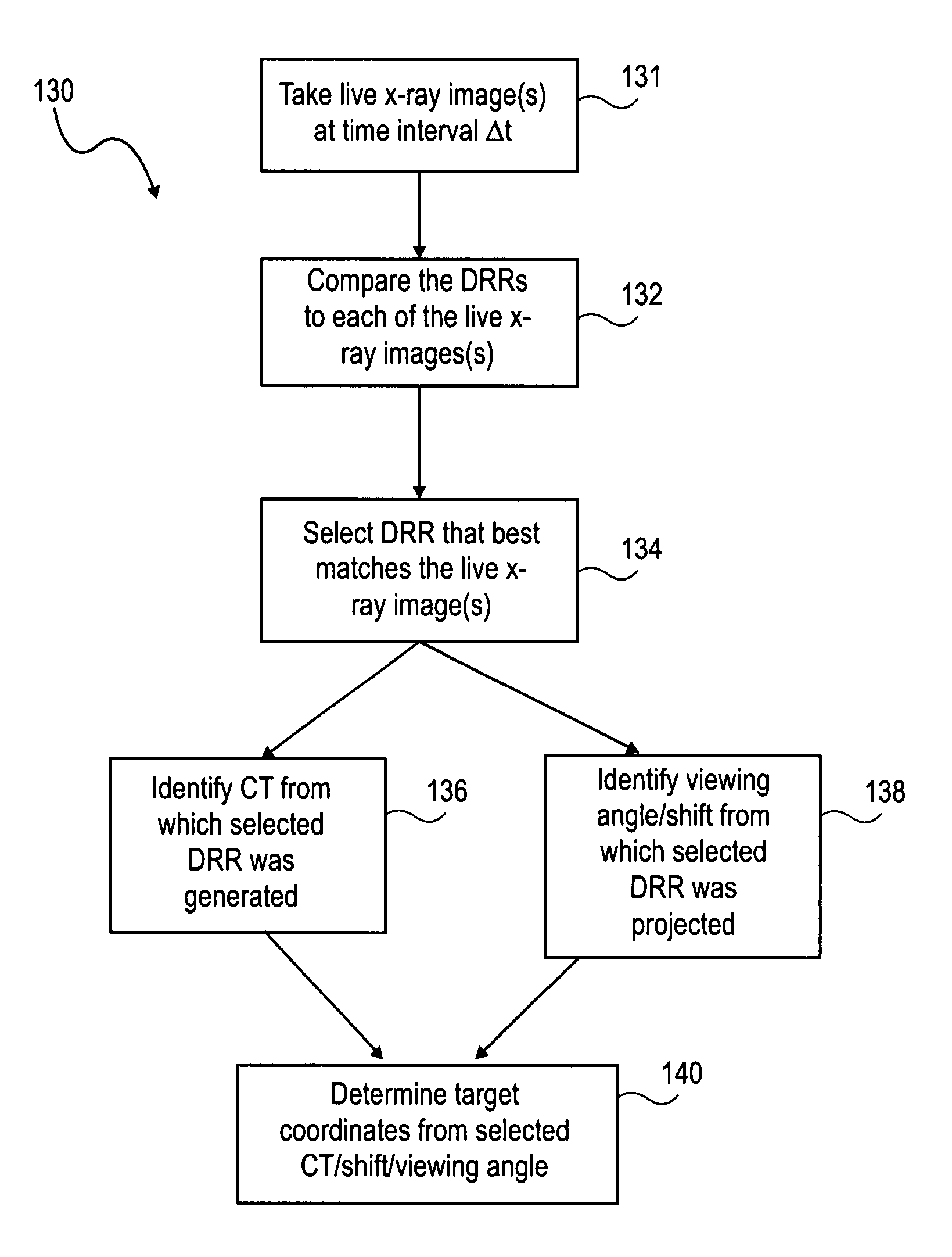
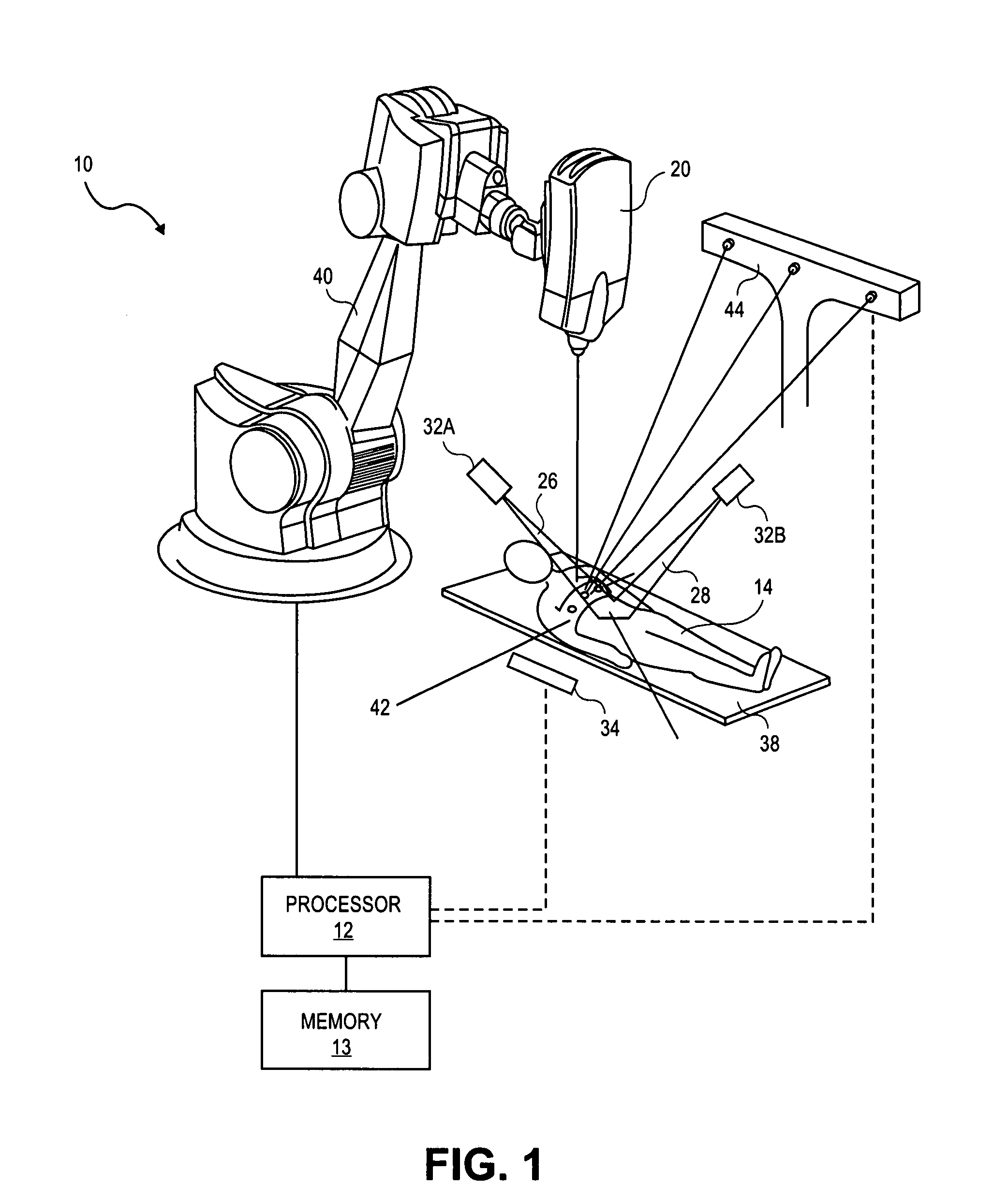
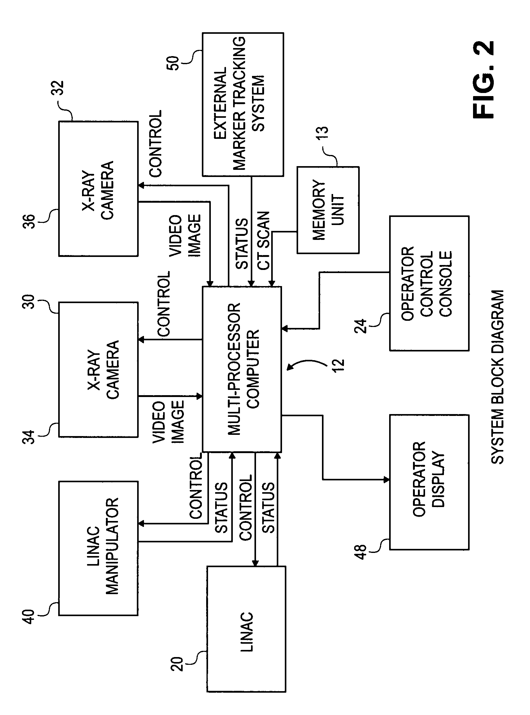
[0021]The invention is particularly applicable to an apparatus and method for directing a radiation beam towards an internal target region without implanting an internal fiducial in or near the target region to determine its location, and it is in this context that the invention will be described. It will be appreciated, however, that the apparatus and method in accordance with the invention has greater utility, such as to other types of medical procedures with other types of medical instruments, such as positioning biopsy needles, ablative, ultrasound or other focused energy treatments, or positioning a laser beam for laser beam treatment. Prior to describing the invention, a typical radiosurgery device will be described to provide a better understanding of the invention.

[0022]As used herein, a “target region” is the region to which a treatment (e.g., radiation) is to be directed. A target region is located in a “relevant volume,” which refers to an internal region surrounding the ...


PUM


Abstract

A method and apparatus for locating an internal target region during treatment without implanted fiducials is presented. The method comprises producing a plurality of first images that show an internal volume including the internal target region, then producing a live image of the internal volume during treatment and matching this live image to one of the plurality of first images. Since the first images show the internal target region, matching the live image to one of the first images identifies the position of the target region regardless of whether the second image itself shows the position of the target region. The first images may be any three-dimensional images such as CT scans, magnetic resonance imaging, and ultrasound. The live image may be, for example, an x-ray image. The invention may be used in conjunction with a real-time sensor to track the position of the target region on a real-time basis.

Description

BACKGROUND OF THE INVENTION[0001]This invention relates generally to an apparatus for improving the accuracy and efficacy of surgical treatments and more particularly to tracking the location of the target region that is displaced during the treatment due to respiratory and other patient motions.[0002]Various treatments require accurate tracking of the location of a target region in order to apply or deliver the treatment to the target region. In radiation therapy and radiosurgery, for example, a tumor may be destroyed by a beam of ionizing radiation that kills the cells in the tumor. As it is desirable to direct the radiation beam only to the tumor and not to the healthy tissues surrounding the tumor, accurate aiming of the beam at the tumor is of paramount importance in radiation treatments. The goal is to focus a high dose of radiation to the tumor while minimizing the exposure of the surrounding healthy tissue to radiation. For adequate distribution of radiation dosage to the tu...

Claims


Application Information

Patent Timeline
Patent Type & Authority Patents(United States)
IPC IPC(8): A61B5/00A61B6/12A61B19/00A61N5/10
CPCA61B6/12A61B6/469A61B19/5244A61N5/1049A61B5/7285A61B6/541A61B8/543A61B19/5212A61B2019/5238A61B2019/524A61B2019/5255A61B2019/5272A61B2019/5289A61N5/1064A61N5/1067A61N2005/1061A61N2005/1062A61B34/20A61B90/361A61B2034/2055A61B2034/2072A61B2090/364A61B2090/376A61B2090/3762
Inventor SCHWEIKARD, ACHIMADLER, JOHN R.
Owner ACCURAY
Why Eureka
Try Eureka