Encapsulated cell indicator system

a cell indicator and cell technology, applied in the direction of genetically modified cells, non-embryonic pluripotent stem cells, skeletal/connective tissue cells, etc., can solve the problems of current cell transplantation methods for cardiac tissue that are clinically inadequate, and the rate of implant incorporation into the host tissue, so as to reduce or alleviate at least one adverse

Inactive Publication Date: 2007-11-20
CARDIO3
View PDF30 Cites 6 Cited by
  • Summary
  • Abstract
  • Description
  • Claims
  • Application Information

AI Technical Summary

Benefits of technology

[0016]By “treating” is meant reducing or alleviating at least one adverse effect or symptom of a disorder characterized by insufficient cardiac function. Adverse effects or symptoms of cardiac disorders are numerous and well-characterized. Non-limiting examples of adverse effects or symptoms of cardiac disorders include: dyspnea, chest pain, palpitations, dizziness, s...

Problems solved by technology

Despite the potential therapeutic value of BMSCs, current cell transplantation methods for cardi...

Method used


Image

  • Encapsulated cell indicator system
  • Encapsulated cell indicator system
  • Encapsulated cell indicator system

Examples


example 1

Induction of Cardiomyogenic Cells from BMSCs

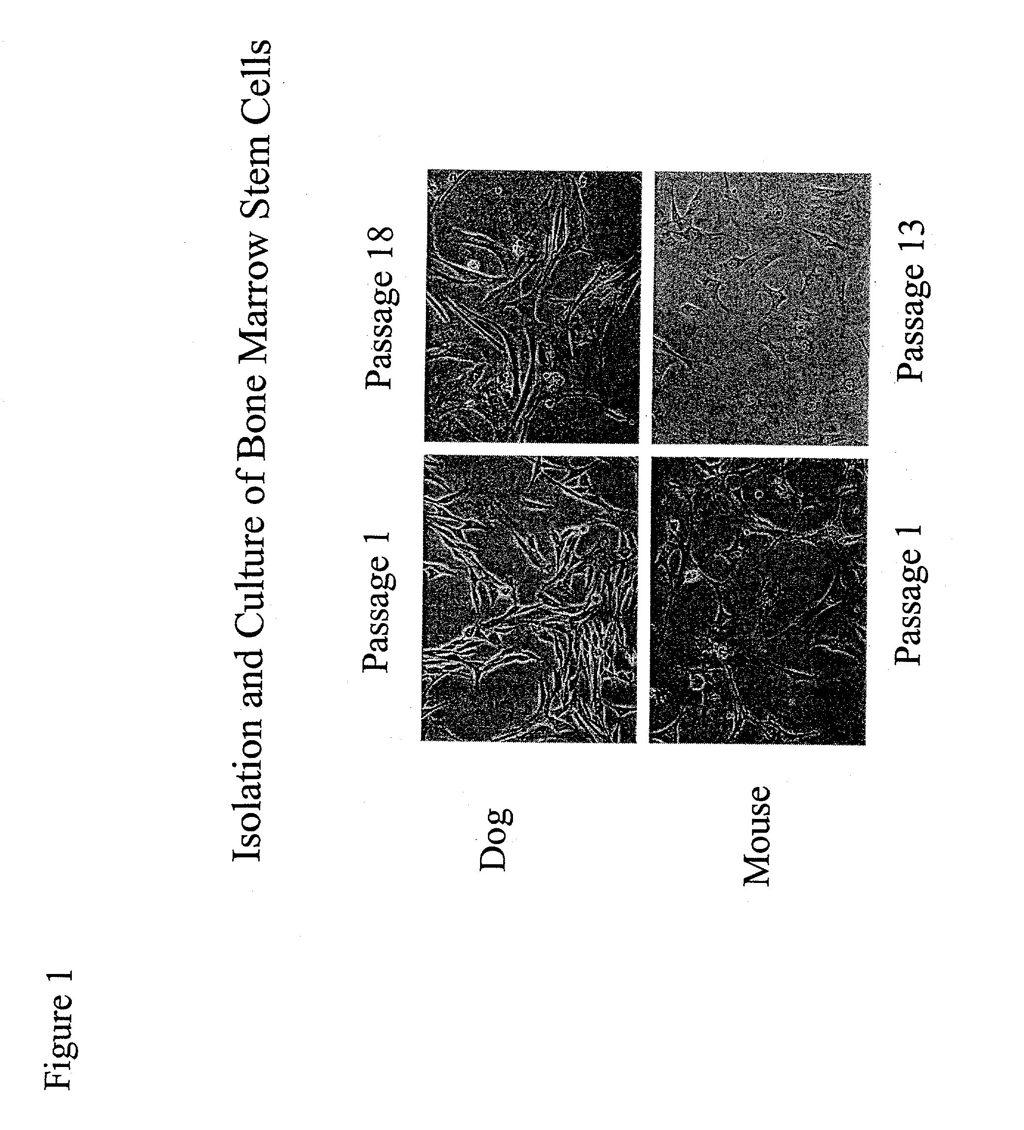
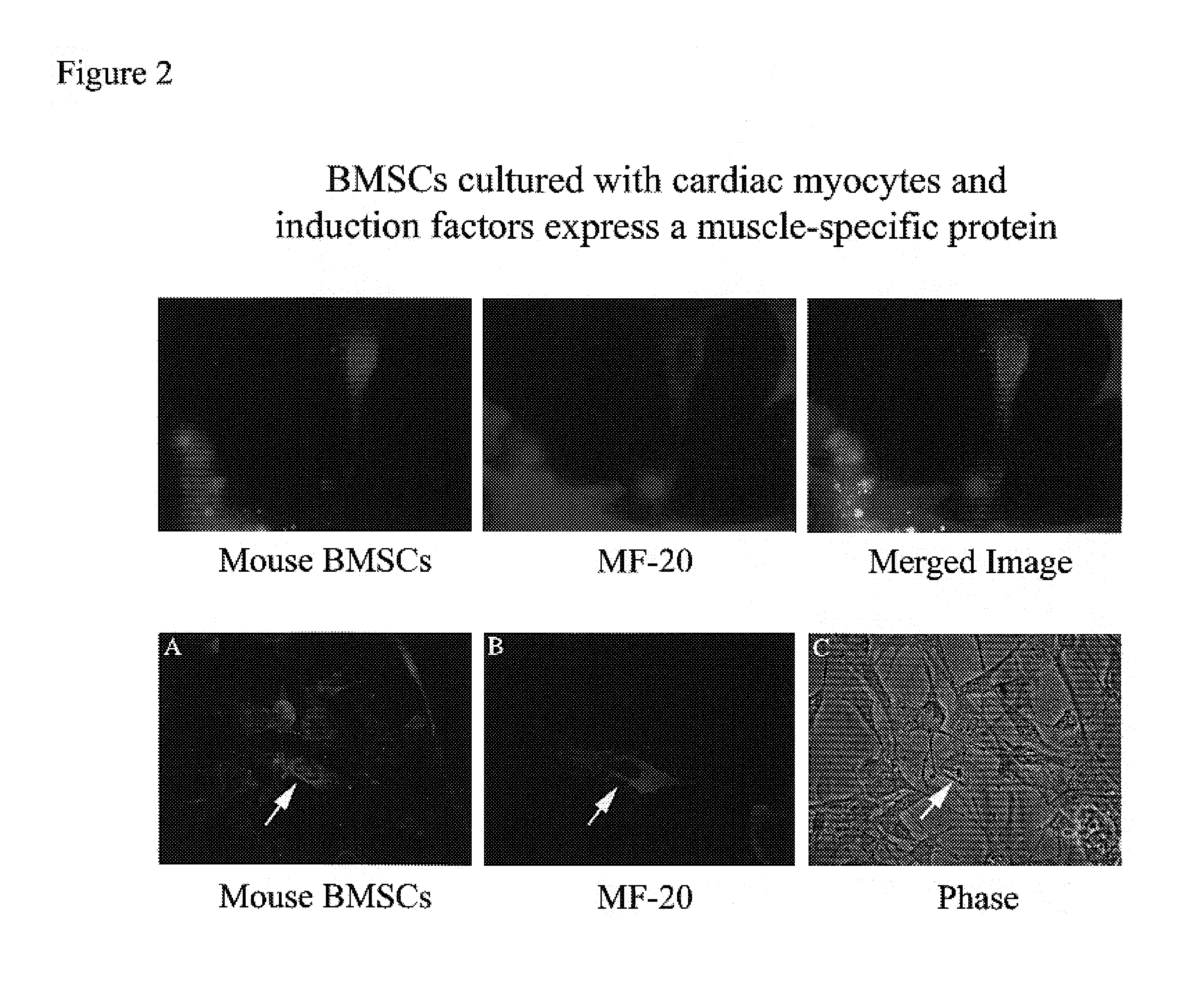
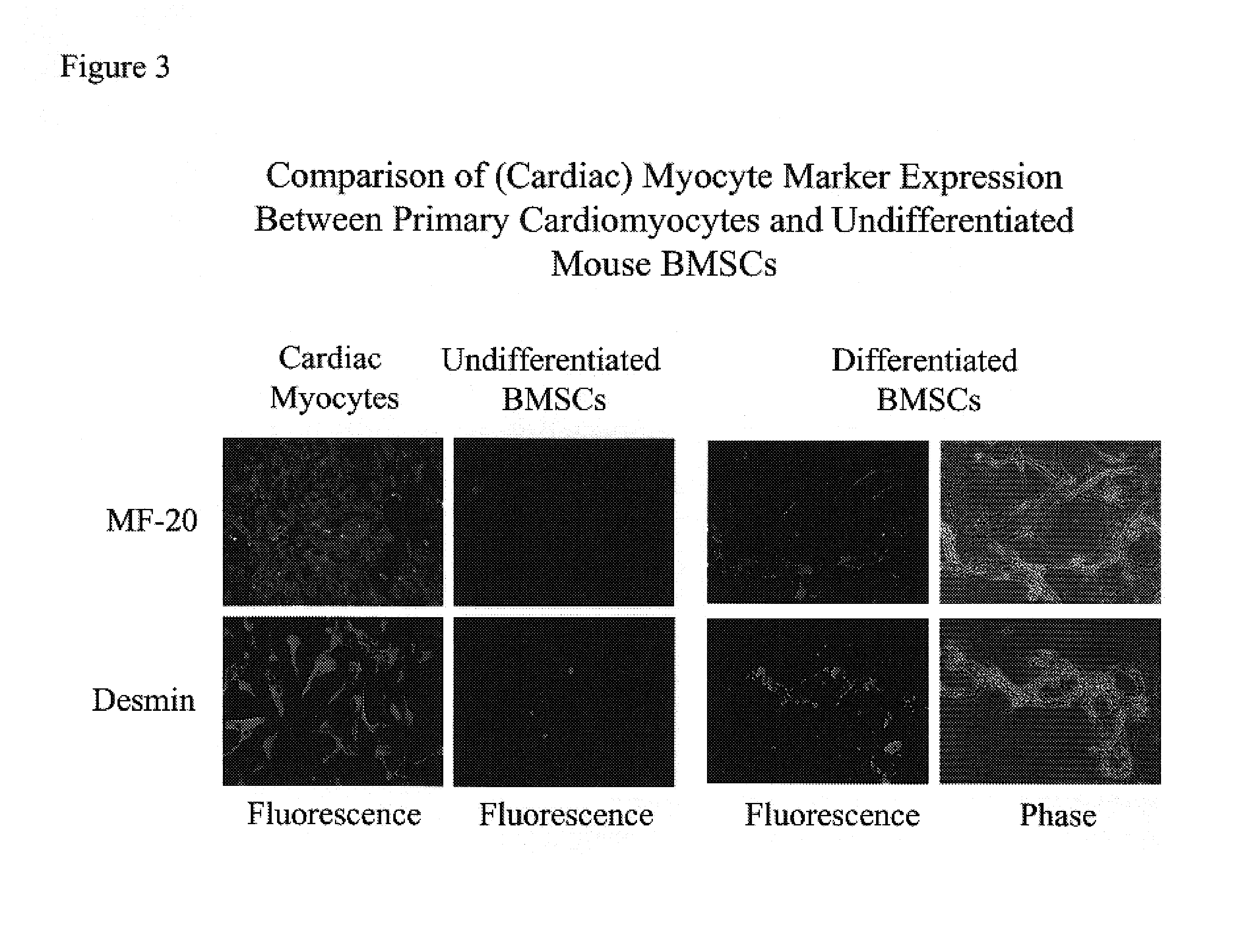
[0073]Marrow was isolated from adult mouse and dog. The BMSCs were isolated and cultured in medium containing 10% fetal bovine serum, 100 μM L-ascorbic acid-2-PO4, 5-15 ng / ml leukemia inhibitory factor (LIF), and 20 nM dexamethasone (for mouse cultures, mouse LIF was used, while for dog cultures, human LIF was used). This in vitro condition allows the BMSCs to maintain their self-renewing character and to expand by passaging without losing responsiveness to the differentiation agents such as growth factors. Further, stem cells cultured through multiple passages maintain a mesenchymal morphology and karyotye (FIG. 1). After 14 days in culture with growth factors (50 ng / ml BMP2, 100 ng / ml bFGF), approximately 80% of the BMSCs were positively stained with Csx / Nkx2.5, MF-20 (a monoclonal antibody specific for sarcomeric myosin), and desmin antibodies, indicating that the cells had differentiated as cardiomyocytes (FIGS. 3 and 4).

[0074]To mimic...

example 2

BMSCs From Humans and Other Mammals

[0076]The foregoing example utilizes mouse BMSCs for illustrative purposes. Human BMSCs are also known in the art to be capable of producing cardiac cells (Pittenger et al., Science 284: 143-147, 1999). BMSCs from other mammals (e.g., humanized pig BMSCs) can also be used in the methods of the invention (Levy et al., Transplantation 69: 272-280, 2000).

example 3

Methods of Inducing BMSCs to Become Cardiomyogenic

[0077]As is described above, co-culturing BMSCs with cardiomyocytes in the presence of BMP2 and / or bFGF results in the induction of cardiomyogenic cells capable of differentiating as cardiomyocytes in culture. The ratio of BMSCs to inducer cells and the concentration of growth factor(s) can each be adjusted to modulate the rate and amount of cardiomyogenic cell induction. For example, the ratio of BMSCs to inducer cells can range from about 1:1 to about 1:1000 or more. The concentration of BMP2 can range from about 0.5 ng / ml to about 1 μg / ml, while the concentration of bFGF can range from about 1 ng / ml to about 5 μg / ml. It is understood that other BMP / TGFβ and FGF family members can be used instead of BMP2 and / or bFGF.

[0078]Other methods known to induce BMSCs to become cardiomyogenic cells can be used in the present invention. Not all methods that induce cardiomyocytes can be used in the invention. For example, 5-azacytidine is used ...


PUM

PropertyMeasurementUnit
Permeabilityaaaaaaaaaa
Fluorescenceaaaaaaaaaa

Abstract

The invention features an encapsulated cell indicator system that includes (a) indicator cells having a signal-responsive element operably linked to a reporter gene; (b) encapsulating material; and (c) a permeable membrane. In this encapsulated cell indicator system, the indicator cells are encapsulated in the encapsulated material and the encapsulated material and the indicator cells are surrounded by the permeable membrane.

Description

CROSS REFERENCE TO RELATED APPLICATIONS[0001]This application claims benefit of the filing date of copending application U.S. Provisional Patent Application No. 60 / 283,838 (filed Apr. 13, 2001), hereby incorporated by reference.BACKGROUND OF THE INVENTION[0002]The invention relates to the field of cell transplantation.[0003]The possibility of bone marrow being an in vivo source of circulating cardiomyocyte progenitors has been suggested. In one experiment, transplanted bone marrow-derived cells were observed to be distributed in a dystrophic mouse heart. Although the molecular characteristics of these cells were not identified, their location in the heart tissue indicated these cells were cardiomyocytes. The ability of bone marrow mesenchymal stem cells (BMSCs) to differentiate as beating cardiomyocytes following introduction of inductive agents such as 5-azacytidine has also been shown. Based on these findings, BMSCs have been proposed to be a source of cells for treatment of cardi...

Claims


Application Information

Patent Timeline
IPC IPC(8): C12N5/00C12N5/02C12N5/08A01K67/027C12N15/09A61K35/12A61K35/28A61P9/00A61P9/10C12N5/071C12N5/074C12N5/077C12N5/0775C12N11/04C12Q1/02C12Q1/66C12R1/91G01N33/50
CPCA01K67/0271G01N33/5005C12N5/0657C12N5/0675C12N5/0691C12N5/0692C12N2501/105C12N2501/115C12N2501/135C12N2501/15C12N2501/155C12N2501/165C12N2503/00C12N2506/21C12N2510/00A61K35/12C12N5/0663C12N2506/1353A61P9/00A61P9/10C12N5/00C12N5/0602
Inventor LEE, IKE W.CROISSANT, JEFFREY D.BALLICA, RABIA
Owner CARDIO3
Why Eureka
Try Eureka