Methods and compositions in breast cancer diagnosis and therapeutics

a breast cancer and composition technology, applied in the field of cancer and molecular genetics, can solve the problems of lack of ability to invade and metastatically spread, lack of molecular markers for breast tissue, and significant risk factors for breast cancer developmen

Inactive Publication Date: 2008-09-30
BAYLOR COLLEGE OF MEDICINE
View PDF6 Cites 5 Cited by
  • Summary
  • Abstract
  • Description
  • Claims
  • Application Information

AI Technical Summary

Problems solved by technology

Although DCIS and LCIS possess some malignant properties, such as loss of growth control, they lack the ability to invade and metastasize and, in this sense, are premalignant.
However, it is likely that estrogens are important, since they are potent mitogens for normal breast epithelial cells, and it is believed that the duration of estrogen exposure to the breast epithelium is a significant risk factor for breast cancer development.
Thus, as has been acknowledged (see, for example, Roger et al., 2000), the art presently lacks a molecular marker for breast tissue, such as a premalignant lesion, which is at risk for breast cancer, particularly for invasive breast cancer, and also lacks a marker for the purpose of improving approaches to risk prediction and treatment strategies.

Method used


Image

  • Methods and compositions in breast cancer diagnosis and therapeutics
  • Methods and compositions in breast cancer diagnosis and therapeutics
  • Methods and compositions in breast cancer diagnosis and therapeutics

Examples


example 1

Materials and Methods—Sample Preparation and Nucleotide Sequence Analysis

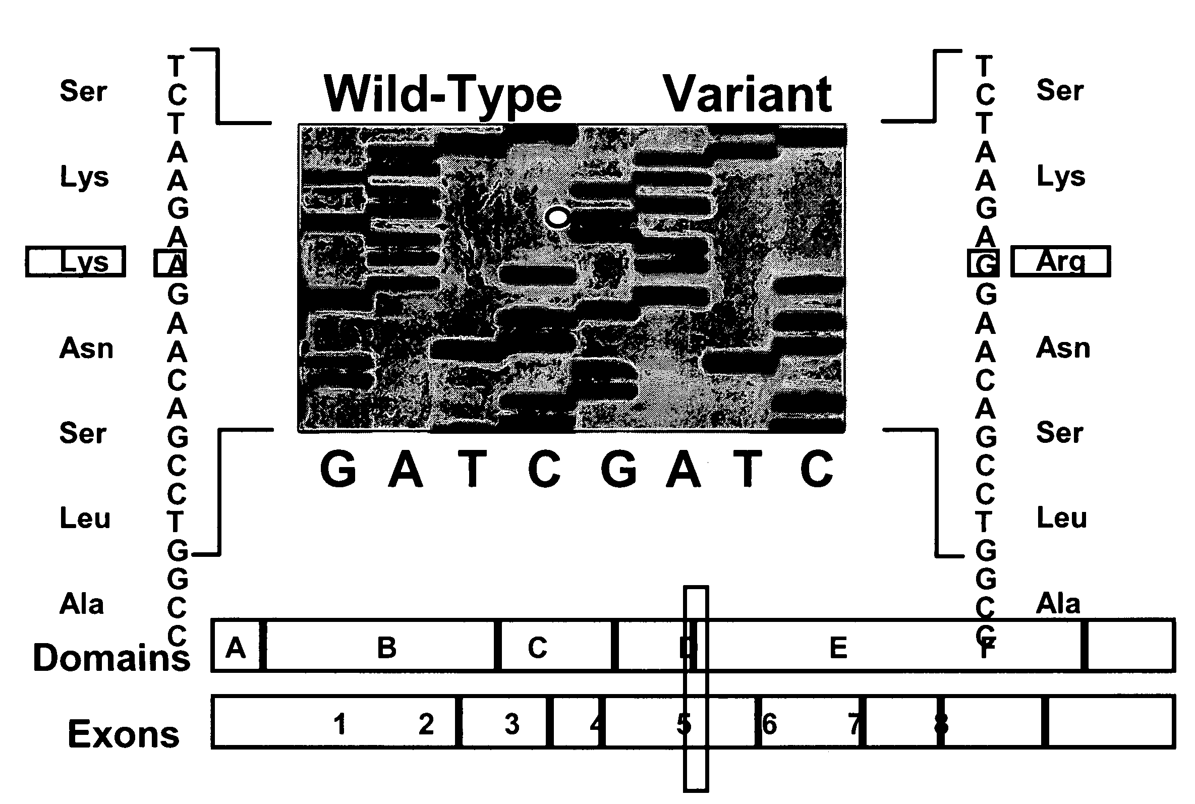
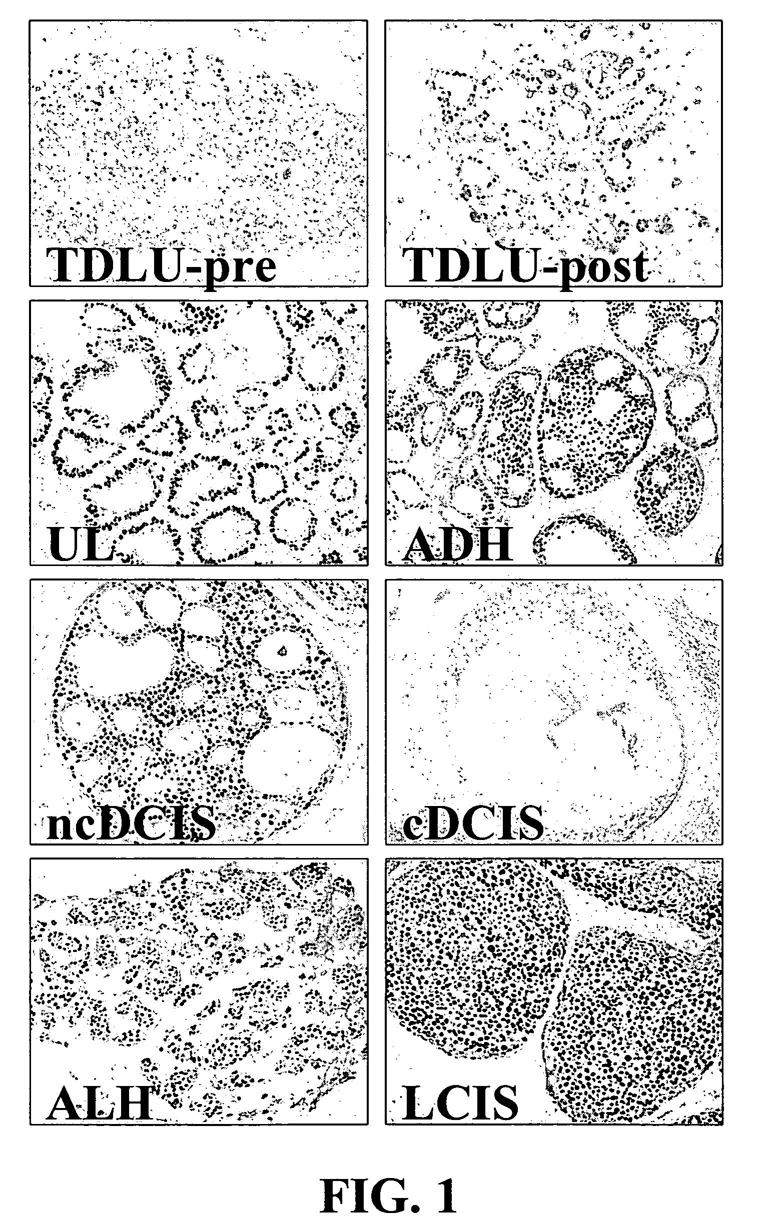
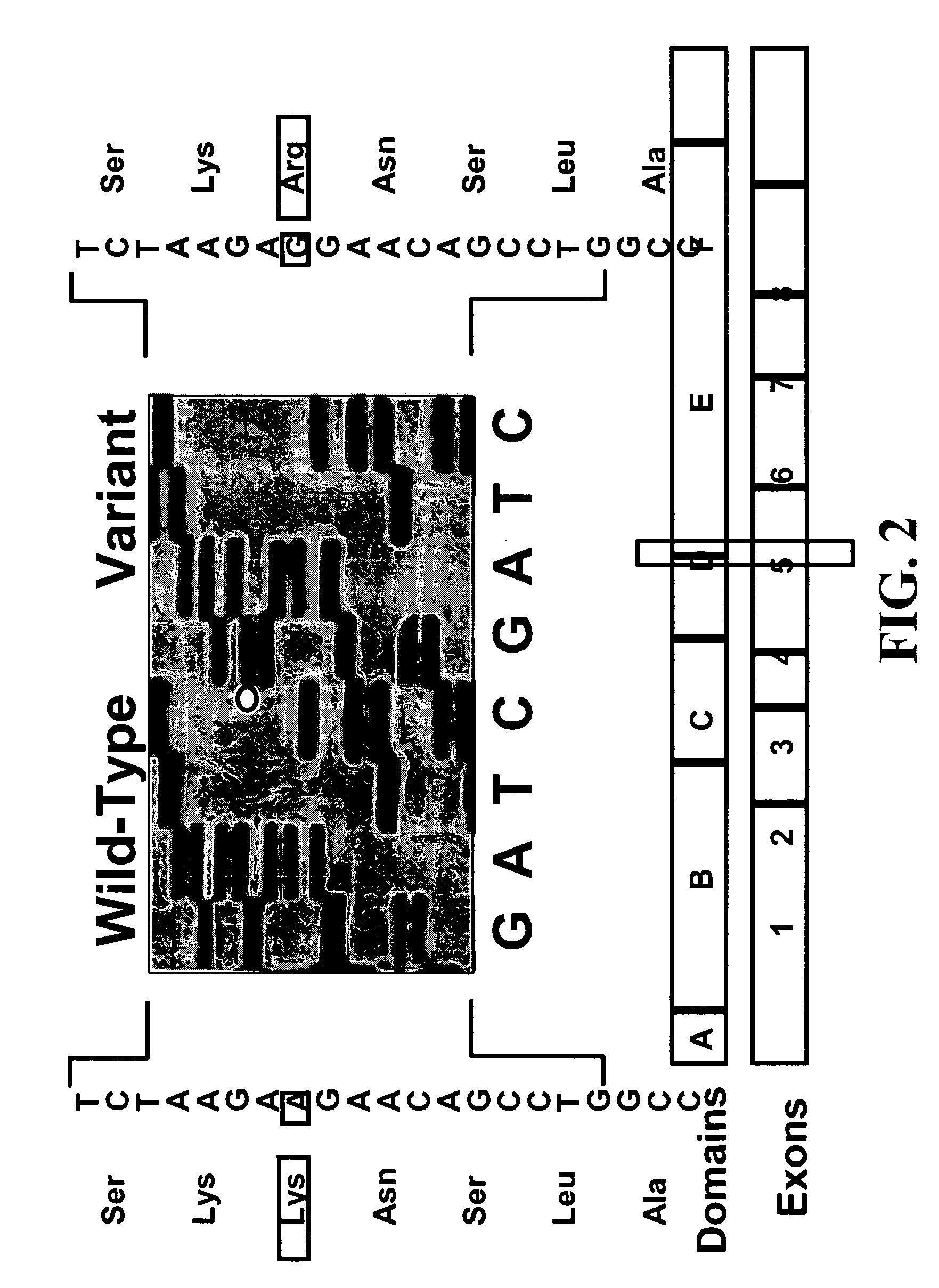
[0331]Histologic slides from archival, clinical specimens were screened microscopically for evidence of hyperplasia. Microdissection of specimens was performed on 55 samples using serial sections from formalin-fixed, paraffin-embedded tissue blocks as described (O'Connell et al., 1999). Briefly, alternative 3- and 10 um-thick sections were cut from the blocks and float mounted on glass slides. The 3-um-thick slides were stained with hematoxylin-eosin and examined under the light microscope to locate regions of normal and hyperplastic tissues; and these areas outlined with a felt-tipped pen. The marked slide was then used as a template to guide manual microdissection from the corresponding regions of the unstained 10-um-thick sections. It was possible to obtain distant normal tissue from 4 of the patients with hyperplasia. A skilled artisan recognizes that there are a variety of methods to isolate desired cells ...

example 2

Materials and Methods—Stable Transfection and Cell Growth Analyses

[0333]The WT ER expression construct was prepared in the pcDNAI vector as described previously (Fuqua et al., 1995). Site directed mutagenesis of this construct was then utilized to generate the A908G transition and the entire coding sequence of ER was verified by dideoxysequence analysis in this clone. The generation of stable transfectants was performed as described by Oesterreich et al. (1993) using cotransfection with the G418-selectable expression vector pSVneo at a ration of 25:1 with the ER plasmids into MCF-7 breast cancer cells. To analyze for expression of both WT or Var sequences, Western blot analyses were performed using the 6F11 antibody (DaKO). Two to three-fold elevated levels of total ER protein were detected in the two WT ER and the three Var clones. In addition, RT / PCR amplification of cDNA from the transfectants (Fuqua et al., 1991) followed by dideoxysequence analysis confirmed that exogenous WT a...

example 3

Statistical Methods

[0334]After taking logarithms to stabilize within group variances, as determined to be appropriate by Box-Cox analysis (Box and Cox, 1996), one-way analysis of variance was used to detect estrogen dose-related differences in growth on Day 8 (i.e. 0 versus 10−12 M versus 10−11 M versus 10−9 M), and to detect differences among estrogen doses (10−12 M versus 10−11 M versus 10−9 M). The Student-Newman-Keuls multiple range test (α=0.02) was used to determine which doses were different from each other. Analyses were preformed using SAS (V6.12, SAS Institute, Cary, N.C.).


PUM

PropertyMeasurementUnit
diametersaaaaaaaaaa
diametersaaaaaaaaaa
temperaturesaaaaaaaaaa

Abstract

The present invention is directed to compositions regarding a specific mutation in estrogen receptor alpha and their use as diagnostic markers in breast tissue, such as premalignant lesions, for the development of breast cancer. More specifically, cells of breast cancer whose nucleic acid comprises the estrogen receptor alpha mutation identify the breast cancer to be an invasive breast cancer.

Description

[0001]This application claims priority to U.S. Ser. No. 60 / 304,018, filed Jul. 9, 2001, U.S. Ser. No. 60 / 262,990, filed Jan. 19, 2001, and U.S. Ser. No. 10 / 052,092, filed Jan. 18, 2002, all of which are incorporated herein in their entirety.[0002]This invention was developed with funds from the United States Government. The United States Government has certain rights in the invention.FIELD OF THE INVENTION[0003]The present invention is directed to the fields of cancer and molecular genetics. Specifically, the present invention is directed to the determination of susceptibility to breast cancer and the diagnosis of invasive breast cancer. More specifically, the present invention is directed to a mutation in estrogen receptor alpha (ER) and its association with breast cancer.BACKGROUND OF THE INVENTION[0004]Invasive breast cancer (IBC) is one of the most common and lethal malignant neoplasms affecting women, especially in Western cultures. The majority of IBCs are thought to develop o...

Claims


Application Information

Patent Timeline
Patent Type & Authority Patents(United States)
IPC IPC(8): C07H21/04G01N33/574G01N33/74
CPCG01N33/57415G01N33/743G01N33/57492G01N2500/00
Inventor FUQUA, SUZANNEO'CONNELL, PETERALLRED, D. CRAIGHOPP, TORSTEN A.
Owner BAYLOR COLLEGE OF MEDICINE
Why Eureka
Try Eureka