Laser microdissection unit

a laser microdissection and laser technology, applied in the field of laser microdissection units, can solve the problems of not being able to achieve simultaneous fluorescence observation and microdissection

Active Publication Date: 2009-02-03
LEICA MICROSYSTEMS CMS GMBH
View PDF28 Cites 3 Cited by
  • Summary
  • Abstract
  • Description
  • Claims
  • Application Information

AI Technical Summary

Benefits of technology

[0009]The laser beam is then directed through the blocking filter and the dichroic beam splitter onto the sample. This allows fluorescence observation of the sample, and cutting of the sample using the laser beam, to be performed simultaneously. For the user, this results in greatly improved capabilities for distinguishing a wide variety of sample regions (e.g. cell types, cell components, etc.) and selecting them for microdissection.
[0012]It proves to be particularly maintenance-friendly and service-friendly if the microdissection beam path is combined into one structural unit that can be placed onto the microscope via at least one alignment surface. It also proves to be advantageous if the microdissection beam path is already optically and mechanically prealigned in the structural unit. The fact that the structural unit and the microscope comprise high-precision alignment surfaces allows the structural unit to be placed in alignment-free fashion onto the microscope.
[0013]The laser microdissection unit according to the present invention can be implemented using both an inverted microscope and an upright microscope. For routine examinations, for example in the field of pathology, it proves to be advantageous if the sample can be stained with multiple fluorescent dyes. This then permits spectral multi-band fluorescence observation. Typical fluorescent dyes are, for example, DAPI, FITC, and Texas Red. Because the dichroic beam splitter and the blocking filter that are spectrally transparent to the laser beam, as well as the excitation filter, exhibit transmissivity properties that permit simultaneous spectral multi-band fluorescence observation, sample recovery by laser microdissection is likewise simultaneously possible.

Problems solved by technology

With an integrated fluorescence axis in the microscope, simultaneous fluorescence observation and microdissection were therefore not hitherto possible.

Method used


Image

  • Laser microdissection unit
  • Laser microdissection unit
  • Laser microdissection unit

Examples


Embodiment Construction

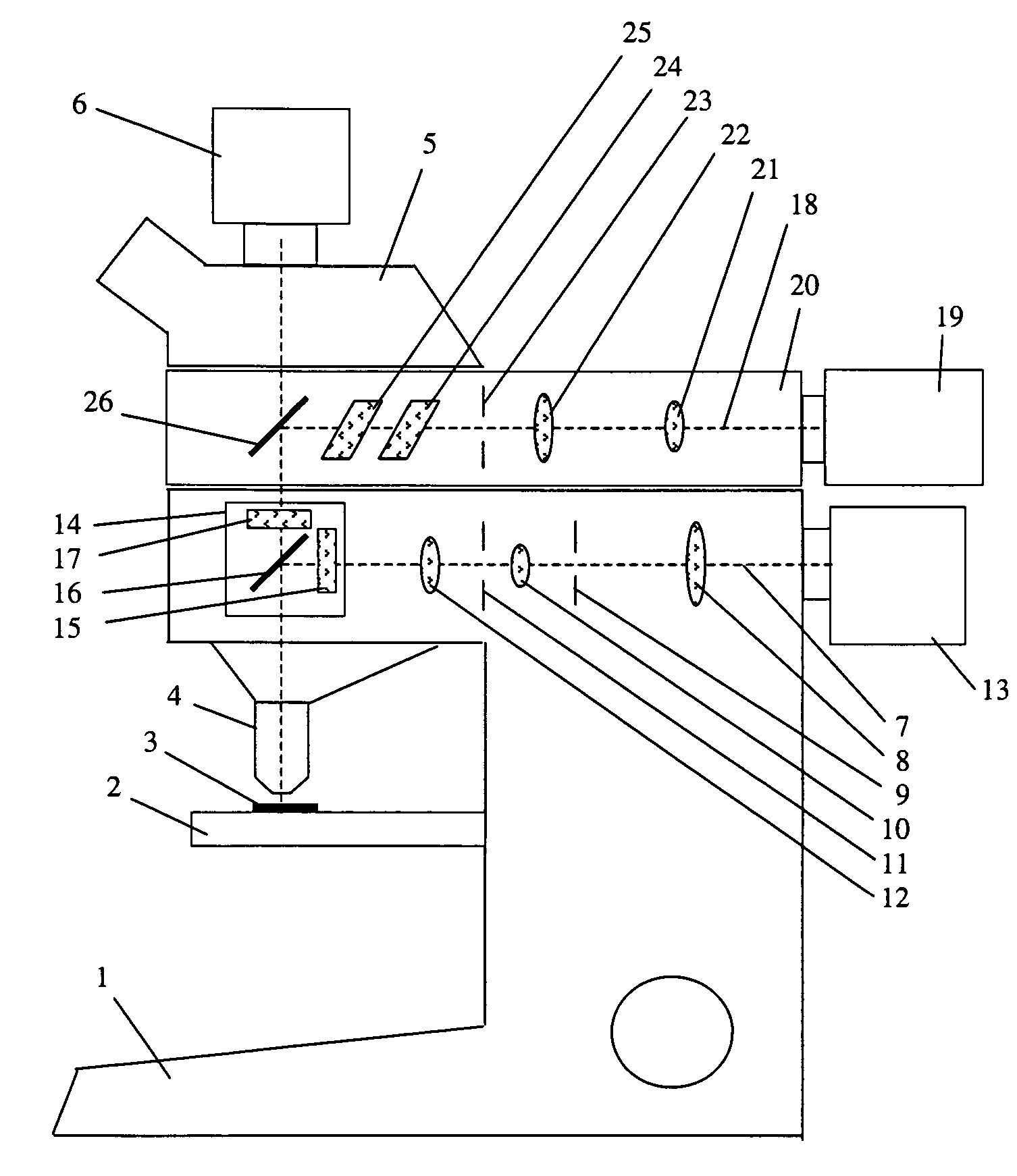
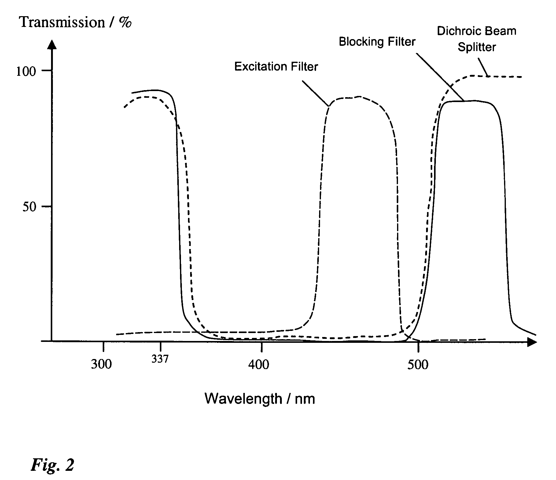
[0018]FIG. 1 shows a laser microdissection unit with a microscope 1. Sample 3 is arranged on microscope stage 2, and is imaged through an objective 4 and a tube 5 onto a camera. Microscope 1 comprises an integrated fluorescence axis 7 in which a lens 8, an aperture diaphragm 9, a lens 10, a field diaphragm 11, and a lens 12 are arranged. The illuminating light proceeding from lamp housing 13 passes along integrated fluorescence axis 7 and strikes fluorescence device 14. The latter encompasses an excitation filter 15, a dichroic beam splitter 16, and a blocking filter 17, which are spectrally matched to at least one fluorescent dye and its fluorescence band.

[0019]A microdissection beam path 18 with a UV laser 19 is arranged above integrated fluorescence axis 7. Microdissection beam path 18 is arranged in a structural unit 20 that is placed onto microscope 1, above integrated fluorescence axis 7, as an attachment.

[0020]The laser beam proceeding from UV laser 19 travels, in microdissec...


PUM


Abstract

A laser microdissection unit for cutting a microscopic sample using a laser beam of a laser includes a microscope and a fluorescence device. The microscope includes an illumination beam path directed onto the sample, and an imaging beam path configured to image the sample. The fluorescence device includes an excitation filter, a dichroic beam splitter, and a blocking filter. The dichroic beam splitter and the blocking filter are spectrally transparent to the laser beam, and the laser beam is directable through the dichroic beam splitter and the blocking filter onto the sample.

Description

CROSS REFERENCE TO RELATED APPLICATIONS[0001]This application claims priority to German patent application 10 2005 008 925.9, filed Feb. 24, 2005, the entire subject matter of which is hereby incorporated by reference herein.FIELD OF THE INVENTION[0002]The invention relates to a laser microdissection unit that cuts a microscopic sample using a laser beam of a laser, which unit includes a microscope having an illumination beam path directed onto the sample and having an imaging beam path that images the sample, and includes a fluorescence device that comprises an excitation filter, a dichroic beam splitter, and a blocking filter.BACKGROUND OF THE INVENTION[0003]“Laser microdissection” refers to a method with which, in the field of medicine and microbiology, a small piece (called the “dissected specimen”) is cut out of a usually planar sample, for example cells or a tissue section, using a finely focused laser beam. The cut-out piece is then available for further biological or medical...

Claims


Application Information

Patent Timeline
Patent Type & Authority Patents(United States)
IPC IPC(8): G01J1/58
CPCG01N1/286G02B21/16G01N2001/2886
Inventor WEISS, ALBRECHT
Owner LEICA MICROSYSTEMS CMS GMBH
Why Eureka
Try Eureka