Devices and methods for the non-invasive detection of spontaneous myoelectrical activity

a non-invasive and bioelectrical technology, applied in the field of devices and methods for the non-invasive detection of spontaneous bioelectrical activity, can solve the problems of serious back pain resulting from radiculopathy, dangerous causes of lower back and neck pain, and need surgery

Inactive Publication Date: 2009-02-17
NEUROMETRIX
View PDF9 Cites 9 Cited by
  • Summary
  • Abstract
  • Description
  • Claims
  • Application Information

AI Technical Summary

Benefits of technology

[0019]FIG. 4 lists the mean relative difference, calculated in dB, between the signal strength of denervated and normal muscle over the muscle activity frequency band referenced to a noise frequency band of 2500-4000 Hz.
[0020]FIG. 5 (Table IV) lists the mean relative difference, calculated in dB, between the signal strength of denervated and normal muscle in various frequency ranges.

Problems solved by technology

Compressive radiculopathies of the lumbosacral and cervical regions are frequent and dangerous causes of lower back and neck pain, respectively.
Back pain resulting from a radiculopathy is serious and may require surgery.
Insertion of the needle electrode results in a burst of electrical activity as the tip of the needle damages muscle fiber membranes, which quickly subsides once the needle ceases to move.
This procedure is consequently an invasive and painful method with a nominal risk of infection or bleeding.
Furthermore, the procedure is expensive and cannot often be obtained for most patients unless the symptoms are severe and surgery is an immediate possibility.
The invasive nature, cost and required expertise of NEMG limits its use to the late confirmation of suspected pathology and makes longitudinal patient monitoring impractical.
Conventional surface electromyography (SEMG) techniques are currently limited in their clinical use due to their lack of spatial resolution.

Method used


Image

  • Devices and methods for the non-invasive detection of spontaneous myoelectrical activity
  • Devices and methods for the non-invasive detection of spontaneous myoelectrical activity
  • Devices and methods for the non-invasive detection of spontaneous myoelectrical activity

Examples


Embodiment Construction

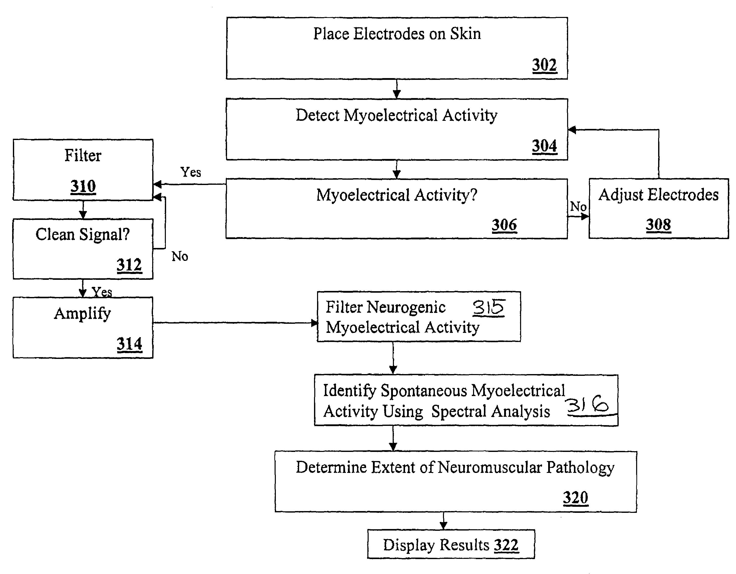
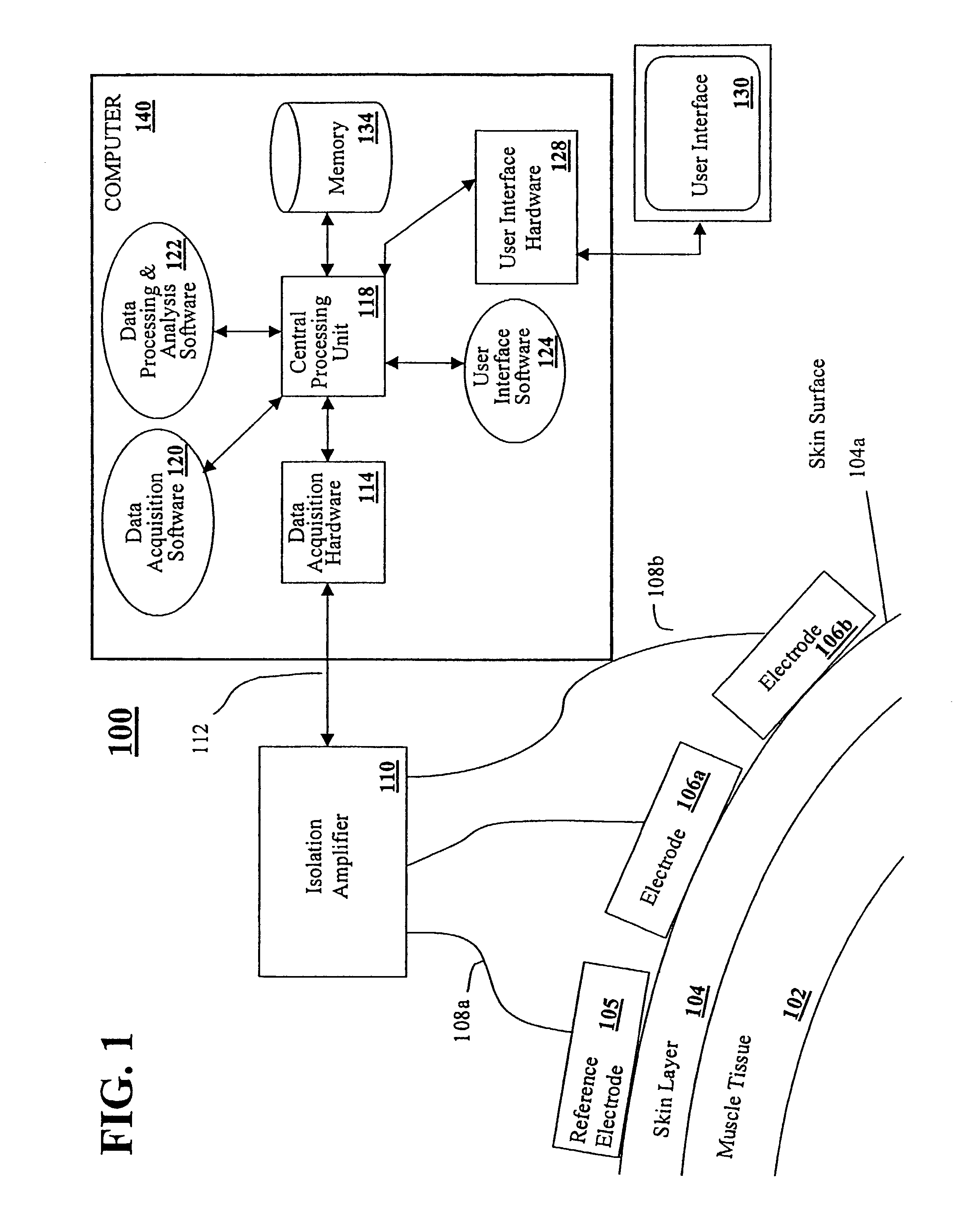
[0029]The devices and methods of the invention provide for the non-invasive detection of spontaneous myoelectrical activity generated from skeletal muscle. The presence of spontaneous myoelectrical activity within muscle is an important diagnostic indicator of neurological or neuromuscular pathology. As the term is used throughout, neuromuscular pathology means the presence of pathology in nerve roots, nerves or muscles. Accordingly, the invention is particularly useful for the non-invasive detection and diagnosis of neuromuscular pathology, such as caused by cervical and lumbrosacral radiculopathy, demyelinating polyneuropathies, intravertebral disc disease, and entrapment neuropathies such as carpal tunnel syndrome. Some causes of neuromuscular pathology can lead to muscle denervation and increased spontaneous myoelectric activity within the affected muscle tissue resulting from muscle fiber fibrillation potentials or positive sharp waves (PSW). Physicians can use the presence and...


PUM


Abstract

A method of detecting neuromuscular pathology in an individual is disclosed comprising the steps of placing a detector on the skin surface of the individual substantially adjacent to a skeletal muscle; obtaining signals from the detector; processing the signals; and determining neuromuscular status in response to the signals. A system for detecting neuromuscular disease in an individual is disclosed, the system comprising at least one means for recording a first signal from a skeletal muscle; a filter in communication with the recording means to generate a second signal consisting substantially of spontaneous myoelectrical activity; and a processor in communication with the filter, wherein the first signal from the skeletal muscle is filtered so as to generate spontaneous myoelectrical signals, and further wherein the processor calculates the power spectral density of the filtered signals and determines the neuromuscular status in response to the power spectral density of the filtered signals.

Description

REFERENCE TO PENDING PRIOR PATENT APPLICATION [0001]This patent application claims benefit of pending prior U.S. Provisional Patent Application Serial No. 60 / 335,061, filed Oct. 24, 2001 by Shai N. Gozani et al. for DEVICES AND METHODS FOR THE NON-INVASIVE DETECTION OF SPONTANEOUS MYOELECTRICAL ACTIVITY, which patent application is hereby incorporated herein by reference.FIELD OF THE INVENTION[0002]This invention relates to devices and methods for the non-invasive detection of spontaneous bioelectrical activity generated from skeletal muscle fiber or nervous tissue, and more particularly to the non-invasive detection of neuromuscular pathology resulting from muscle full or partial denervation.BACKGROUND OF THE INVENTION[0003]Fibrillation potentials are a form of spontaneous muscle activity arising from single muscle fibers. The presence of these potentials are consistent with muscle denervation. The detection of fibrillation potentials can indicate underlying neuromuscular pathologi...

Claims


Application Information

Patent Timeline
Patent Type & Authority Patents(United States)
IPC IPC(8): A61B5/04
CPCA61B5/04004A61B5/30A61B5/313
Inventor GOZANI, SHAI N.KELLER, STEVEN P.
Owner NEUROMETRIX
Why Eureka
Try Eureka