Method and device for examining the skin

a skin and epiluminescence microscope technology, applied in the field of skin examination, can solve the problems of difficult diagnosis, difficult technique, and limited application of conventional epiluminescence microscopes, and achieve the effect of easy management of data format and convenient handling

Active Publication Date: 2009-02-24
SIEMENS HEALTHCARE GMBH
View PDF5 Cites 10 Cited by
  • Summary
  • Abstract
  • Description
  • Claims
  • Application Information

AI Technical Summary

Benefits of technology

[0016]The integrated device produces a substantial simplification in terms of handling for the examining physician. The reason for this is firstly that the handling of the two examination methods is standardized as a result of the common control unit. Secondly, the common control unit offers in a straightforward way a simple means of merging the data generated by both examination methods for collective analysis or archiving, of overlaying said data or of using it synergetically in some other way. Furthermore, the integration of epiluminescence microscopy and OCT in a common device provides a substantial cost benefit compared with two individual devices.
[0017]The device is provided with a first handle applicator as part of the epiluminescence microscopy device, which applicator is equipped with a microscopic lens, at least one incident light source and means for transmitting an epiluminescence microscopy image to an image processing unit. In terms of its external form the handle applicator is embodied in particular in the shape of a pen or pistol in order to enable the examining physician to handle it easily. The means for transmitting the epiluminescence microscopy image preferably comprise a digital image detector disposed in the handle applicator, e.g. a CCD camera and an electronic data line via which the image data is transmitted electronically from the image detector to the image processing unit. In this arrangement the image processing unit is used for editing the image data with regard to contrast, sharpness etc. as well as, where applicable, for converting the image data into an easily manageable data format.

Problems solved by technology

However, conventional epiluminescence microscopes are only suitable to a limited extent for a fast and efficient examination for skin cancer.
However, this technique is problematical particularly in clinical use, especially as it is virtually impossible to avoid entrapping air in the immersion oil.
Air bubbles caused in this way can make the diagnosis more difficult and lead in particular to a distortion of the results of the analysis.
Although digital epiluminescence microscopes (incident light microscopes) equipped with polarization filters and used without immersion oil are known for example from EP 0 794 731 A1, when an epiluminescence microscope of this kind is used for a relatively long time there is frequently thermal stress on the skin tissue caused as a result of the incident light sources used.
Moreover, a digital epiluminescence microscope typically has only a comparatively low local resolution, particularly in deeper lying layers of the dermis.
A disadvantage of OCT resides in the greatly reduced viewing field of this technology.
For this reason OCT is not suitable for examining a relatively large area of skin.

Method used


Image

  • Method and device for examining the skin
  • Method and device for examining the skin
  • Method and device for examining the skin

Examples


Embodiment Construction

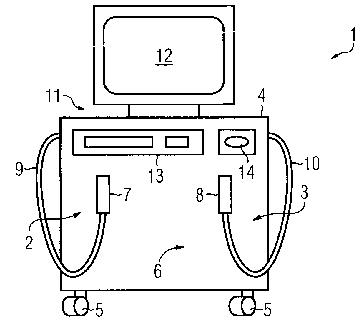
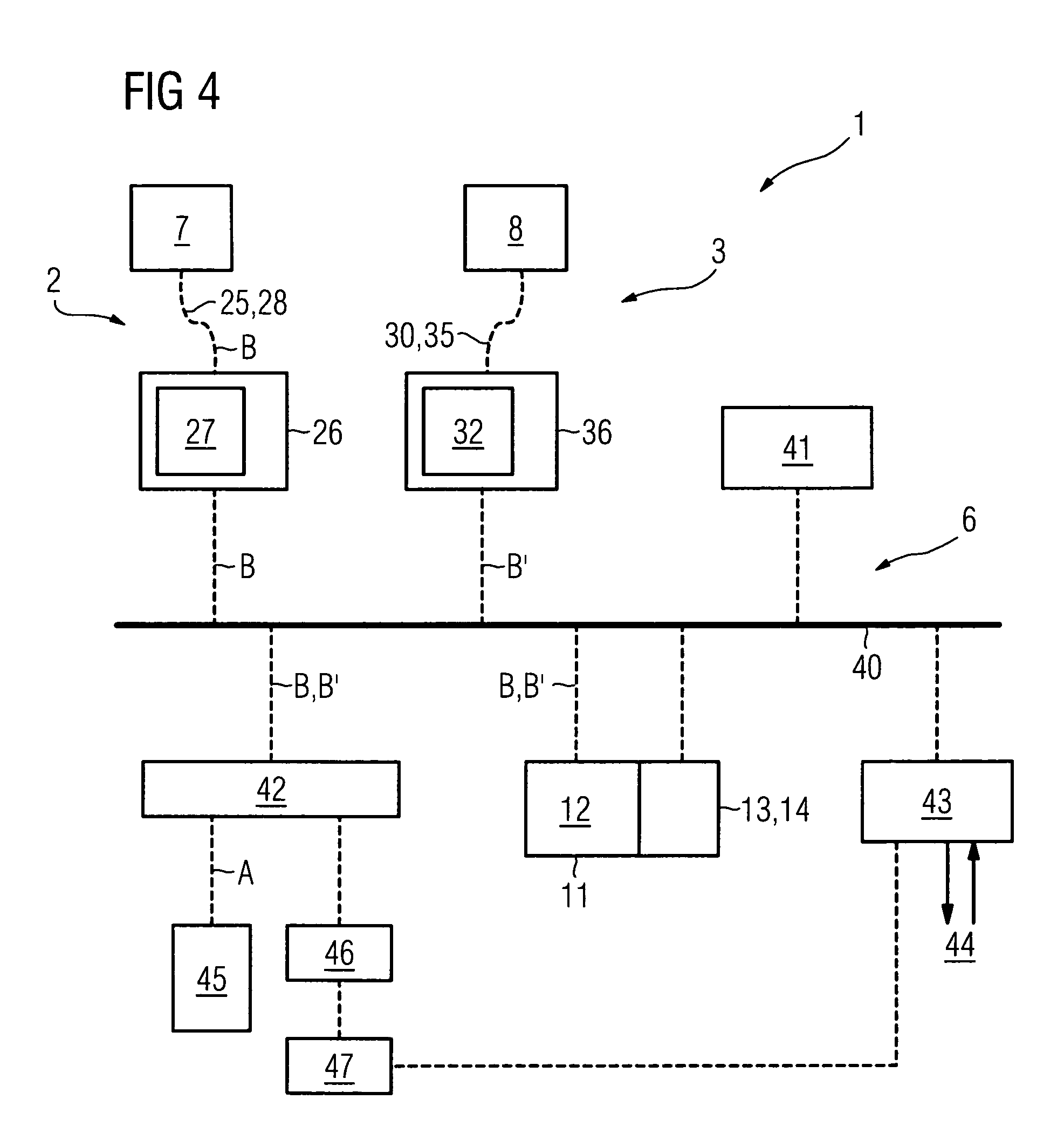
[0027]The device 1 shown in FIG. 1 comprises an epiluminescence microscopy device 2 and an optical coherence tomography device, abbreviated below to OCT device 3, which are integrated in a common housing 4. The housing 4 is preferably mounted on rollers 5 so as to be mobile and movable. It further comprises a control unit 6 (hidden in the housing 4 in the illustration according to FIG. 1) that is common to both devices 2 and 3 and that in particular comprises a data processing system. The epiluminescence microscopy device 2 and the OCT device 3 are each equipped with a handle applicator 7 and 8 respectively.

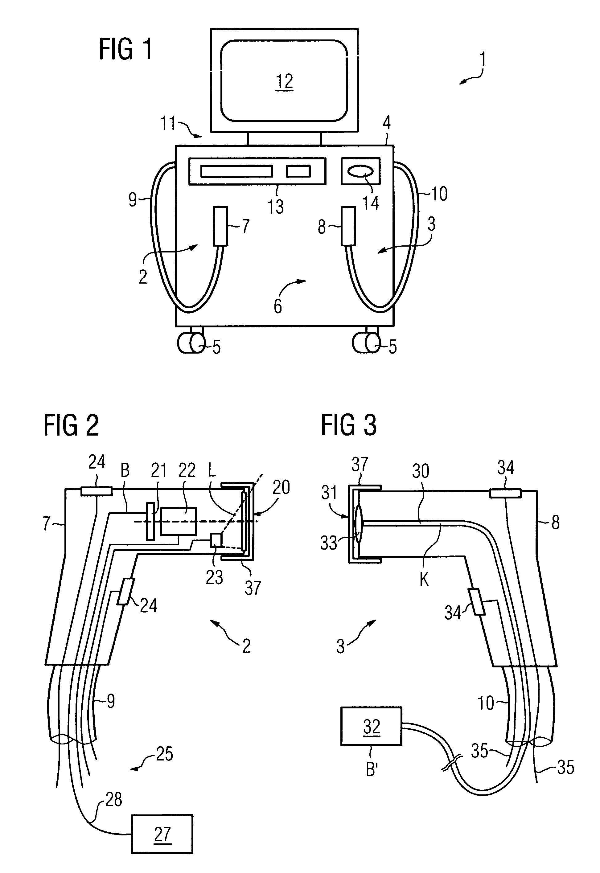
[0028]Each handle applicator 7,8 is—as can be clearly seen from FIGS. 2 and 3—embodied in the form of a pistol so as to be easy to handle for an examining physician and is connected to the housing 4 in each case by means of an associated supply tube 9 and 10 respectively.

[0029]The housing 4 also carries input / output means 11 for operating the control unit 6. Said input / output mea...


PUM

PropertyMeasurementUnit
penetration depthaaaaaaaaaa
digital epiluminescence microscopyaaaaaaaaaa

Abstract

For an improved method for examining the skin of a proband, in particular as part of a skin cancer screening examination or a skin cancer therapy, it is provided that an epiluminescence microscopy image (B) is first taken of a distinctive skin site and by analysis of the image (B) the skin site is rated as suspect or inconsequential with regard to the presence of a disease. In a further method step it is provided that an image (B′) of a skin site rated as suspect is recorded by means of optical coherence tomography and the diagnosis of disease is verified or refuted on the basis of this image (B′). A device particularly suited to the performance of the method comprises an epiluminescence microscopy device (2) and an optical coherence tomography device (3) together with a common control unit (6).

Description

CROSS REFERENCE TO RELATED APPLICATIONS[0001]This application claims priority to the German application No. 10 2004 002 918.0, filed Jan. 20, 2004 which is incorporated by reference herein in its entirety.FIELD OF INVENTION[0002]The invention relates to a method for examining the skin of a proband, in particular as part of a skin cancer screening examination or a skin cancer therapy. The invention further relates to a device for performing the method.BACKGROUND OF INVENTION[0003]It has been possible to observe a significant increase in the incidence of skin cancer diseases in recent times. The principal cause of this is the increasing trend for people to expose themselves to intensive solar radiation. The most common skin disease having fatal consequences is that which is known as malignant melanoma, which occurs in the Central European region, for example, with an incidence of approximately 12 / 100,000 of the population / year (Fortschritte der Medizin (title of German medical journal...

Claims


Application Information

Patent Timeline
Patent Type & Authority Patents(United States)
IPC IPC(8): A61B5/00A61B5/103A61B5/107G01N21/17A61B10/00
CPCA61B5/0066A61B5/444
Inventor MASCHKE, MICHAEL
Owner SIEMENS HEALTHCARE GMBH
Why Eureka
Try Eureka