Method and device for skin cancer screening

a skin cancer and screening technology, applied in the field of dermatology, can solve the problems of insufficient training of primary care physicians to perform it well, too many unnecessary biopsies, and inability to detect early melanomas, so as to improve the access of potential patients and improve the efficiency and cost-effective screening of large patient populations

Inactive Publication Date: 2009-04-21
KENET ROBERT
View PDF10 Cites 16 Cited by
  • Summary
  • Abstract
  • Description
  • Claims
  • Application Information

AI Technical Summary

Benefits of technology

[0009]It is an object of the present invention to improve access of potential patients to skin cancer screening experts using the Internet, thus enabling more efficient and cost-effective screening of large patient populations. It is a further object of this invention to empower consumers to assist in screening and monitoring their own skin and that of their family members, with guidance from skin cancer experts.
[0010]It is a further object of this invention to provide for consumers a digital epiluminescence microscopy (ELM) camera or a digital ELM lens attachment or lens stand that they may used with a digital or analog camera at home to obtain ELM photographs of pigmented lesions on their skin. ELM can improve the accuracy of screening and can help identify early, curable melanoma and benign lesions that mimic melanoma.
[0011]In the present invention, consumers may submit digital photographs of their pigmented lesions for clinical inspection by experts using digital cameras that they have at home. Alternatively, for a more accurate skin cancer risk assessment, they may submit photographs obtained with a special optical technique called epiluminescence microscopy (ELM). In this invention, two methods of performing ELM at home will be available to the consumer. In the first technique, the consumer will be instructed to wet their skin using water, alcohol, or oil. This will permit a crude type of ELM image by decreasing refractive-index mismatch at the air-skin interface, allowing visualization of some pigmented structures below the skin surface. The second technique requires a special ELM lens attachment that is also a subject of the present invention.

Problems solved by technology

In 1992 a Consensus Conference sponsored by the National Institutes of Health recommended that skin cancer screening be initiated in the United States, but recognized that primary care physicians do not have sufficient training to perform it well.
If screening is attempted by those without sufficient experience, early melanomas may be missed and too many unnecessary biopsies may be performed.

Method used


Image

  • Method and device for skin cancer screening
  • Method and device for skin cancer screening
  • Method and device for skin cancer screening

Examples


Embodiment Construction

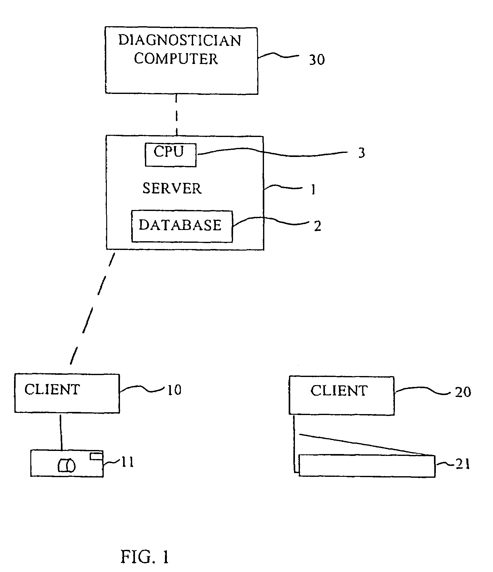
[0059]FIG. 1 shows a server 1 having a database 2 and a central processing unit 3. The database 2 may be for example MICROSOFT ACCESS and the CPU a PENTIUM III processor.

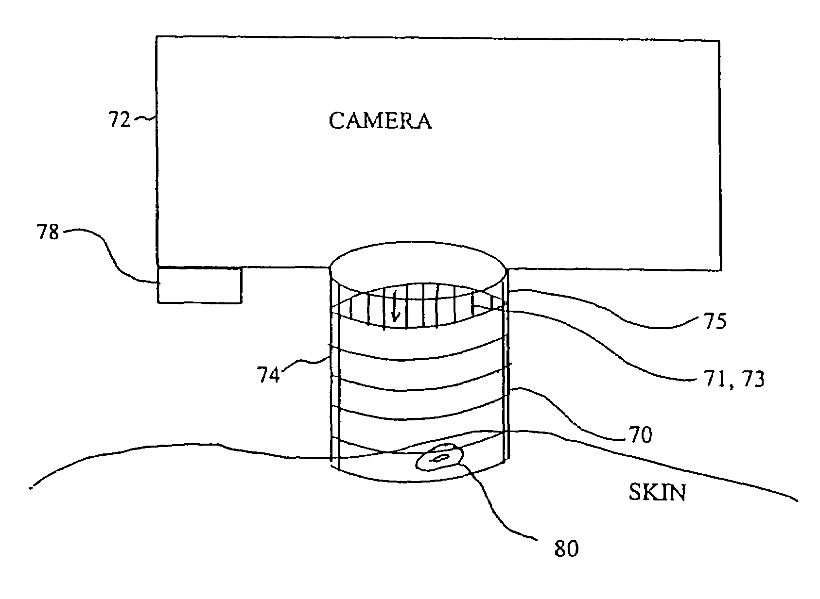
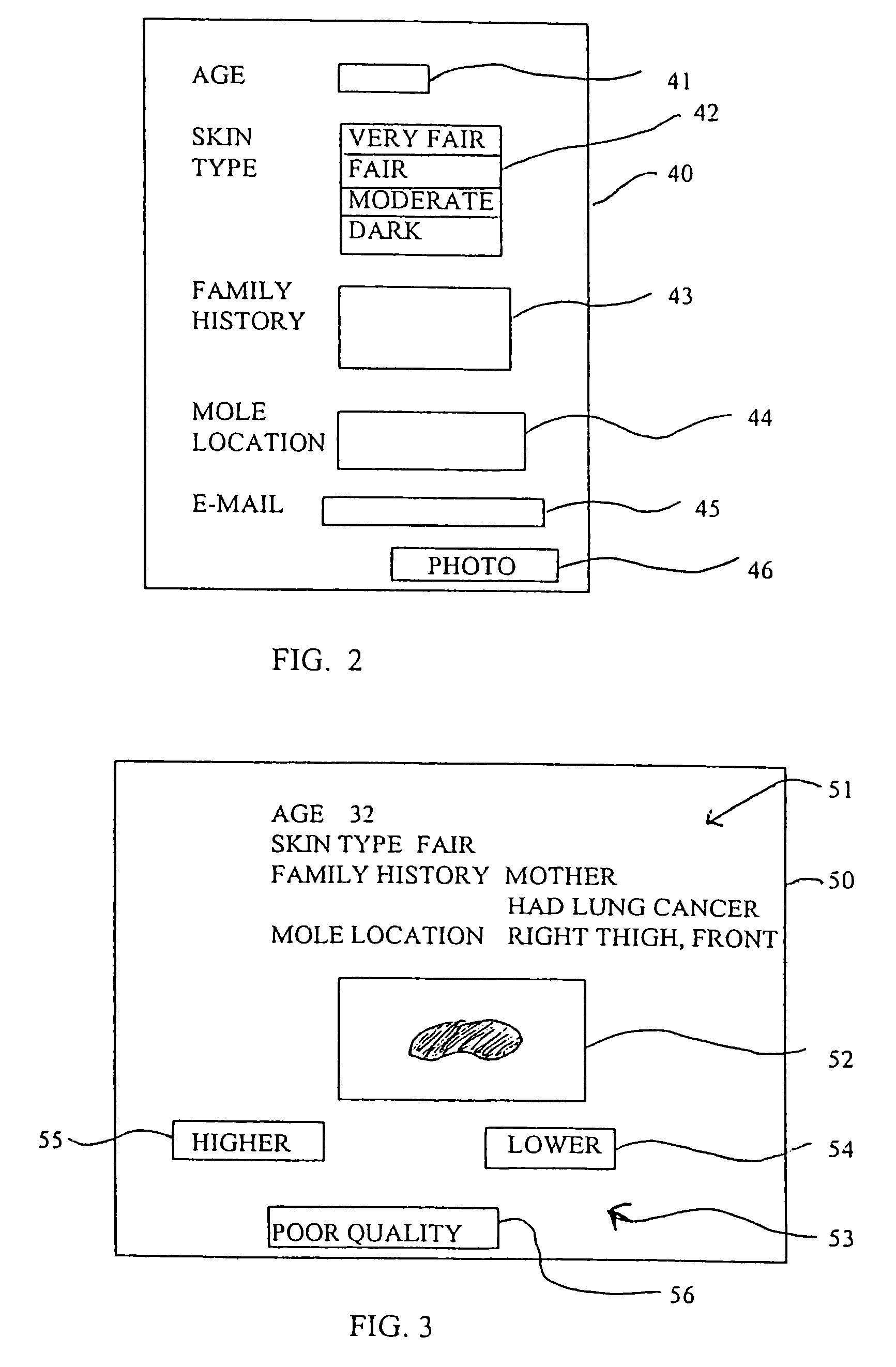
[0060]Server 1 is accessible, for example through the Internet and ISPs by clients 10, 20. Each client may view a page of information stored on the server 1, such as first submission page 40 shown in FIG. 2. As defined herein a page of information may be single or more than one page of information viewable on a screen. First submission page 40 includes data input fields 41, 42, 43, 44 and 45, for submitting age, skin type, family and personal history, pigmented lesion location and an e-mail address, respectively. Risk factor questions may be asked, as can questions about the lesion, such as whether the lesion has changed color or is growing. The data input fields may be blank spaces or may be a selection area with predefined choices, such as shown with skin type data input field 42. Other data input fields or differ...


PUM


Abstract

A method for providing a preliminary diagnosis of skin cancer, more specifically a screening risk assessment of pigmented lesions receiving digital photographs of skin abnormalities from a plurality of consumers at a server, receiving medical information related to each of the plurality of consumers at the server, assigning an identification to at least one of the consumers and the digital photographs, reviewing the digital photographs and categorizing the digital photographs into at least three categories so as to define category information, the at least three categories including a first category of a first risk, a second category of risk lower than the first category and a third category of insufficient photograph quality, and providing the category information as a function of the identification. The digital photographs may be of pigmented skin lesions and may be clinical photographs or may utilize the optical technique of epiluminescence microscopy (ELM) to visualize pigmented or capillary structure below the skin surface that are not visible to the naked eye.

Description

RELATED APPLICATION[0001]The present application is a divisional of U.S. application Ser. No. 09 / 782,851, filed Feb. 14. 2001, which claimed priority from U.S. Provisional Application 60 / 183,551, filed Feb. 18, 2000, the entire disclosure of which is hereby incorporated by reference. The present application claims priority from U.S. Provisional Application No. 60 / 183,551 filed Feb. 18, 2000, the entire contents of which are hereby incorporated by reference herein.BACKGROUND OF THE INVENTION[0002]1. Field of the Invention[0003]The present invention relates generally to dermatology and more particularly to a method and device for aiding in screening populations of patients for skin cancer.[0004]2. Background Information[0005]Skin cancer, the most deadly form of which is melanoma, typically is diagnosed by a dermatologist examining pigmented skin lesions (colloquially known as “moles”) and / or other skin abnormalities on a patient. Typically, a dermatologist makes a determination based ...

Claims


Application Information

Patent Timeline
Patent Type & Authority Patents(United States)
IPC IPC(8): G03B29/00G01J4/00A61B5/00A61B5/103G06T7/00G16H30/20G16H40/60
CPCA61B5/0002A61B5/0059A61B5/444A61B5/445G06F19/321G06F19/322G06F19/3418G06F19/3487G06F19/363G06T7/0012G16H10/20G16H10/60G16H15/00G16H30/20G16H40/60
Inventor KENET, ROBERT
Owner KENET ROBERT
Why Eureka
Try Eureka