
Method for assaying the activity of lysosomal enzymes

Inactive Publication Date: 2009-07-21
GENZYME CORP
View PDF6 Cites 35 Cited by
  • Summary
  • Abstract
  • Description
  • Claims
  • Application Information

AI Technical Summary

Benefits of technology

[0034]The present invention provides an inexpensive and simple method for the determination of LSDs. The claimed method offers several advantages, including a simple and expedite sample collection, minimal invasiveness, reduced sample volume, easy sample handling and storage for extended periods of time. The claimed method makes economically viable the massive screening of the population for LSDs. In addition, the present invention may be used for assaying the activity of any lysosomal enzyme, in any sample containing lysosomal enzymes.

Problems solved by technology

In the absence of hexosaminidase A, the GM2 ganglioside lipid accumulates abnormally in nervous cells causing progressive and irreparable damage to the brain.
There is not therapy available for these disorders at the present.
Other LSDs, like for instance, late stage GM1 gangliosidosis, demand risky and costly therapeutical procedures such as bone marrow transplants.
However, the application of these techniques to LSD detection has been restricted by the following limitations:1) In hybridization assays the test is limited to known mutations of the lysosomal enzyme under study.
For sequencing assays it is unfeasible to detect carrier individuals.
The lysosomal enzyme activity assays known in the art not based in DNA technology have also several restrictions in their application.
One of their principal limitations is the minimum sample volume (usually between 5 to 10 ml of blood) necessary for testing.
This volume is too high to allow infants to stand neonatal screening for these disorders.
The consequences of this limitation are substantial.
Another inconvenience of the enzymatic activity assays is that the sources of samples more commonly utilized for these tests (whole blood, plasma and serum) must be stored under controlled conditions and only for limited periods of time (no more than 3 days).
These restrictions limit considerably the time that may lapse between the sample collection and the enzymatic activity assay.
Two additional limitations of the known lysosomal enzyme activity assays are their complexity and unavailability.
Leukocyte isolation and purification from blood is a very specialized and tedious laboratory procedure.
In addition, these assays are not performed in routinary clinical testing.
These conditions restrict considerably the access of the general population to this type of tests.
However, this method does not provide a conclusive LSD diagnosis.
The method is unable to identify the specific deficient lysosomal enzyme.
In practice this method is complemented with other conventional enzyme activity assays for LSDs diagnosis because of its unreliability.
However, this method is not used in routine clinical practice, especially for newborns.
This procedure is extremely uncomfortable to patients, especially infants, and sample collection is difficult.
In addition, a relative high volume of tears is required for testing.
It is frequently necessary to repeat sampling when using his method because the amount of sample collected is insufficient for assay.
However, this method fails to distinguish clearly between healthy and diseased individuals and has been found to be irreproducible in practice.

Method used


Image

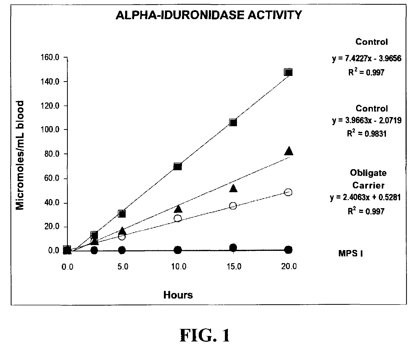
  • Method for assaying the activity of lysosomal enzymes
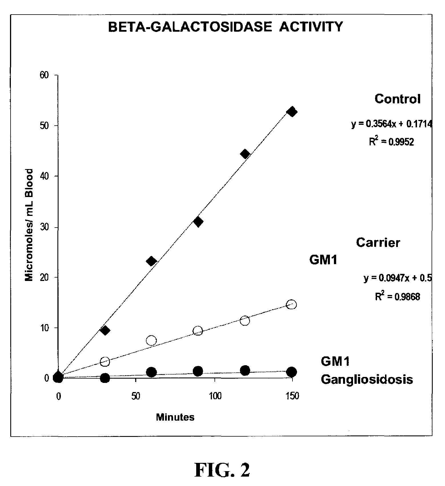
  • Method for assaying the activity of lysosomal enzymes
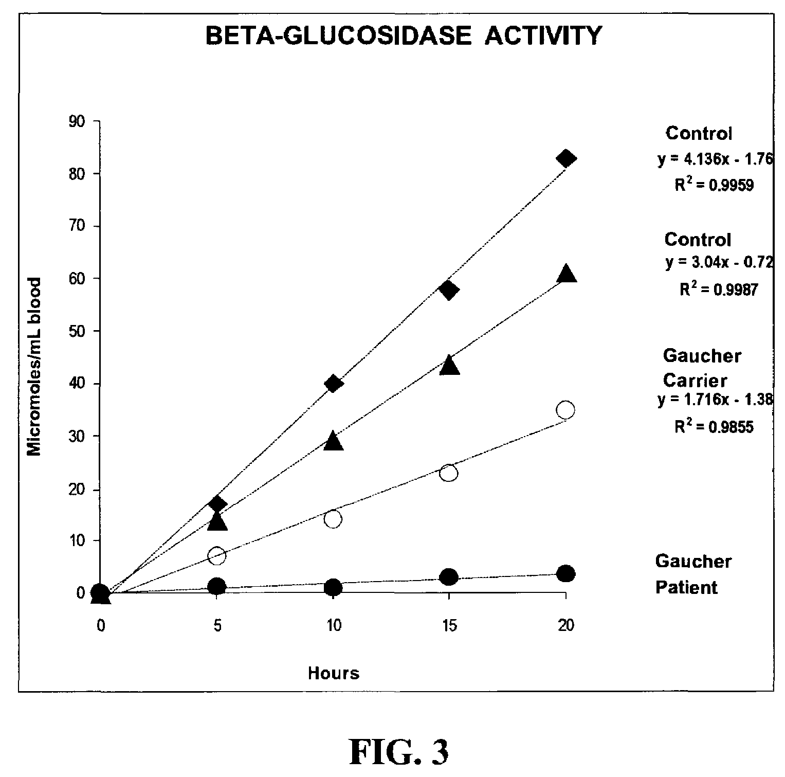
  • Method for assaying the activity of lysosomal enzymes

Examples


example 1

Preparation of Dried Blood Samples

[0101]A drop of blood obtained by venepuncture was spotted on filter paper (Schleicher and Schuell N° 903, Keene, N.H., USA) and allowed to dry at room temperature (22° C.) overnight on a flat non-absorbing surface. The dried blood spots on filter paper were stored on plastic bags at 4° C. until analysis.

[0102]A sample for analysis was prepared by punching out a 3 mm-diameter circle (about 5.5 μl of whole blood) with a standard paper punch from the dried blood spots on the filter paper. A sample for analysis was prepared by punching out a 1.5 mm-diameter circle (about 2 μl of whole blood) with a standard paper punch from the dried blood spots on the filter paper.

[0103]This protocol was also performed by utilizing a heelprick finger to obtain the blood.

example 2

Preparation of Dried Chorionic Villae Samples

[0104]A chorionic villae sample (about 10 mg) was sonicated twice by 20 seconds in 50 μl of cold distilled water (Heat Systems-Ultrasonics, Inc., model W225R).

[0105]After removing 10 μl for protein determination, the sonicated cells were spotted on filter paper (Schleicher and Schuell N° 903, Keene, N.H., USA) and allowed to dry for 6 hours at room temperature (22° C.) on a flat non-absorbing surface. See Lowry et al., J. Biol. (hem. 193:265-275 (1951). The spotted filter paper was stored on plastic bags at −20° C. until analysis.

[0106]A sample for analysis was prepared by punching out a 3mm-diameter circle with a standard paper punch from the dried chorionic villae spots on the filter paper.

example 3

Preparation of Dried Cultured Amniocytes Samples

[0107]Cultured amniocytes were suspended in 500 μl of cold phosphate saline buffer (pH 7.4). After centrifugation at 1,200 g for 5 minutes at 4° C. the supernatant was removed by aspiration. The cell pellet was resuspended in 40 μl of cold distilled water and sonicated (Heat Systems-Ultrasonics, Inc., model W225R).

[0108]After removing 10 μl for protein determination, the sonicated cells were spotted on filter paper (Schleicher and Schuell N° 903, Keene, N.H., USA) and allowed to dry for 6 hours at room temperature (22° C.) on a flat non-absorbing surface. See Lowry et al., supra. The spotted filter paper was stored on plastic bags at −20° C. until analysis.

[0109]A sample for analysis was prepared by punching out a 3 mm-diameter circle with a standard paper punch from the dried amniocytes spots on the filter paper.


PUM


Abstract

A method, and associated kit, for assaying the activity of lysosomal enzymes present in dried bodily fluids and cell tissue samples, such as α-L-iduronidase, β-D-galactosidase, β-D-glucosidase, chitotriosidase, total α-D-galactosidase and α-D-galactosidase A, hexosaminidase A and B, α-D-mannosidase, β-D-mannosidase, α-L-fucosidase, N-acetyl-α-galactosaminidase, arylsulfatases, sphingomyelinase, β-galactocerebrosidase, iduronate-2-sulfatase and β-D-glucuronidase. The method includes: (a) combining with a dried bodily fluid or cell tissue sample containing at least one type of lysosomal enzyme: (1) an eluent, (2) an incubation buffer and (3) a substrate or substrates capable of reacting with the assayed lysosomal enzymes and producing their corresponding enzyme product or products, (b) allowing the dried bodily fluid or cell tissue sample to react with the eluent, incubation buffer and substrate or substrates for an adequate time and temperature, and (c) applying measuring means to the enzyme product to determine the activities of the lysosomal enzymes present.

Description

CROSS-REFERENCE TO RELATED APPLICATIONS[0001]This application is a continuation, under 35 USC 120 and 35 USC 365(c), of International Application PCT / US01 / 26259, filed Aug. 24, 2001 and claims, under 35 USC 119(e) and 35 USC 365, the benefit of U.S. Provisional Application No. 60 / 227,573, filed Aug. 25, 2000. The teachings of U.S. Provisional Application No. 60 / 227,573 and PCT / US01 / 26259 are incorporated herein by reference in their entirety.TECHNICAL FIELD OF THE INVENTION[0002]This invention relates to a method of assaying lysosomal enzymes present in dried samples of bodily fluids and cell tissues. The invention also relates to a diagnostic kit for assaying lysosomal enzymes present in dried samples of bodily fluids and cell tissues.BACKGROUND OF THE INVENTION[0003]The lysosome is an organelle founded in the cytoplasm of eukaryotic cells, which serves as storage for many hydrolytic enzymes and as a center for degrading and recycling cellular components. This organelle contains se...

Claims


Application Information

Patent Timeline
IPC IPC(8): C12Q1/34G01N33/48
CPCC12Q1/34
Inventor CHAMOLES, NESTOR A.
Owner GENZYME CORP
Learn More