Optical spectroscopy for the detection of ischemic tissue injury

a technology of ischemic tissue injury and optical spectroscopy, which is applied in the direction of diagnostics using spectroscopy, instruments, fluorescence/phosphorescence, etc., can solve the problems of variable amount of additional warm ischemia, difficult to quantify how much warm ischemic organ damage has occurred, and the organs of cadaveric donors are often difficult to transplan

Inactive Publication Date: 2009-09-08
LAWRENCE LIVERMORE NAT SECURITY LLC
View PDF8 Cites 29 Cited by
  • Summary
  • Abstract
  • Description
  • Claims
  • Application Information

AI Technical Summary

Problems solved by technology

Transplant surgeons often face the difficult question of whether organs from a cadaveric donor are suitable for transplantation.
It is currently difficult to quantify how much warm ischemic organ damage has occurred, especially if a donor has experienced significant hemodynamic instability.
Organ procurement in brain-dead and non-heart-beating donors can cause a variable amount of additional warm ischemia.
Transplanting organs that have sustained significant pre-implantation warm ischemic damage may leave the recipient with unacceptably poor allograft function and adversely affect graft and patient survival.
Post transplant monitoring is currently the only reliable way of assessing ischemic organ damage; unfortunately, at that stage the organ is already in the recipient and may have to be removed if found to be nonviable.
First, how much ischemic damage has an organ sustained?
However, such approaches are hindered by clinical practicality as well as by hypothermic preservation.

Method used


Image

  • Optical spectroscopy for the detection of ischemic tissue injury
  • Optical spectroscopy for the detection of ischemic tissue injury
  • Optical spectroscopy for the detection of ischemic tissue injury

Examples


Embodiment Construction

[0021]Referring now to the drawings, specific embodiments of the invention are shown. The detailed description of the specific embodiments, together with the general description of the invention, serves to explain the principles of the invention.

General Description

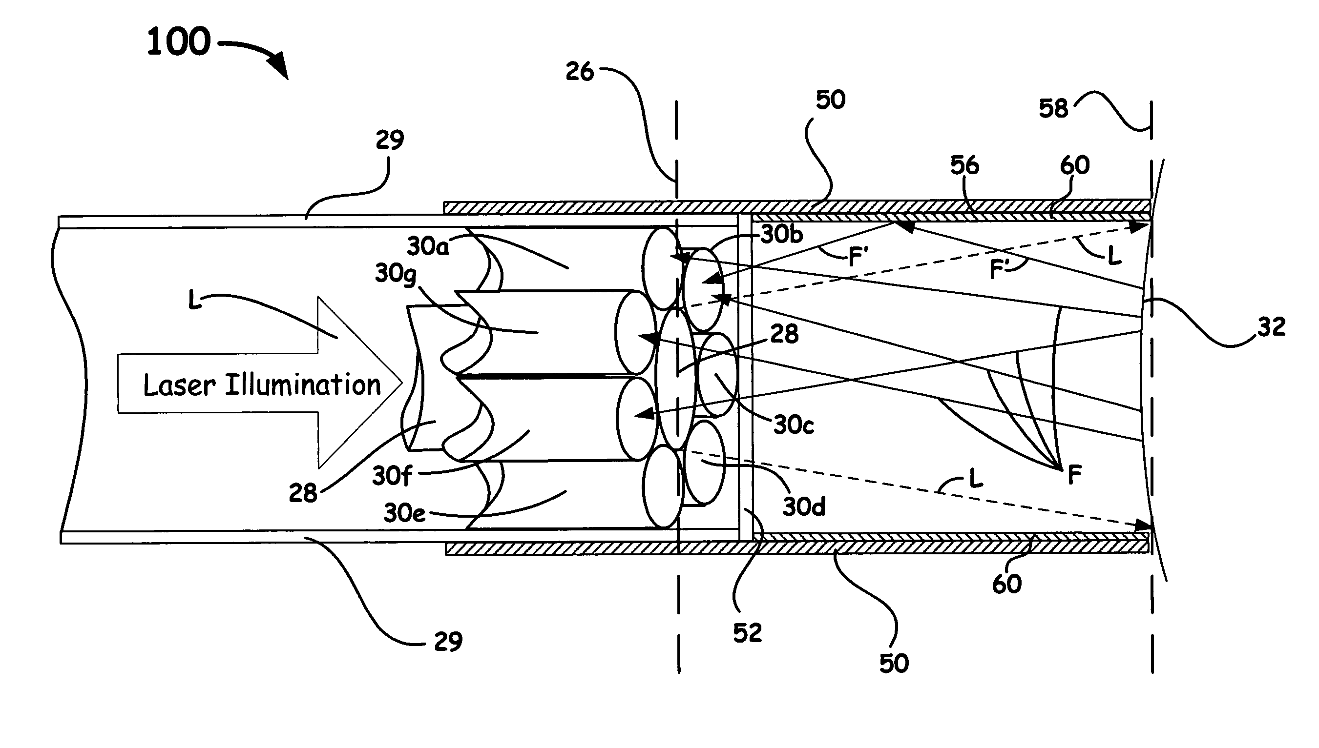
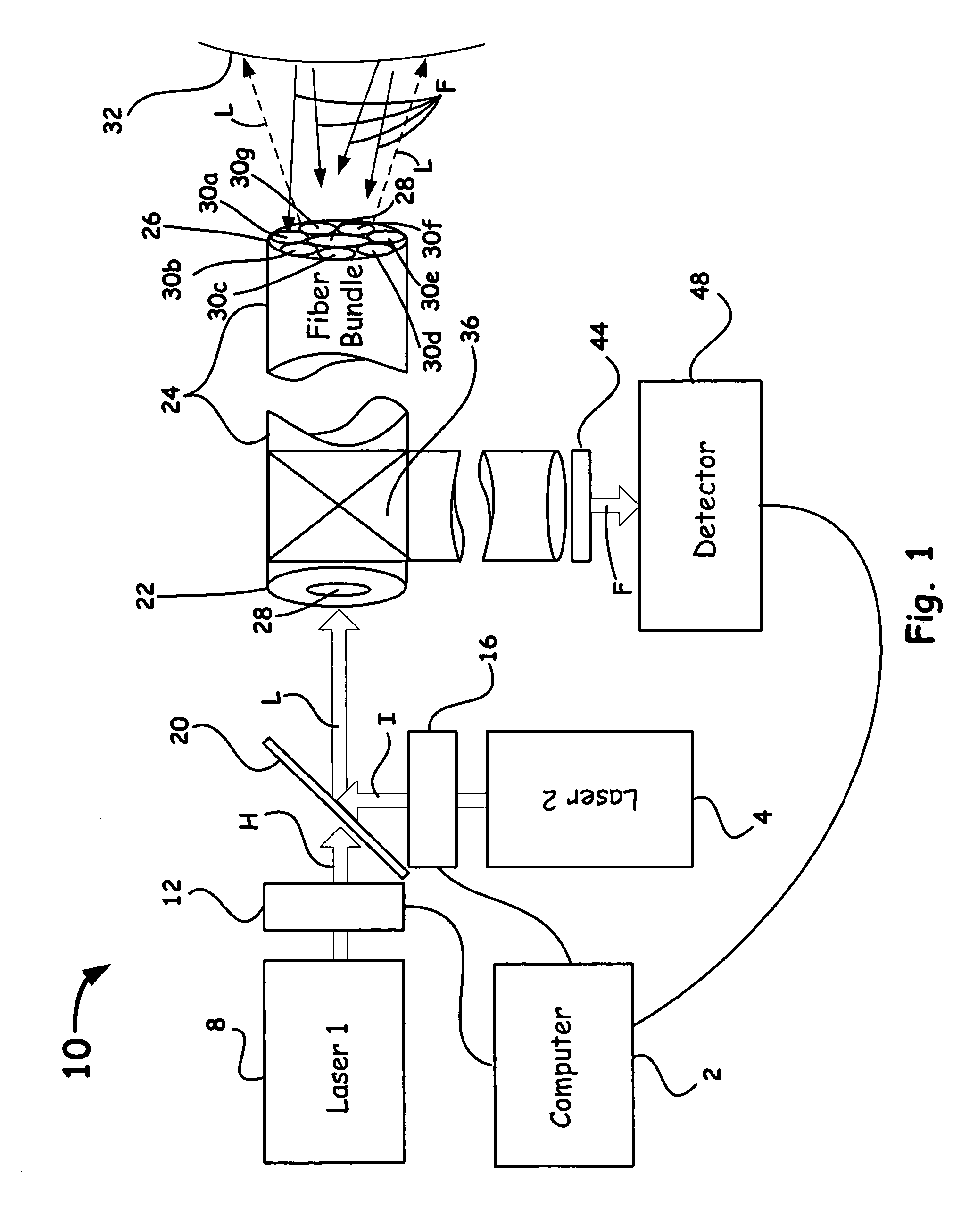
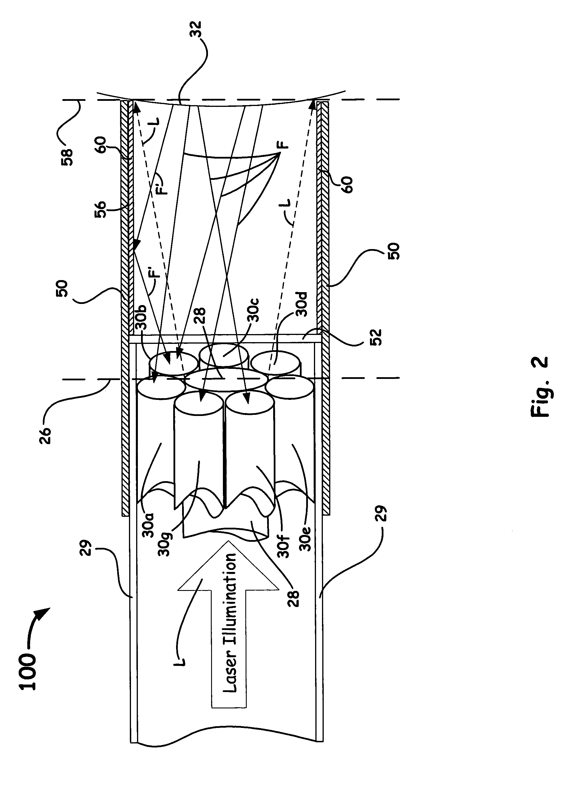
[0022]The present invention provides apparatus and methods for real-time determination and quantification of the state of tissue components. In general, the apparatus as disclosed herein, can include an optical probe having at least one illumination optical fiber, a plurality of collection optical fibers, at least one radiation source so as to cause scattering and / or photoexcitation so as to induce autofluorescence in a targeted tissue component; a detector system that acquires such autofluorescence and scattered spectra of polarized and unpolarized light; and a processor primarily arranged for comparing received autofluorescence signals so as to assess in real-time tissue properties, the metabolic state, injury, and tissu...


PUM

PropertyMeasurementUnit
surface areaaaaaaaaaaa
wavelengthaaaaaaaaaa
diameteraaaaaaaaaa

Abstract

An optical method and apparatus is utilized to quantify ischemic tissue and / or organ injury. Such a method and apparatus is non-invasive, non-traumatic, portable, and can make measurements in a matter of seconds. Moreover, such a method and apparatus can be realized through optical fiber probes, making it possible to take measurements of target organs deep within a patient's body. Such a technology provides a means of detecting and quantifying tissue injury in its early stages, before it is clinically apparent and before irreversible damage has occurred.

Description

RELATED APPLICATION[0001]This application claims the benefit of U.S. Provisional Application No. 60 / 535,585, filed Jan. 8, 2004, and U.S. Provisional Application No. 60 / 559,887, filed Apr. 5, 2004, both entitled, “Optical Spectroscopy for the Detection of Ischemic Tissue Injury,” which is incorporated herein by this reference.[0002]The United States Government has rights in this invention pursuant to Contract No. W-7405-ENG-48 between the United States Department of Energy and the University of California for the operation of Lawrence Livermore National Laboratory.BACKGROUND OF THE INVENTION[0003]1. Field of the Invention[0004]The present invention relates to a medical diagnostic for investigating tissue components. More particularly, the present invention relates to an apparatus and a method that utilizes autofluorescence emission and / or polarized elastic light scattering as an interrogation means for investigating tissue states for medical applications.[0005]2. Description of Rela...

Claims


Application Information

Patent Timeline
Patent Type & Authority Patents(United States)
IPC IPC(8): A61B6/00A61B5/00G01N21/49G01N21/64
CPCA61B5/0071A61B5/0084G01N21/6456G01N21/6486G01N21/49A61B5/0075A61B5/413G01N2021/6484G01N2021/6419
Inventor DEMOS, STAVROSFITZGERALD, JASONTROPPMANN, CHRISTOPHMICHALOPOULOU, ANDROMACHI
Owner LAWRENCE LIVERMORE NAT SECURITY LLC
Why Eureka
Try Eureka