Expansion of definitive endoderm cells
a technology of endoderm cells and endoderm cells, which is applied in the field of cell biology and medicine, can solve the problems of limited islet cell donor pancreases, unique challenges, and inability to generate insulin-producing -cells from hescs,
- Summary
- Abstract
- Description
- Claims
- Application Information
AI Technical Summary
Benefits of technology
Problems solved by technology
Method used
Image
Examples
example 1
Human ES Cells
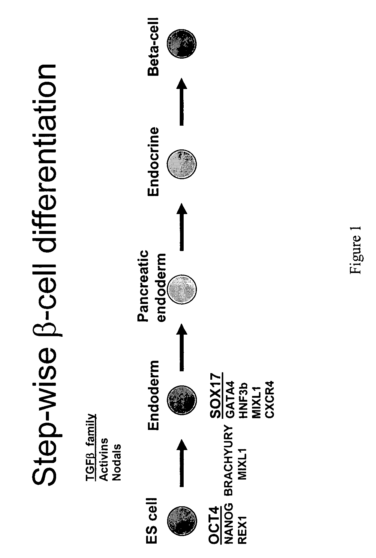
[0187]For our studies of endoderm development we employed human embryonic stem cells, which are pluripotent and can divide seemingly indefinitely in culture while maintaining a normal karyotype. ES cells were derived from the 5-day-old embryo inner cell mass using either immunological or mechanical methods for isolation. In particular, the human embryonic stem cell line hESCyt-25 was derived from a supernumerary frozen embryo from an in vitro fertilization cycle following informed consent by the patient. Upon thawing the hatched blastocyst was plated on mouse embryonic fibroblasts (MEF), in ES medium (DMEM, 20% FBS, non essential amino acids, beta-mercaptoethanol, and FGF2). The embryo adhered to the culture dish and after approximately two weeks, regions of undifferentiated hESCs were transferred to new dishes with MEFs. Transfer was accomplished with mechanical cutting and a brief digestion with dispase, followed by mechanical removal of the cell clusters, washing an...
example 2
hESCyt-25 Characterization
[0189]The human embryonic stem cell line, hESCyt-25 has maintained a normal morphology, karyotype, growth and self-renewal properties over 18 months in culture. This cell line displays strong immunoreactivity for the OCT4, SSEA-4 and TRA-1-60 antigens, all of which, are characteristic of undifferentiated hESCs and displays alkaline phosphatase activity as well as a morphology identical to other established hESC lines. Furthermore, the human stem cell line, hESCyt-25, also readily forms embryoid bodies (EBs) when cultured in suspension. As a demonstration of its pluripotent nature, hESCyT-25 differentiates into various cell types that represent the three principal germ layers. Ectoderm production was demonstrated by Q-PCR for ZIC1 as well as immunocytochemistry (ICC) for nestin and more mature neuronal markers. Immunocytochemical staining for β-III tubulin was observed in clusters of elongated cells, characteristic of early neurons. Previously, we treated EB...
example 3
Production of SOX17 Antibody
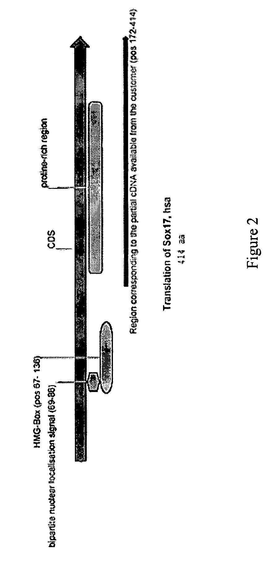
[0190]A primary obstacle to the identification of definitive endoderm in hESC cultures is the lack of appropriate tools. We therefore undertook the production of an antibody raised against human SOX17 protein.
[0191]The marker SOX17 is expressed throughout the definitive endoderm as it forms during gastrulation and its expression is maintained in the gut tube (although levels of expression vary along the A-P axis) until around the onset of organogenesis. SOX17 is also expressed in a subset of extra-embryonic endoderm cells. No expression of this protein has been observed in mesoderm or ectoderm. It has now been discovered that SOX17 is an appropriate marker for the definitive endoderm lineage when used in conjunction with markers to exclude extra-embryonic lineages.
[0192]As described in detail herein, the SOX17 antibody was utilized to specifically examine effects of various treatments and differentiation procedures aimed at the production of SOX17 positiv...
PUM
| Property | Measurement | Unit |
|---|---|---|
| concentration | aaaaa | aaaaa |
| concentration | aaaaa | aaaaa |
| concentration | aaaaa | aaaaa |
Abstract
Description
Claims
Application Information
 - R&D
- Intellectual Property
- Life Sciences
- Materials
- Tech Scout
- Unparalleled Data Quality
- Higher Quality Content
- 60% Fewer Hallucinations
Browse by: Latest US Patents, China's latest patents, Technical Efficacy Thesaurus, Application Domain, Technology Topic, Popular Technical Reports.
© 2025 PatSnap. All rights reserved.Legal|Privacy policy|Modern Slavery Act Transparency Statement|Sitemap|About US| Contact US: help@patsnap.com


