
Devices for minimally invasive pelvic surgery

a pelvis and pelvis technology, applied in the field of pelvis surgery devices, can solve the problems of affecting one or more biological functions of the tissue, urine leaking out of the urethra during stressful activity, and weakened or damaged anatomical tissues, so as to simplify the delivery of the implan

Inactive Publication Date: 2009-12-29
BOSTON SCI SCIMED INC
View PDF69 Cites 65 Cited by
  • Summary
  • Abstract
  • Description
  • Claims
  • Application Information

AI Technical Summary

Benefits of technology

This approach reduces surgical trauma by eliminating the need for transvaginal incisions and excessive dilation, providing a more versatile method for sling implantation suitable for various patient conditions and anatomical sites.

Problems solved by technology

Anatomical tissues may become weakened or damaged by age, injury, or disease.
Even in the absence of tissue necrosis, weakening of an anatomical structure may impair one or more of the biological functions of the tissue.
In ISD, the urinary sphincter valve, located within the urethra, fails to close properly (coapt), causing urine to leak out of the urethra during stressful activity.
As a result, the patient's response time becomes insufficient to promote urethral closure and, consequently, the patient suffers from urine leakage and / or flow.
Despite the numerous advances in sling design, implantation methodologies, and delivery devices, no single method and / or device is appropriate for every situation.

Method used


Image

  • Devices for minimally invasive pelvic surgery
  • Devices for minimally invasive pelvic surgery
  • Devices for minimally invasive pelvic surgery

Examples


Embodiment Construction

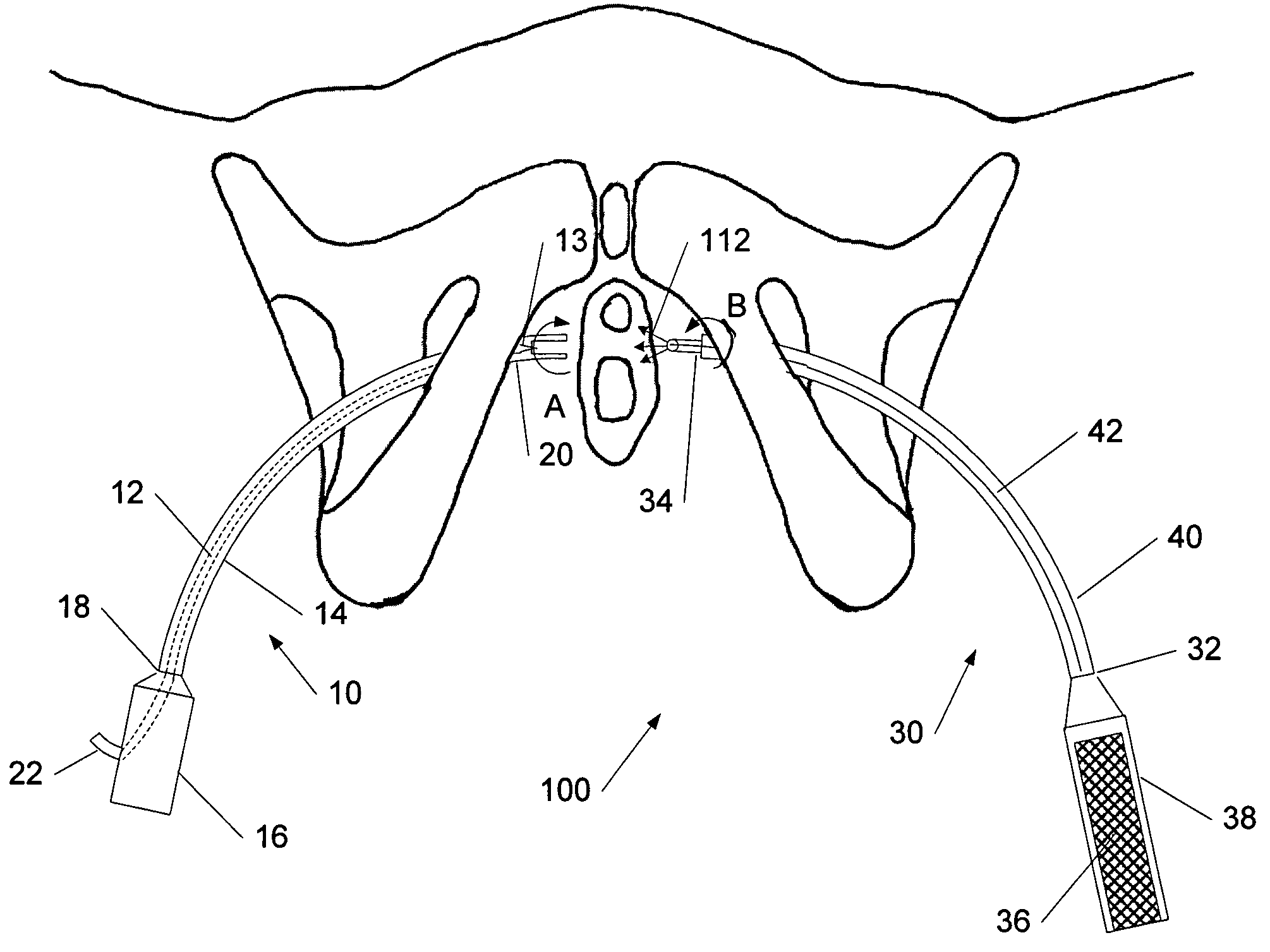
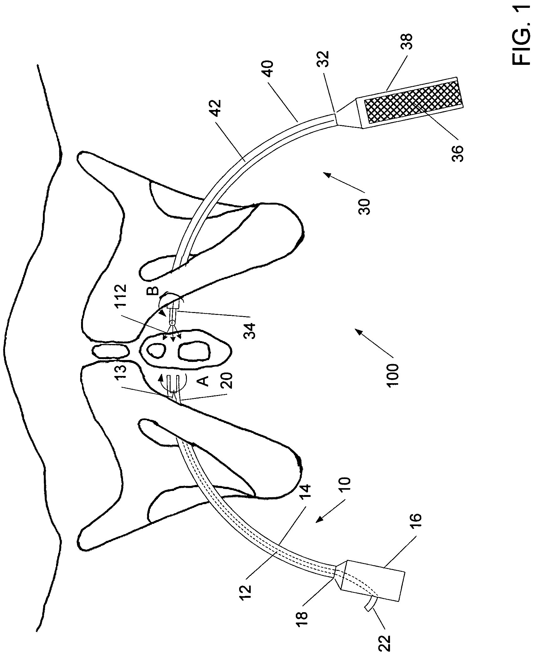
[0026]As described above in summary, the invention in various illustrative embodiments is directed to systems, devices, and methods employing a delivery device, which can be segmented or in one piece, to deliver a sling to the periurethral tissues of a patient. The delivery device of the present invention may inserted in the ischiopubic region and passed through an obturator foramen, without making a transvaginal incision.

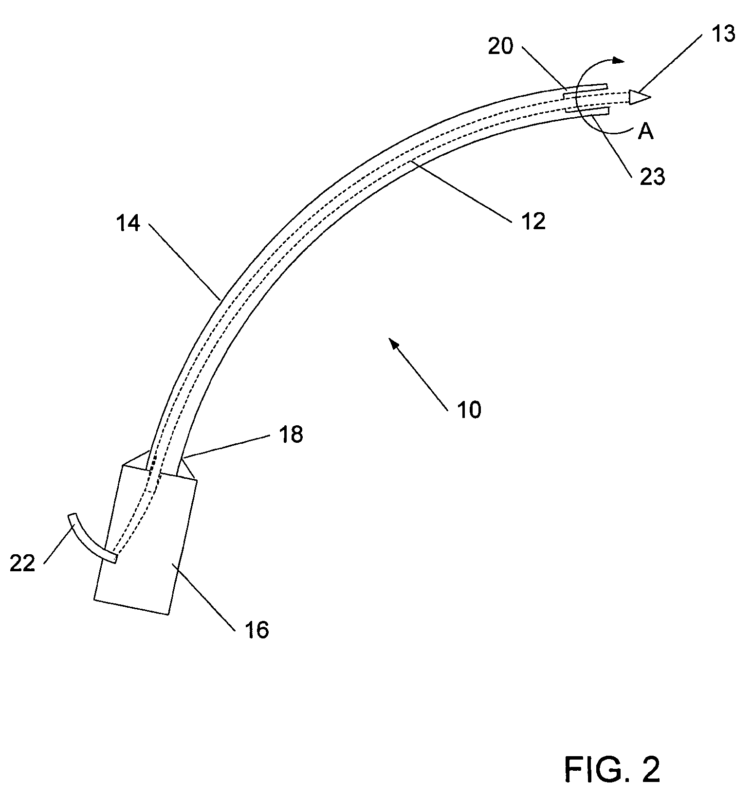
[0027]FIG. 1 shows a perspective front view of one embodiment of a cooperating sling delivery assembly 100 according to the invention. As shown, the cooperating sling delivery assembly 100 in accordance with one aspect of the present invention includes a delivery device 10 and a sling assembly 30. The delivery device 10 includes a shaft 12, which may have a needle-shaped or blunt tip 13, a guide tube 14, and a handle 16. The proximal end 18 of the guide tube 14 may be attached to the distal part of the handle 16 in any variety of manners, including brazing, threadi...


PUM


Abstract

Systems and methods employing a sling delivery assembly having a sling delivery device couplable to a sling assembly to deliver an implant through a transobturator incision point are disclosed. Successful coupling between the sling delivery device and the sling assembly can be verified through changes in an optical or electrical signal produced at the coupling location.

Description

FIELD OF THE INVENTION[0001]The invention generally relates to systems and methods for delivering an implantable sling to an anatomical location in a patient. In various embodiments, the invention is directed to systems and methods relating to the use of a segmented or continuous sling delivery device for delivering an implantable sling.BACKGROUND OF THE INVENTION[0002]Anatomical tissues may become weakened or damaged by age, injury, or disease. This decrease in the structural integrity of anatomical tissues may have significant medical consequences. Even in the absence of tissue necrosis, weakening of an anatomical structure may impair one or more of the biological functions of the tissue. To help alleviate this impact on biological function, implantable, supportive slings have been developed. These slings can be implanted into a patient to provide support for the weakened or damaged tissue. The support provided by the sling mimics the natural position and structure of the tissue, ...

Claims


Application Information

Patent Timeline
Patent Type & Authority Patents(United States)
IPC IPC(8): A61F2/02
CPCA61B17/0469A61B17/0482A61B17/06109A61B17/3415A61B17/3468A61B2017/06104A61B2017/00349A61B2017/00477A61B2017/00805A61B2017/06042A61B2017/06052A61B19/5202A61B90/30
Inventor MACLEAN, BRIAN
Owner BOSTON SCI SCIMED INC
Learn More