Joint replica models and methods of using same for testing medical devices

a technology of replica models and medical devices, applied in the field of joint replica models and methods of using same for testing medical devices, can solve the problems of difficult quantitatively describing a device, general complexity of models that are needed, and limited models, so as to reduce product quality, increase development costs, and prolong product timeliness

Active Publication Date: 2010-04-20
SAKEZLES CHRISTOPHER
View PDF12 Cites 21 Cited by
  • Summary
  • Abstract
  • Description
  • Claims
  • Application Information

AI Technical Summary

Benefits of technology

[0015]The use of a poorly conceived model in research and development testing will lead to reduced product quality, increased development costs, and greatly lengthened product timeliness Fortunately, these failures may be avoided by employing an intelligent development scheme in conjunction with a high quality model. Accordingly, the subject invention pertains to complex synthetic anatomical models that are designed to enable simulated use testing by medical device companies, medical device designers, individual inventors, or any other entity interested in the performance of medical devices. These models are unique in possessing a level of complexity that allows them to be substituted for either a live animal, an animal cadaver, or a human cadaver in the testing of these devices. These models are further characterized by a similarity of geometry, individual component physical properties, and component-to-component interfacial properties with the appropriate target tissue and anatomy.
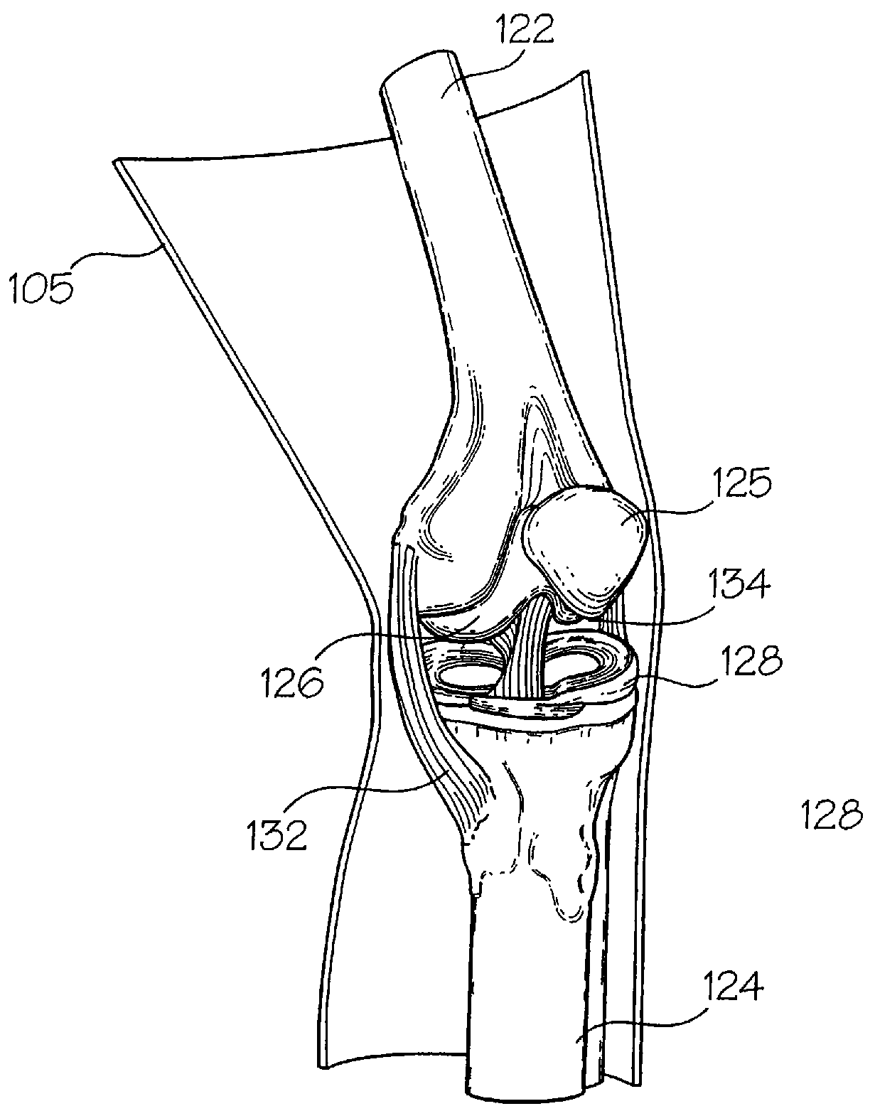
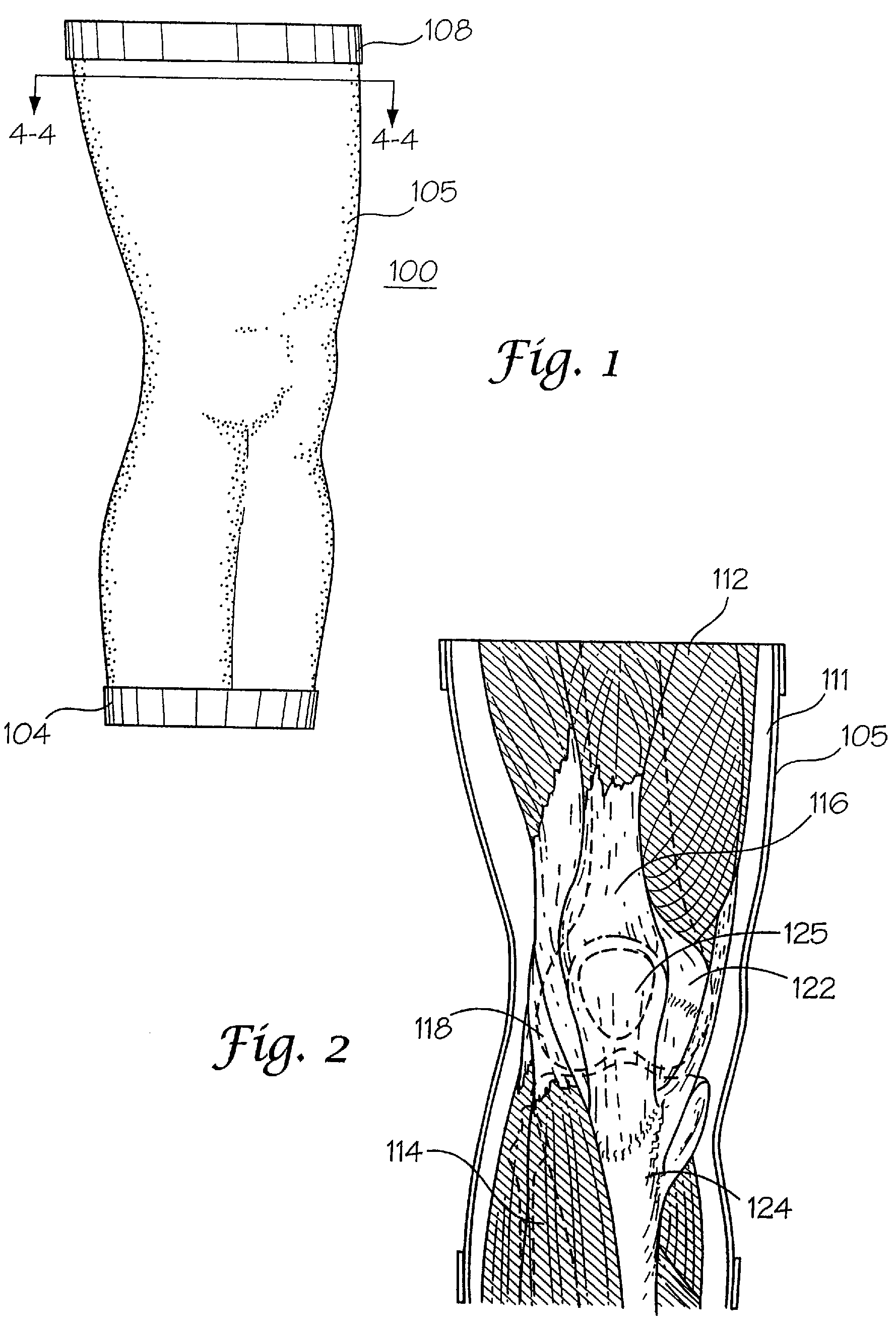
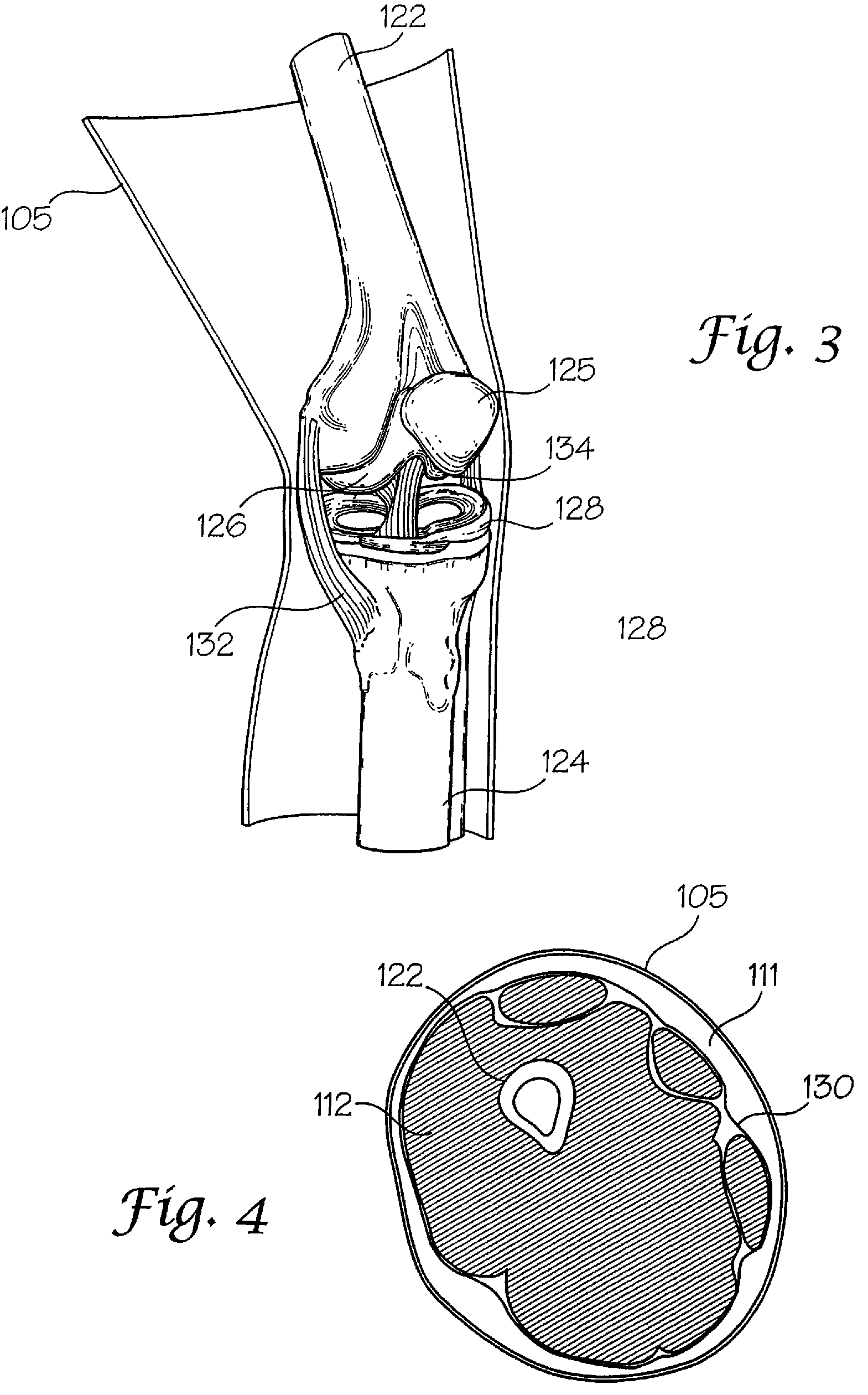
[0018]In certain embodiments, the subject invention pertains to joint replica models that comprise components made of tissue analog materials. The models may be used for development, experimentation, or training in the field of orthopedic surgical devices, and / or implant devices. In exemplary embodiments, joint replica models comprise joint structures such as cartilage, tendons, ligaments, and muscles that are made of hydrogel materials. The joint replica models provide for real feedback helpful in designing surgical equipment and / or implants that minimize deleterious effects on sensitive joint tissues.
[0019]Model embodiments of the subject invention may be nearly as simple to use as a bench top fixture, but provide feedback that is superior in many respects to cadaver tests, animal studies, and even human clinical trials. In fact, a prototype device may be tested not just in terms of device performance, but also in terms of effect on the target anatomy. In certain embodiments, components of the joint replica models may be removed, allowing a quasi-histological examination of the target anatomy after each use. In addition, because the models are artificial and mass produced, multiple tests may be performed either under identical conditions or by altering only the test parameters (temperature, flow, contact angle, etc) desired. This capability helps to eliminate the statistically confounding effect of model variation that plagues cadaver, animal, and human subject studies, and also enables the use of designed experiments to explore device-tissue interactions and interactions between various design parameters.
[0021]In contrast, some embodiments of the subject invention enable a potentially large number of tests to be completed in an environment that is both geometrically and mechanically similar to the target anatomy. These tests may be performed by an engineering technician on a lab bench, but the tests still produce very high quality data. Also, because this data may be generated early in the development cycle, design errors are discovered sooner, leading to a shorter cycle and a reduced development budget. Further, unlike traditional bench top testing, use of embodiments of the subject invention allows the user to predict how a device will actually function in the human body, and since the effect of the device on the target tissue can be predicted by way of the quasi-histological examination, the risk to the patient may be predicted from the beginning of the process.
[0023]In contrast, use of embodiments of the subject invention enables the generation of animal study quality data (in a much greater quantity) using a simple bench top setup that may be used by an engineering technician. The need to contract with research facilities, employ costly medical practitioners, and also any exposure to biohazards is eliminated. In addition, these models may be used at body temperature in the presence of any real or simulated physiologic fluid, and since the device contacting portions of the model may be removed and replaced, an unlimited number of tests may be performed.
[0025]In effect, the inclusion of models according to the subject invention in the development process allows the collection of animal study quality performance data (Table II) at a risk level that is normally associated with bench top studies (see ARM representing models according to the subject invention). In fact, by employing this technology early on in the development process, vital feedback on device performance may be collected before erroneous assumptions can adversely affect the design. This capability not only reduces the probability of costly late stage design changes, but also shortens the project timeline and reduces the overall cost of development. In addition, these models may be used in an ordinary laboratory by engineering personnel. The need to own or contract with research facilities, pay for costly medical practitioners, and absorb risks associated with biohazard exposure are all eliminated. An innocent life (the animal) is also spared.

Problems solved by technology

Of course, these models are limited by the assumptions made in their derivation and the computational power of the computer.
Unfortunately, it may be difficult to quantitatively describe a device, and more importantly the actual use environment, in sufficient detail to yield realistic results.
Once again, human subjects are also employed in the development of many medical devices, but since humans represent the actual use environment, these tests (clinical studies) are not considered simulation tests.
However, since this process is iterative, as the design matures the models that are needed generally become more complex.
Unfortunately, human subjects are unavailable for use until late in the development cycle due to risk, regulatory, and ethical considerations.
Unfortunately, these studies are expensive because of the staff and facilities required to support the work.
These studies can easily exceed $100,000 in total costs, and grow even more costly as the number of animals is increased.
The inability to test prototype devices on human subjects is the reason medical device developers resort to animal studies in the first place.
Still, animal models suffer from a whole range of unique problems, including the many deviations between human and animal anatomy and physiology, the confounding effects of variation between individual animals, and the unpredictability that arises from using a model that is extraordinarily complex.
The great expense and specialized facilities required limit their in-house use.
Reproducibility may also be an issue as both inter- and intrasubject variability are difficult to control.
Additional considerations include contention with the Animal Welfare Act, the significant expense associated with contracting regulated facilities and medical practitioners, and the risks related to handling biohazardous materials.
Unfortunately, there tends to be an inverse relationship between the usefulness and complexity of the model employed.
For example, cadaver tissues provide an accurate representation of anatomical geometry, but the required chemical preservation greatly alters the physical properties of the tissues.
In addition, biological temperatures and flows cannot generally be simulated, subjects are difficult to source and maintain in useful quantities, and an educational institution must almost always be contracted (at considerable expense) to perform the study.
Unfortunately, these models are typically designed by individuals lacking an understanding of anatomy and physiology, and are usually fabricated from typical engineering materials such as metal, glass, and plastic.
While an argument may be made that these models are better than nothing, they are certainly not representative of actual use conditions.
A poor model is therefore more likely to yield misleading data, and a design based at an early stage upon this data is more likely to require correction at a later stage in development.
The use of a poorly conceived model in research and development testing will lead to reduced product quality, increased development costs, and greatly lengthened product timeliness Fortunately, these failures may be avoided by employing an intelligent development scheme in conjunction with a high quality model.
These models are unique in possessing a level of complexity that allows them to be substituted for either a live animal, an animal cadaver, or a human cadaver in the testing of these devices.
However, the use of engineering materials in the construction of these models make them dissimilar to the target anatomy in a profound way.
In addition, these models may only be used to predict device performance, not the effect of the device on the target tissue.
Also, because this data may be generated early in the development cycle, design errors are discovered sooner, leading to a shorter cycle and a reduced development budget.
It is impossible to use these models at normal body temperature or in the presence of fluids, and they cannot be employed to accurately predict the physical effect of the device on the target tissue.
An educational institution must almost always be contracted (along with a principal investigator) to perform the study, and since the specimens are difficult to source it is common to run only a single test.
Biohazards are an additional risk.
Needless to say, these studies are very expensive and grow ever more costly as the number of animals is increased.
The cost of discovering a design flaw at this stage is very high, possibly causing modification, termination, or repetition of the study.
Biohazards are also a significant risk.
In fact, by employing this technology early on in the development process, vital feedback on device performance may be collected before erroneous assumptions can adversely affect the design.
This capability not only reduces the probability of costly late stage design changes, but also shortens the project timeline and reduces the overall cost of development.

Method used


Image

  • Joint replica models and methods of using same for testing medical devices
  • Joint replica models and methods of using same for testing medical devices
  • Joint replica models and methods of using same for testing medical devices

Examples


Embodiment Construction

[0073]The interaction of a foreign body with living tissues results in complications that are related to, among other things, shear forces, normal forces, abrasive action, blunt trauma, pressure necrosis, or other physical insults caused by the invading device. Not only are studies to predict the long-term effect of this invasion difficult and expensive to conduct, but when live patients are involved the studies often yield inconclusive results. As an alternative to using these patients, a bench top model may be employed to physically simulate the insult to the tissue as a relatively inexpensive, easily repeatable, and logical first step before resorting to animal studies and clinical trials. However, for this approach to be productive, the model employed must be representative of the actual target anatomy in which the medical device will normally be used.

[0074]The subject invention pertains to complex synthetic joint replica models that are designed to enable simulated use testing ...


PUM


Abstract

Disclosed herein are joint replica models that are designed to enable simulated use testing by medical device companies, medical device designers, individual inventors, or any other entity interested in the performance of medical devices. These models are unique in possessing a level of complexity that allows them to be substituted for either a live animal, an animal cadaver, or a human cadaver in the testing of these devices. These models are further characterized by a similarity of geometry, individual component physical properties, and component-to-component interfacial properties with the appropriate target tissue and anatomy.

Description

CROSS-REFERENCE TO RELATED APPLICATIONS[0001]This application is a continuation in part application to U.S. patent application Ser. No. 11 / 172,575; filed Jun. 30, 2005 now U.S. Pat. No. 7,507,092; which is a continuation in part application to U.S. patent application Ser. No. 11 / 098,248; filed Apr. 4, 2005 now U.S. Pat. No. 7,272,766, which is a continuation in part application to U.S. patent application Ser. No. 11 / 050,161, filed Feb. 3, 2005 now U.S. Pat. No. 7,427,199. This application is also related to U.S. Provisional Application Nos. 60 / 749,971 and 60 / 764,491; filed Dec. 13, 2005 and Feb. 2, 2006, respectively. This application claims the benefit of priority to foregoing under 35 USC §§ 119 and 120, and the teachings of such applications are hereby incorporated herein.BACKGROUND OF THE INVENTION[0002]During the development of any new medical device, various tests may be required, including the characterization of physical properties (geometric, mechanical, electrical, electro...

Claims


Application Information

Patent Timeline
Patent Type & Authority Patents(United States)
IPC IPC(8): G09B23/28
CPCG09B23/28G09B23/34G09B23/32G09B23/30B33Y80/00
Inventor SAKEZLES, CHRISTOPHER
Owner SAKEZLES CHRISTOPHER
Why Eureka
Try Eureka