Digital karyotyping

a digital karyotype and genome technology, applied in the field of genetics, can solve the problems of limited sequence number, inability to detect small alterations, and hidden abnormalities at the cellular and organismal level

Active Publication Date: 2010-04-27
THE JOHN HOPKINS UNIV SCHOOL OF MEDICINE
View PDF12 Cites 84 Cited by
  • Summary
  • Abstract
  • Description
  • Claims
  • Application Information

AI Technical Summary

Problems solved by technology

Somatic and hereditary variations in gene copy number can lead to profound abnormalities at the cellular and organismal levels.
However, methods employing metaphase chromosomes have a limited mapping resolution (˜20 Mb) (15) and therefore cannot be used to detect smaller alterations.
Recent implementation of comparative genomic hybridization to microarrays containing genomic or transcript DNA sequences provide improved resolution, but are currently limited by the number of sequences that can be assessed (16) or by the difficulty of detecting certain alterations (9).

Method used


Image

  • Digital karyotyping
  • Digital karyotyping
  • Digital karyotyping

Examples


example 1

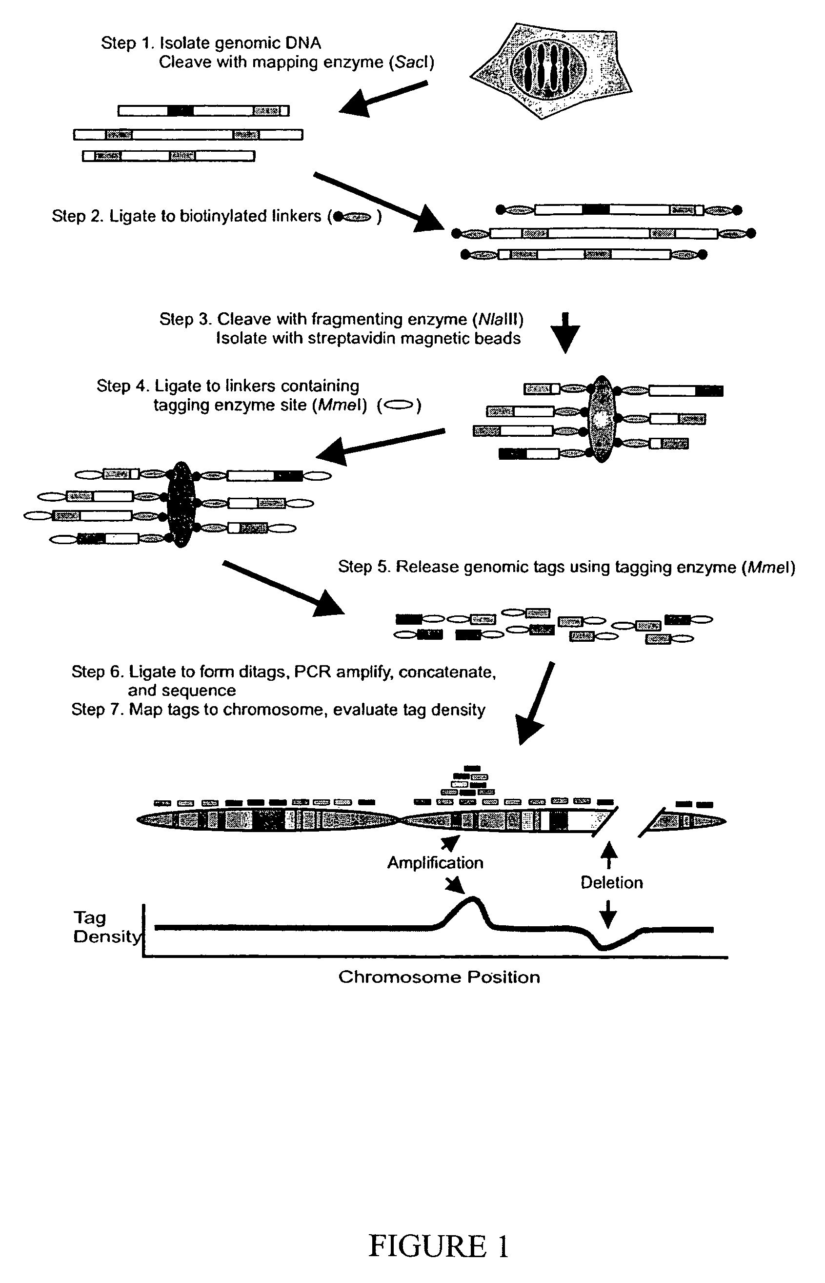
Principles of Digital Karyotyping

[0025]These concepts are practically incorporated into Digital Karyotyping of human DNA as described in FIG. 1. Genomic DNA is cleaved with a restriction endonuclease (mapping enzyme) that is predicted to cleave genomic DNA into several hundred thousand pieces, each on average 6 / 44), the majority of DNA molecules in the template are expected to be cleaved by both enzymes and thereby be available for subsequent steps. DNA fragments containing biotinylated linkers are separated from the remaining fragments using streptavidin-coated magnetic beads (Step 3). New linkers, containing a 5-bp site recognized by MmeI, a type IIS restriction endonuclease (18), are ligated to the captured DNA (Step 4). The captured fragments are cleaved by MmeI, releasing 21 bp tags (Step 5). Each tag is thus derived from the sequence adjacent to the fragmenting enzyme site that is closest to the nearest mapping enzyme site. Isolated tags are self-ligated to form ditags, PCR am...

example 2

Analysis of Whole Chromosomes

[0028]We characterized 210,245 genomic tags from lymphoblastoid cells of a normal individual (NLB) and 171,795 genomic tags from the colorectal cancer cell line (DiFi) using the mapping and fragmenting enzymes described above. After filtering to remove tags that were within repeated sequences or were not present in the human genome (see Materials and Methods), we recovered a total of 111,245 and 107,515 filtered tags from the NLB and DiFi libraries, respectively. Tags were ordered along each chromosome, and average chromosomal tag densities, defined as the number of detected tags divided by the number of virtual tags present in a given chromosome, were evaluated (Table 2). Analysis of the NLB data showed that the average tag densities for each autosomal chromosome was similar, ˜0.16+ / −0.04. The small variations in tag densities were likely due to incomplete filtering of tags matching repeated sequences that were not currently represented in the genome da...

example 3

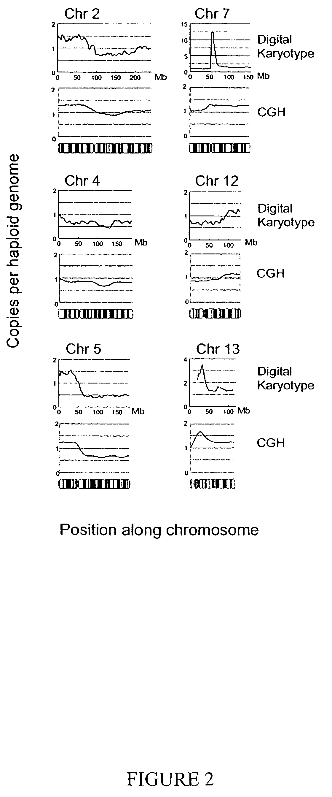
Analysis of Chromosomal Arms

[0030]We next evaluated the ability of Digital Karyotyping to detect subchromosomal changes, particularly gains and losses of chromosomal arms. Tag densities were analyzed along each chromosome using sliding windows containing 1000 virtual tags (˜4 Mb), as windows of this size were predicted to reliably detect such alterations (Table 1). For the NLB sample, tag density maps showed uniform content along each chromosome, with small variations (<1.5 fold) present over localized regions, presumably due to overrepresentation of tags matching repeated sequences (data not shown). In contrast, the DiFi tag density map (normalized to the NLB data) revealed widespread changes, including apparent losses in large regions of 5q, 8p and 10q, and gains of 2p, 7q, 9p, 12q, 13q, and 19q (FIG. 2 and FIG. 5). These changes included regions of known tumor suppressor genes (21) and other areas commonly altered in colorectal cancer (11, 12, 22). These alterations were confirme...


PUM

PropertyMeasurementUnit
sizeaaaaaaaaaa
densitiesaaaaaaaaaa
densityaaaaaaaaaa

Abstract

Alterations in the genetic content of a cell underlie many human diseases, including cancers. A method called Digital Karyotyping provides quantitative analysis of DNA copy number at high resolution. This approach involves the isolation and enumeration of short sequence tags from specific genomic loci. Analysis of human cancer cells using this method identified gross chromosomal changes as well as amplifications and deletions, including regions not previously known to be altered. Foreign DNA sequences not present in the normal human genome could also be readily identified. Digital Karyotyping provides a broadly applicable means for systematic detection of DNA copy number changes on a genomic scale.

Description

[0001]This application claims the benefit of provisional application Ser. No. 60 / 426,406 filed Nov. 15, 2002, the contents of which are expressly incorporated herein.[0002]The work underlying this invention was supported in part by the U.S. government. Thus the U.S. government retains certain rights in the invention according to the provisions of grant nos. CA 43460, CA 57345, CA 62924 of the National Institutes of Health.[0003]A portion of the disclosure of this patent document contains material which is subject to copyright protection. The copyright owner has no objection to the facsimile reproduction by anyone of the patent document or the patent disclosure, as it appears in the Patent and Trademark Office patent file or records, but otherwise reserves all copyright rights whatsoever.FIELD OF THE INVENTION[0004]The invention relates to the field of genetics. In particular, it relates to the determination of karyotypes of genomes of individuals.BACKGROUND OF THE INVENTION[0005]Som...

Claims


Application Information

Patent Timeline
Patent Type & Authority Patents(United States)
IPC IPC(8): C12Q1/68C12P19/34C40B40/08G01N33/48G06F19/00C12Q
CPCC12Q1/6841C12Q2565/102C12Q2537/157C12Q2521/313
Inventor WANG, TIAN-LIVELCULESCU, VICTORKINZLER, KENNETHVOGELSTEIN, BERT
Owner THE JOHN HOPKINS UNIV SCHOOL OF MEDICINE
Why Eureka
Try Eureka