
CSF biomarker dilution factor corrections by MRI imaging and algorithm

a biomarker and dilution factor technology, applied in the field of cerebrospinal fluid biomarker analysis, can solve the problems of slow process for analyzing biomarkers in csf, inability to provide accurate results, and impede the research and development of effective drugs, so as to achieve rapid and inexpensive measurement of increased ventricular size.

Inactive Publication Date: 2010-06-15
NEW YORK UNIV MEDICAL CENT
View PDF0 Cites 3 Cited by
  • Summary
  • Abstract
  • Description
  • Claims
  • Application Information

AI Technical Summary

Benefits of technology

[0011]The present invention discloses a novel method for correcting biomarker dilution in ventricular CSF that greatly reduces the time needed to make the corrections, thus permitting relatively inexpensive and accurate diagnosis and monitoring of biomarkers that heretofore were previously cost-prohibitive to be used as a routine test. The application of the invention can be to correct any biomarker found in CSF, and not just the markers associated with MCI and Alzheimer's Disease.
[0012]The present invention permits a fast and accurate method for detection of ventricular size so as to adjust the level of biomarkers according to the CSF volume that reduces the time to make the correction to a matter of minutes, and does not require highly trained specialists in brain anatomy to study the scans. The costs to perform the correction allow for a fast and inexpensive way (so as to be covered by health insurance) to test for cases of MCI, Alzheimer's Disease, both for those who show signs of such illnesses, as well as for those who do not, by combining MRI and CSF markers to improve the sensitivity and specificity for the prediction of cognitive decline.
[0013]The benefits of the present invention include the ability to detect a problem and permit the prescription of drugs to slow / inhibit the progression of some of the aforementioned brain disorders before the patient's illness has advanced to a stage where it has a severe impact.

Problems solved by technology

However, heretofore, the process for analyzing biomarkers in CSF has been slow, cost prohibitive, and has not always provided accurate results.
In fact, the sometimes inaccurate results of the prior art can hinder research and development of effective drugs, as well as increase the likelihood that a proper diagnosis of a particular illness may not be made in a timely fashion, or not at all for that matter.
Heretofore, neuroimaging methods have not been used to correct the diluted CSF biomarker.
The prior art methods using MRI scans to calculate the CSF compartment heretofore have been cost prohibitive because of the need for highly trained persons to spend sometimes as much as several hours to calculate CSF volume for a single patient by methods that require manual identification and dissection of areas of the brain, with the ventricular size and other CSF containing structures being determined by the number of pixels counted in each of the areas identified.
In particular, defining the anatomical boundaries of the ventricular system from MRI scans is complex and time consuming.
The MRI scans can number as many as fifty or more “slices” of the ventricle being scanned, thus making the calculations a rather tedious procedure, and subject to error even by highly trained personnel.
The prior art has had attempts to provide a semi-automated program to measure the whole brain CSF, but there was absolutely no disclosure, suggestion or motivation that the determination of ventricular size could be used to correct biomarkers in CSF volumes.

Method used


Image

  • CSF biomarker dilution factor corrections by MRI imaging and algorithm
  • CSF biomarker dilution factor corrections by MRI imaging and algorithm

Examples


Embodiment Construction

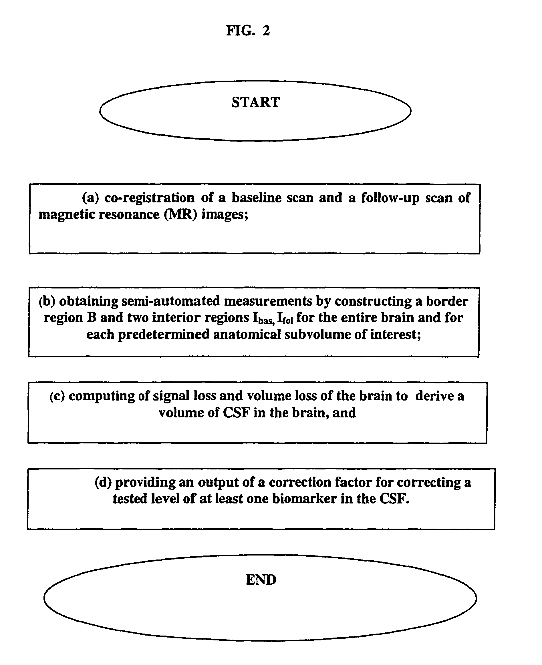
[0040]The present invention is not limited to any particular type of MRI data, as it can be a single sequence, such as high resolution fast gradient recalled echo (GRE or MPRAGE) data. This high resolution sequence for anatomical work can be acquired at the baseline and follow up in any plane (sagittal, axial or coronal). An example of this sequence is defined as TR=35 ms, TE=9 ms, 60 degree flip angle, 256×192 acquisition matrix, 1.1 mm section thickness, 124 sections, 24 cm FOV, and a 1 NEX, for a total acquisition time of 12 minutes. In addition, there can be a multi-sequence, including but not in any way limited to two IR sequences, multiple echo MR sequences, etc., that can be used to segment the brain into cerebral gray matter, white matter and cerebrospinal fluid (as known in the art, exemplified by Rusinek, H, de Leon M J, George A et al: Alzheimer Disease: Measuring Loss of cerebral gray matter with MR Imaging, Radiology 178: 109-114, 1991, the contents of which are herein ...


PUM

PropertyMeasurementUnit
thicknessaaaaaaaaaa
thicknessaaaaaaaaaa
MRIaaaaaaaaaa

Abstract

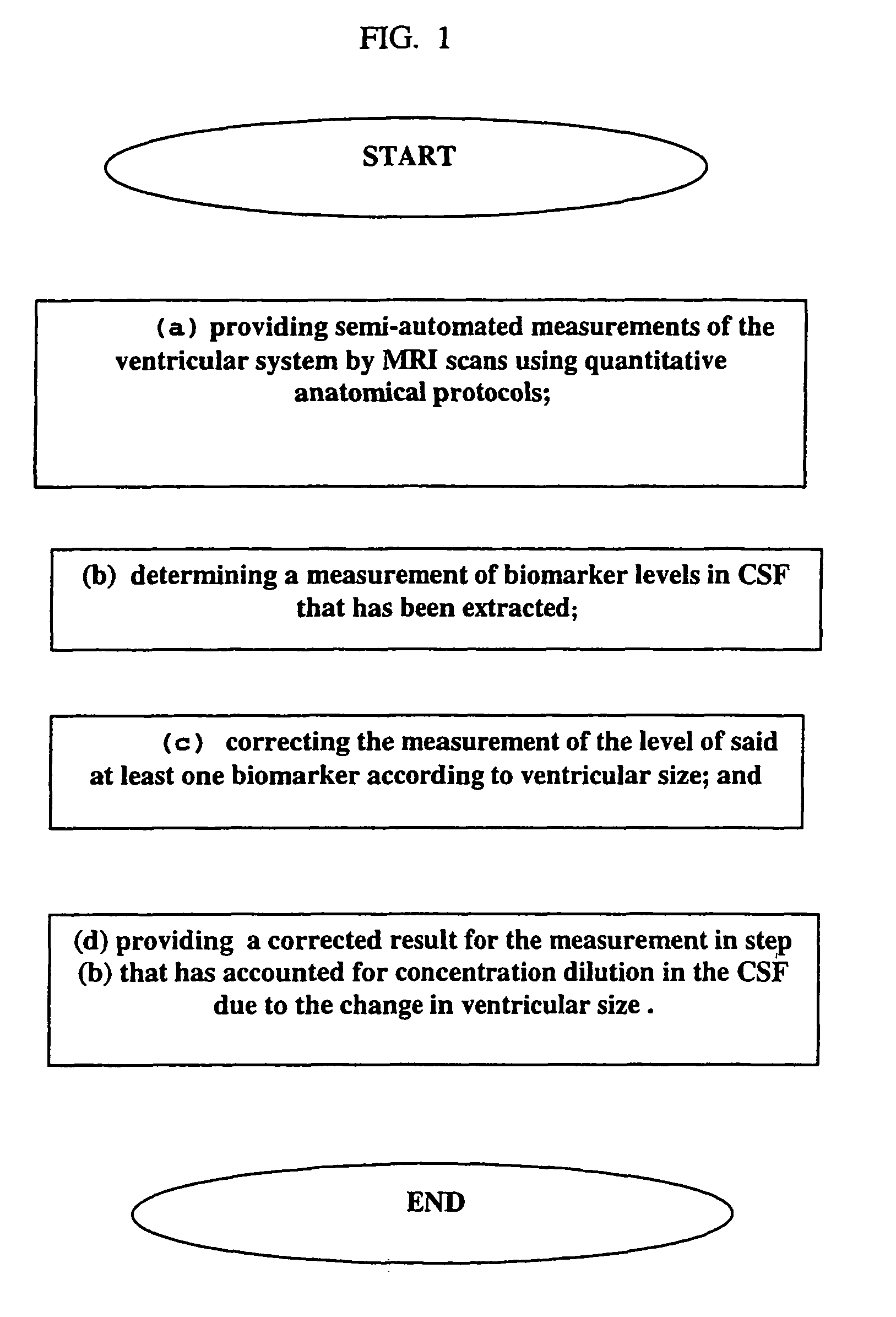
A method is disclosed for providing a correcting factor for the dilution of measurements of at least one biomarker in cerebrospinal fluid (CSF). The method comprises providing semi-automated measurements of the ventricular system by MRI scans using quantitative anatomical protocols, determining a measurement of biomarker levels in CSF that has been extracted, correcting the measurement of the level of said at least one biomarker according to the ventricular size, and providing a corrected result of the measurement determined in step (b), said corrected result accounting for concentration dilution due to the change in ventricular size. The method is particularly suited for the measurement of all biomarkers found in the CSF, such as those associated with mild cognitive impairment (MCI) and Alzheimer's Disease.

Description

BACKGROUND OF THE INVENTION[0001]1. Field of the Invention[0002]The present invention relates to the analysis of biomarkers in cerebrospinal fluid (CSF). More particularly, the present invention relates to (1) discovering the importance of correcting CSF biomarker levels for their dilution in CSF and (2) correcting CSF measures for their dilution using a semi-automated procedure for determining CSF volumes in patients with variable and disease-related increases in CSF volume. This invention is important for diagnosing and monitoring the course of Alzheimer's disease, but the scope of the invention reaches far beyond diagnosing and monitoring of Alzheimer's disease. For example, the invention can be applied to monitor other disorders, such as vascular dementia, boxer's syndrome, multiple sclerosis, amyotrophic lateral sclerosis (ALS), and stroke, just to name a few.[0003]2. Description of the Related Art[0004]Cerebrospinal Fluid (CSF) is a liquid that fills the ventricles of the brai...

Claims


Application Information

Patent Timeline
Patent Type & Authority Patents(United States)
IPC IPC(8): A61B5/055C12Q1/00G01N33/53G01N33/542G01N33/566A61B5/05A61B5/107G01N33/68
CPCA61B5/055A61B5/48G01N33/6896A61B5/1075A61B5/4088
Inventor DE LEON, MONYRUSINEK, HENRY
Owner NEW YORK UNIV MEDICAL CENT
Learn More