Biochip for the detection of phosphorylation and the detection method using the same

a biochip and phosphorylation technology, applied in the field of biochip for detecting phosphorylation and a detection method using the same, can solve the problems of inconvenient use, high labor intensity, and slow method, and achieve the effects of simple process, increased sensitivity, and time saving

Inactive Publication Date: 2010-09-14
KOREA ATOMIC ENERGY RES INST
View PDF0 Cites 0 Cited by
  • Summary
  • Abstract
  • Description
  • Claims
  • Application Information

AI Technical Summary

Benefits of technology

[0021]The biochip for detecting phosphorylation and the method for detecting phosphorylation using the same of the present invention increase sensitivity by using a radio-isotope, by which a smaller amount of a sample can be used by simpler processes, compared with the conventional method. In addition, the method of the present invention produces a very clear result owing to a simplified procedure excluding blocking process for preventing non-specific fixation on the chip. So, using the biochip of the present invention has advantages of saving time and having economical efficiency. According to the present invention, only a small amount of a sample is required, suggesting that the size of a spot is very small, indicating the number of samples that can be integrated on the surface can be increased, which enables fast analysis of a large amount of samples within a short period. Therefore, the method of the present invention can be effectively used for the analysis of kinase activity.

Problems solved by technology

Fluorometry based on DNA chip analysis has been most widely used so far, but each method has been recognized to have merits and demerits, and which protein chip analysis method is most appropriate for disease is still undecided.
The conventional method to study the activity of kinase is to examine cell membrane using a radio-isotope, but this method is very slow-going and requires a great labor.
ELISA is a comparatively accurate method but takes a lot of time and requires a huge amount of samples.
The method using an antibody facilitates mass-analysis but requires high costs and the processes are very complicated.
However, this kit is limited in mass-analysis.
But, this method requires high costs for mass-analysis.
Except for the said four protein chip related techniques, there is no method available for screening the activity of protein kinase in a large scale by using protein chip or peptide chip.

Method used


Image

  • Biochip for the detection of phosphorylation and the detection method using the same
  • Biochip for the detection of phosphorylation and the detection method using the same
  • Biochip for the detection of phosphorylation and the detection method using the same

Examples


example 1

Preparation of Elevated Protein-Substrate Fusion Protein

[0066] Cloning of SrMep45-Substrate Fusion Protein

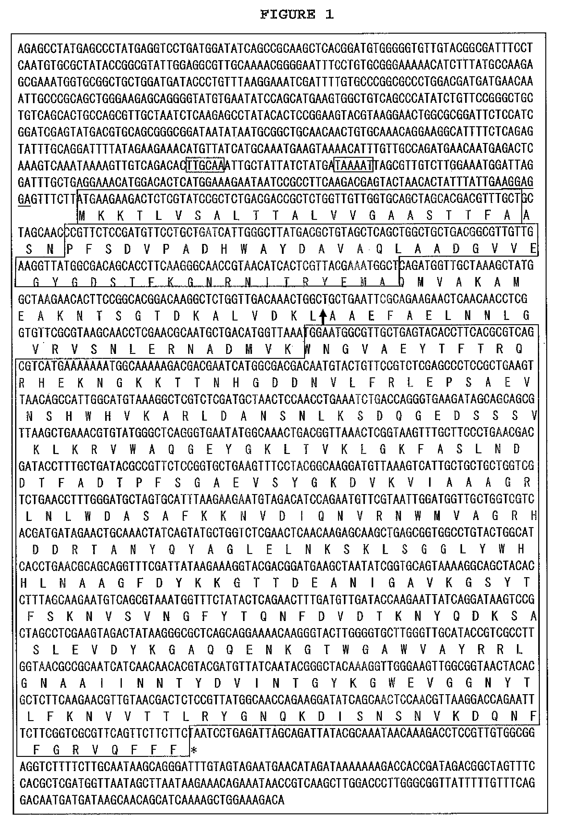
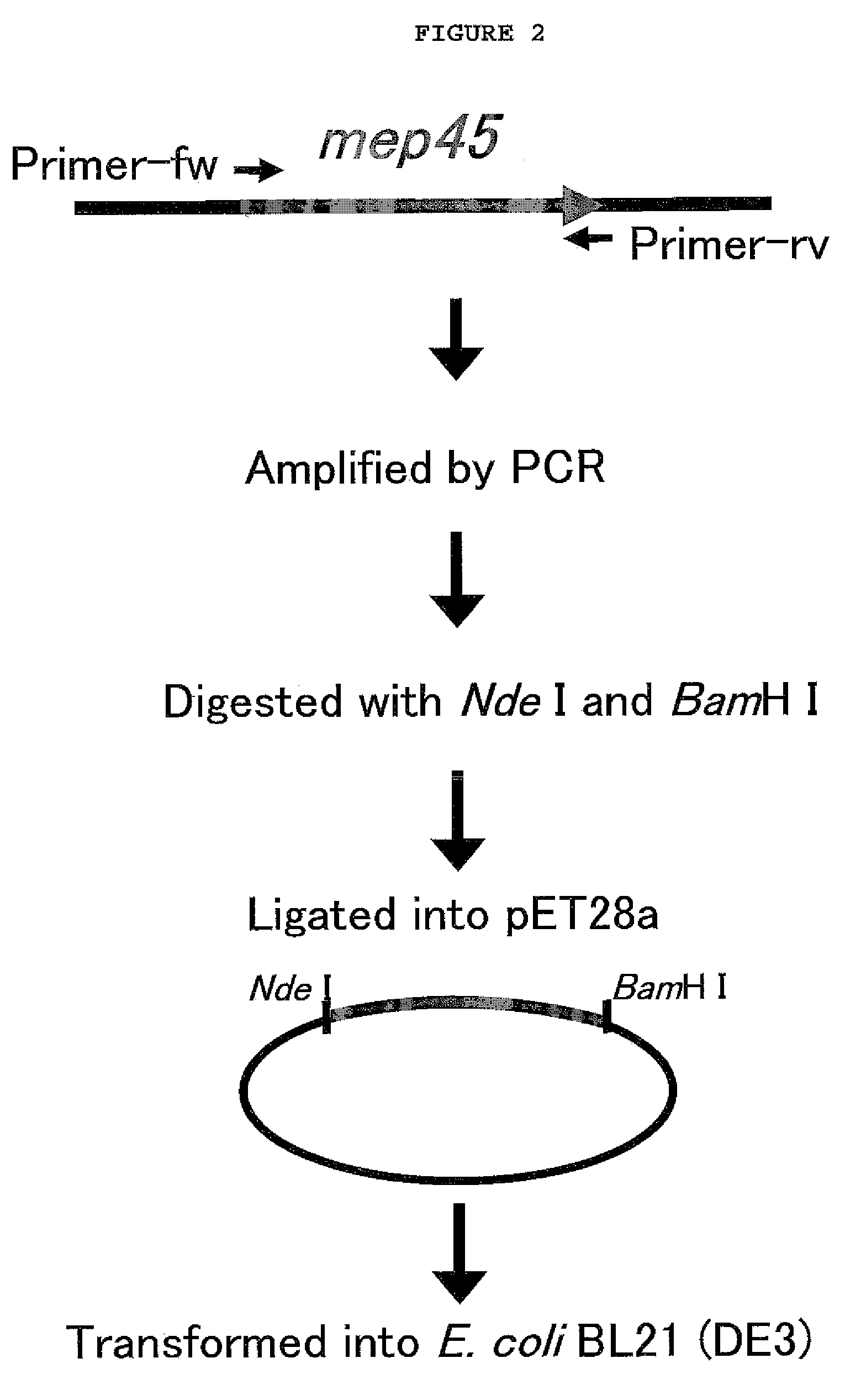
[0067]To prepare SrMep45 (Selenomonas ruminantium membrane protein)-substrate fusion protein, the PCR product obtained by PCR amplification with chromosome DNA of Selenomonas ruminantium subsp. lactilytica, ATCC 19205) (Kanegasaki, S., and Takahashi, H., J. Bacteriol. 93, 456-463, 1967) and plasmid were cloned (FIG. 1).

[0068]For the cloning of each substrate (AAKIQASFRGHMARKK; SEQ. ID. NO: 1, PKTPKKAKKL; SEQ. ID. NO: 2, or EPPLSQQAFADLWKK; SEQ. ID. NO: 3) for PKC (Protein Kinase C), cdc2-PK (cdc2 Protein Kinase) and DNA-PK (DNA-dependent Protein Kinase), PCR was performed using pSrMep45 as a template with primers PKC-Fw-Nde (5′-CATCATATGGCTGCTAAAATTCAAGCTTCTTTTCGTGGTCATATGGCTCGTAAAAAAGCTAGCAACCCGTTCTCCGATG-3′; SEQ. ID. NO: 4), PKC-Rv-Bam (5′-GACGGATCCTTATTTTTTACGAGCCATATGACCACGAAAAGAAGCTTGAATTTTAGCAGCGAAGAAGAACTGAACGCGACCGAAG-3′; SEQ. ID. NO: 5), cdc2-MP-Fw-Nde (5′-CATCATATGCCTA...

example 2

Construction of Biochip

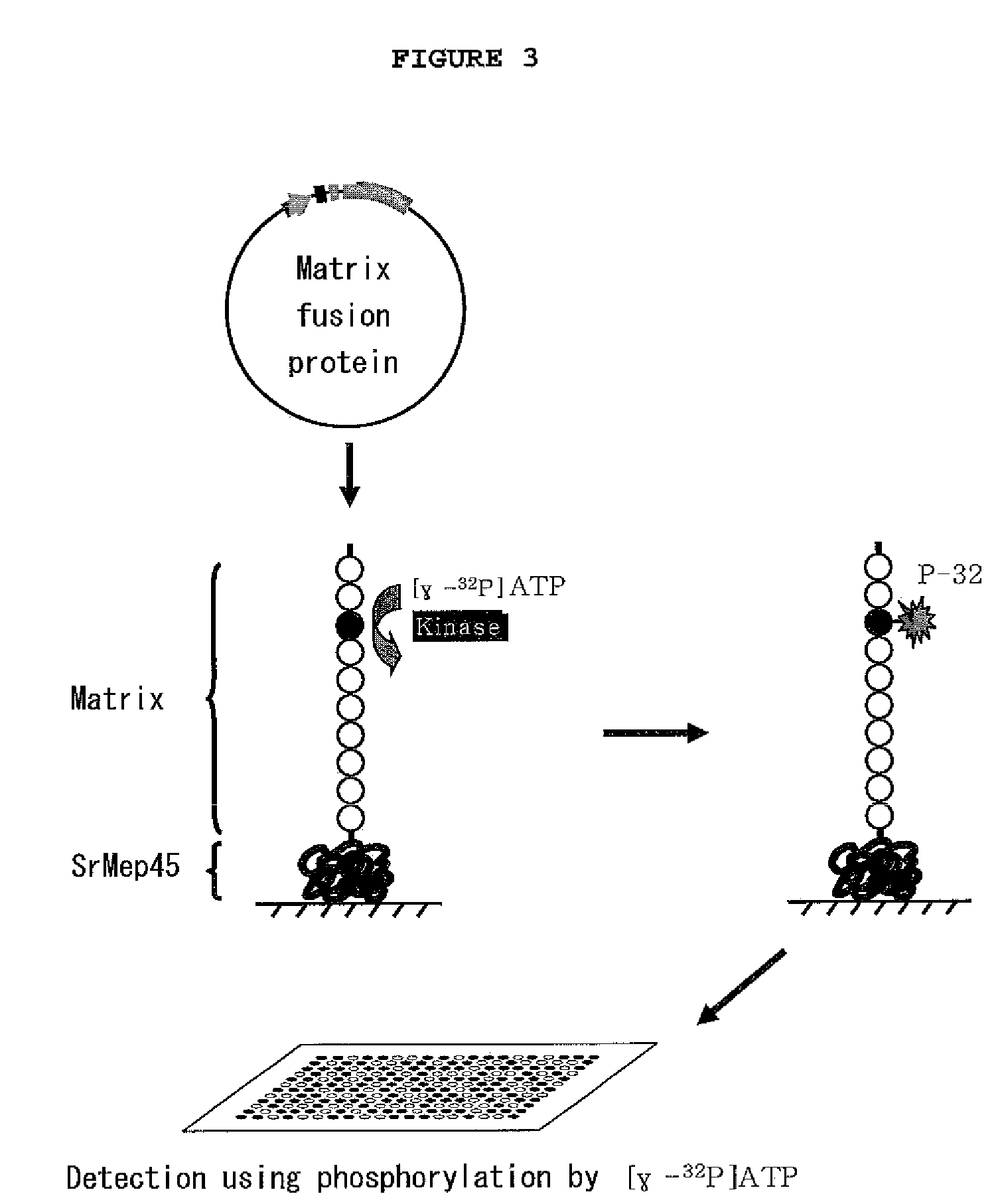
[0075]To fix substrate on the aldehyde-treated slide glass (Nuricell Inc., Korea), 0.1 mg / ml of the Mep45-kinase substrate fusion protein or 1.25 μg / ml of peptide substrate (Promega, Madison, Wis.) was integrated. Particularly, the recombinant Mep45-kinase substrate fusion protein solution (10% glycerol, PBS, pH 7.5) was prepared at the concentration of 0.1 mg / ml, and this substrate solution was integrated on the aldehyde-treated slide glass at the spot intervals of 300 μm by using microarray device (Genetix Ltd, UK). The size of the spot was regulated to be 300 μm. The integrated biochip was reacted in a humid chamber at room temperature for one hour, leading to fixation.

example 3

Confirmation of Phosphorylating Conditions between kinase and substrate using [γ-32P]ATP

[0076]The biochip constructed in Example 2 was washed three times with PBS (200 mM NaCl, 3 mM KCl, 2 mM KH2PO4, 1 mM Na2HPO4, pH 7.5), followed by reaction of the kinase and substrate on the chip. Particularly, the chip was washed with kinase buffer (40 mM Tris-HCl, 20 mM MgCl2, 0.1 mg / ml BSA, pH 7.5) once. Then, 50 μl of kinase reaction solution (kinase buffer containing 100 μM ATP, [γ-32P]ATP (0.1˜0.6 μCi) (GE Healthcare Life Sciences, UK) and 0.01˜50 unit / ml kinase of recombinant Mep45-substrate fusion protein) was distributed on the surface of the biochip. The biochip was covered with cover well, followed by reaction for one hour. One hour later, the biochip was washed with washing buffer three times, followed by washing again with PBS. Centrifugation was performed at 200×g for one minute to eliminate remaining moisture completely. The reacted biochip was sensitized on X-ray film or screen of...


PUM

PropertyMeasurementUnit
diameteraaaaaaaaaa
diameteraaaaaaaaaa
diameteraaaaaaaaaa

Abstract

The present invention relates to a biochip for detecting phosphorylation and a method for detecting phosphorylation using the same, more precisely a biochip prepared by integrating a protein produced from the recombination of a substrate of kinase selected from the group consisting of PKC (Protein Kinase C), cdc2-PK (cdc2 Protein Kinase) and DNA-PK (DNA-dependent Protein Kinase) and the elevated protein such as Selenomonas ruminantium membrane protein on a matrix surface coated with an active group, a kit for detecting phosphorylation composed of the said biochip and a cofactor labeled with a radio-isotope and a method for detecting phosphorylation using the same. The biochip for detecting phosphorylation of the present invention using a radio-isotope facilitates the detection of phosphorylation with a minimum amount of a sample by simple processes, compared with the conventional method using an antibody. Since this method can analyze a large amount of samples in a shorter period of time, it can be effectively used for the analysis of kinase activity.

Description

CROSS-REFERENCES TO RELATED APPLICATION[0001]This patent application claims the benefit of priority under 35 U.S.C §119 of Korean Patent Application No. 10-2008-0059603 filed on Jun. 24, 2008, the contents of which are incorporated herein by reference.BACKGROUND OF THE INVENTION[0002]1. Field of the Invention[0003]The present invention relates to a biochip for detecting phosphorylation and a method for detecting phosphorylation using the same, more precisely a biochip prepared by integrating a fusion protein produced from the recombination of a substrate of a kinase selected from the group consisting of PKC (Protein Kinase C), cdc2-PK (cdc2 Protein Kinase) and DNA-PK (DNA-dependent Protein Kinase), and the elevated protein such as Selenomonas ruminantium membrane protein on a matrix surface coated with an active group, a kit for detecting phosphorylation composed of the said biochip and a cofactor labeled with a radio-isotope and a method for detecting phosphorylation using the same...

Claims


Application Information

Patent Timeline
Patent Type & Authority Patents(United States)
IPC IPC(8): C12Q1/48C07K14/00
CPCC12Q1/485G01N33/54353G01N33/60G01N33/6803G01N2333/912G01N2440/14C07K2319/00
Inventor PARK, SANG-HYUNKO, KYONG CHEOL
Owner KOREA ATOMIC ENERGY RES INST
Why Eureka
Try Eureka