Medical X-ray examination apparatus for performing K-edge imaging

a medical x-ray and imaging technology, applied in tomography, instruments, applications, etc., can solve the problems of large number of potential clinical applications that suffer from this limited sensitivity or are completely impossible, and achieve the effect of increasing the sensitivity of selective imaging of a k-edge absorption imag

Active Publication Date: 2011-02-22
KONINKLIJKE PHILIPS ELECTRONICS NV
View PDF10 Cites 5 Cited by
  • Summary
  • Abstract
  • Description
  • Claims
  • Application Information

AI Technical Summary

Benefits of technology

[0008]It is an object of the invention to provide a medical X-ray examination apparatus of the kind mentioned in the opening paragraph with an increased sensitivity of the selective imaging of a K-edge absorption image, so that the medical X-ray examination apparatus according to the invention can be used for more clinical applications than the known medical X-ray examination apparatus.
[0010]By using a spatial reducer to reduce the spatial resolution of the K-edge absorption image, a plurality of interconnected data elements containing image information of the K-edge absorption image is combined. By combining these data elements, the signal to noise ratio (SNR) of the K-edge absorption image is improved. Since the sensitivity of the selective imaging of a K-edge absorption image is at least partially determined by the SNR of the K-edge absorption image, an improved SNR directly contributes to a better sensitivity of the selective imaging of a K-edge absorption image. With an increased sensitivity of the selective imaging of a K-edge absorption image, the medical X-ray examination apparatus according to the invention can be used for more clinical applications than the known medical X-ray examination apparatus.
[0011]A further advantage of the medical X-ray examination apparatus according to the invention is that when the SNR of the K-edge absorption image is improved, the quantitative information of the local densities of the K-edge material that has bound to an object of interest becomes more accurate (i.e. when the SNR of the K-edge absorption image is improved, the quantitative information of the local densities of targeted contrast agents having a payload of K-edge material, like gadolinium-based targeted contrast agents, that has bound to an object of interest (e.g. a tumor, soft plaque in coronaries, a lesion etc.) becomes more accurate). This corresponds well to the clinical evolution towards clinical requests for quantitative measures that inform physicians about how much K-edge material has bound to an object of interest.
[0014]A further preferred embodiment of the medical X-ray examination apparatus according to the invention is characterized in that the spatial resolution reducer comprises a replacement combiner unit for combining the quantitative information represented by the clusters of individual data elements into a single quantitative measure. The replacement combiner unit combines the quantitative information represented by all clusters within an object of interest into a single quantitative measure, which reduces the clinical output. Because of the reduced clinical output, i.e. the single quantitative measure together with information of the position of the clusters within the anatomical background and the shape of the clusters which are shown in the conventional X-ray absorption image, physicians will be able to come to their diagnostic decisions in a more efficient way than with the known medical X-ray examination apparatus.
[0015]An even further preferred embodiment of the medical X-ray examination apparatus according to the invention is characterized in that the cluster-defining unit is configured to define clusters of a plurality of individual data elements which are interconnected in the K-edge absorption image, based on model constraints. To optimize the process of defining clusters so that the clusters represent a (part of a) real anatomical object (e.g. a tumor, soft plaque in coronaries, a lesion etc.), the cluster-defining unit is configured to define clusters based on model constraints, wherein a large number of different models or heuristics can be used to provide good clustering results.

Problems solved by technology

A disadvantage of the known medical X-ray examination apparatus is that the sensitivity of the selective imaging of a K-edge absorption image is relatively low.
When using the known medical X-ray examination apparatus, a large number of potential clinical applications either suffer from this limited sensitivity or are completely impossible because of the low sensitivity.

Method used


Image

  • Medical X-ray examination apparatus for performing K-edge imaging
  • Medical X-ray examination apparatus for performing K-edge imaging

Examples


Embodiment Construction

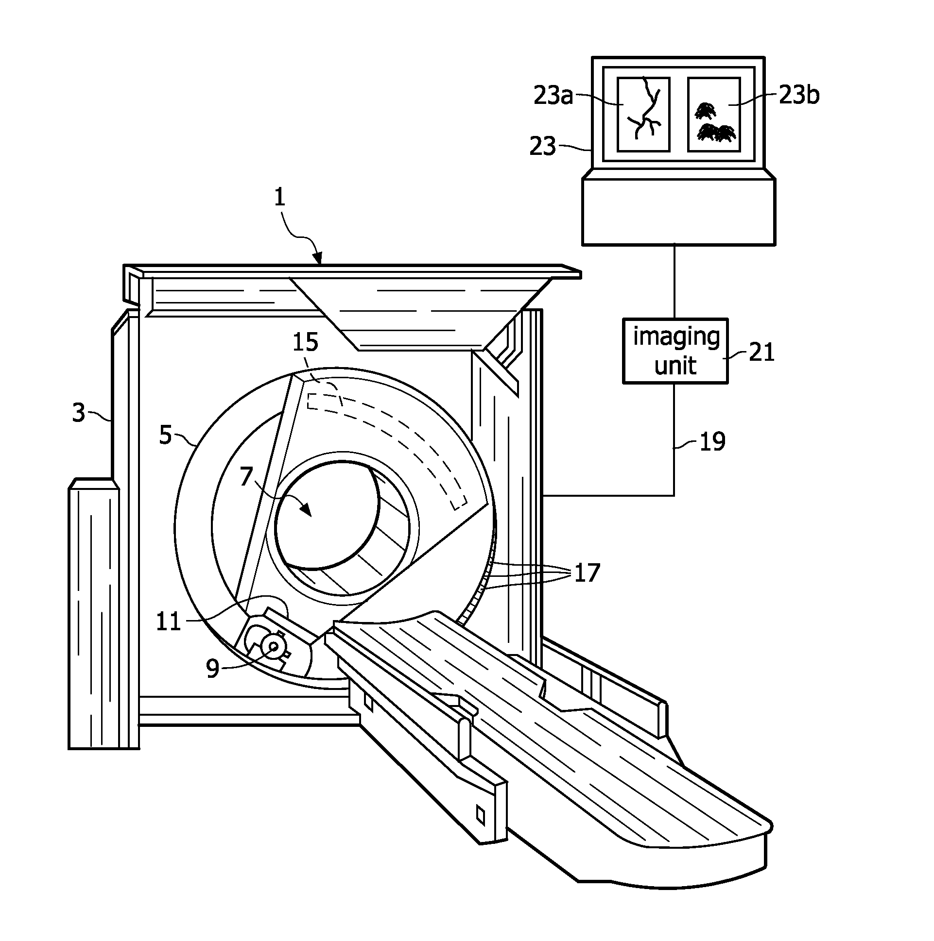
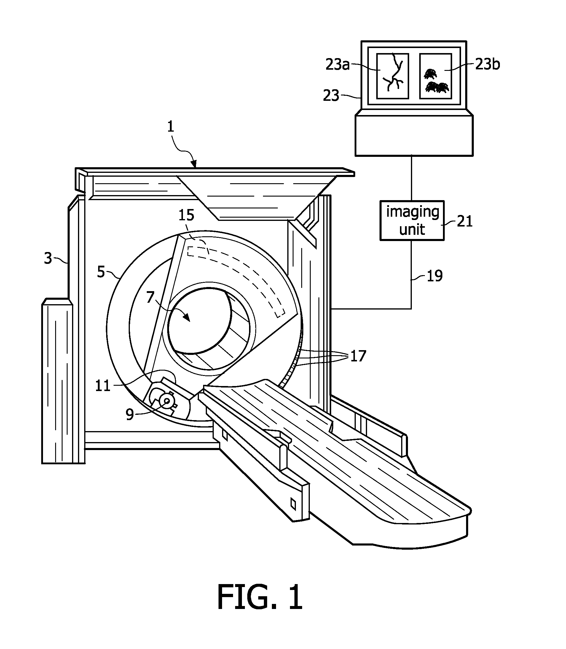
[0019]FIG. 1 is diagrammatic illustration of a CT scanner 1 according to the invention. The CT scanner 1 includes a stationary gantry 3 and a rotating gantry 5 which define an examination region 7. The rotating gantry 5 is suspended from the stationary gantry 3 for rotation about the examination region 7. A radiation source 9, such as an X-ray tube, is arranged on the rotating gantry 5 for rotation therewith. The radiation source 9 produces a beam of penetrating radiation that passes through the examination region 7 as the rotating gantry 5 is rotated by an external motor (not shown) about a longitudinal axis of the examination region 7. A collimator and shutter assembly 11 forms the beam of penetrating radiation into a cone shape and selectively gates the beam on and off. Alternately, the radiation beam is gated on and off electronically at the source 9. A patient support table 13 supports an object or a patient to be examined. The patient support table 13 is positioned such that t...


PUM


Abstract

The invention relates to a medical X-ray examination apparatus (1) for performing K-edge imaging. The medical X-ray examination apparatus (1) comprises an imaging unit (21), which is configured to spectrally decompose an X-ray absorption spectrum to image the X-ray absorption spectrum as a conventional X-ray absorption image (23a) and a K-edge absorption image (23b). The conventional X-ray absorption image (23a) includes data elements representing the anatomical background of an object of interest. The K-edge absorption image (23b) includes data elements representing quantitative information of local densities of material showing K-edge absorption within the object of interest. The imaging unit (21) comprises a spatial resolution reducer for reducing the spatial resolution of the K-edge absorption image, so that with a medical X-ray examination apparatus according to the invention an increased sensitivity of the selective imaging of a K-edge absorption image is achieved as compared to the sensitivity of the selective imaging of a K-edge absorption image of a known medical X-ray examination apparatus.

Description

FIELD OF THE INVENTION[0001]The invention relates to a medical X-ray examination apparatus for performing K-edge imaging, comprising an imaging unit which is configured to spectrally decompose an X-ray absorption spectrum to image the X-ray absorption spectrum as:[0002]a conventional X-ray absorption image, including data elements representing the anatomical background of an object of interest, and[0003]a K-edge absorption image, including data elements representing quantitative information of local densities of material showing K-edge absorption within the object of interest.BACKGROUND OF THE INVENTION[0004]Such a medical X-ray examination apparatus is known from the scientific article “K-edge imaging in X-ray computed tomography using multi-bin photon counting detectors” (Phys. Med. Biol. 52, (2007) 4679-4696). The known medical X-ray examination apparatus is a computed tomography (CT) scanner comprising an imaging unit with a multi-bin X-ray photon counting detector. This imaging...

Claims


Application Information

Patent Timeline
Patent Type & Authority Patents(United States)
IPC IPC(8): A61B6/00
CPCA61B6/032A61B6/482A61B6/4042A61B6/481
Inventor PROKSA, ROLAND
Owner KONINKLIJKE PHILIPS ELECTRONICS NV
Why Eureka
Try Eureka