Imaging and therapeutic targeting of prostate and bladder tissues
a prostate and bladder tissue technology, applied in the field of molecular biology and medicine, can solve the problems of no effective no standard therapeutic treatment of bone metastases, and negative results that do not prove the absence of metastasis, so as to facilitate selective uptake and overcome deficiencies in the art
- Summary
- Abstract
- Description
- Claims
- Application Information
AI Technical Summary
Benefits of technology
Problems solved by technology
Method used
Image
Examples
example 1
Materials and Methods
[0249]Cell culture and peptide incubation. All PCa cell lines (LNCaP, C4-2, LAPC4 and PC3) were maintained as described previously (Zhou et al., 2005). To determine the peptide uptake, 1×10⁵ cells per well were plated in a 12-well plate. Next day, the culture medium was replaced with RPMI medium supplemented with 0.5% FBS (Invitrogen, Carlsbad, Calif.) containing different concentrations fluorescence (FITC)-tagged peptides and incubation was carried out at the indicated time. For determining cellular location of peptide, cells were cultured overnight in slide chamber at a density of 25×104 / cm2. The FITC tagged peptides were incubated for 30 minutes in the presence of RPMI medium supplemented with 0.5% of FBS prior to fluorescence microscopy.
[0250]Peptide synthesis. All peptides TAT (FITC-G-RKKRRQRRR (SEQ ID NO:1)), PENE (FITC-G-RQIKIWFQNRRMKWKK (SEQ ID NO:2)), KALA (FITC-G-KLALKLALKALKAALKLA (SEQ ID NO:3)), homopolyers of L-arginine R11 (FITC-G-R11; SEQ ID NO:6)...
example 2
R11-PPL Inhibits Cancer Cell Growth
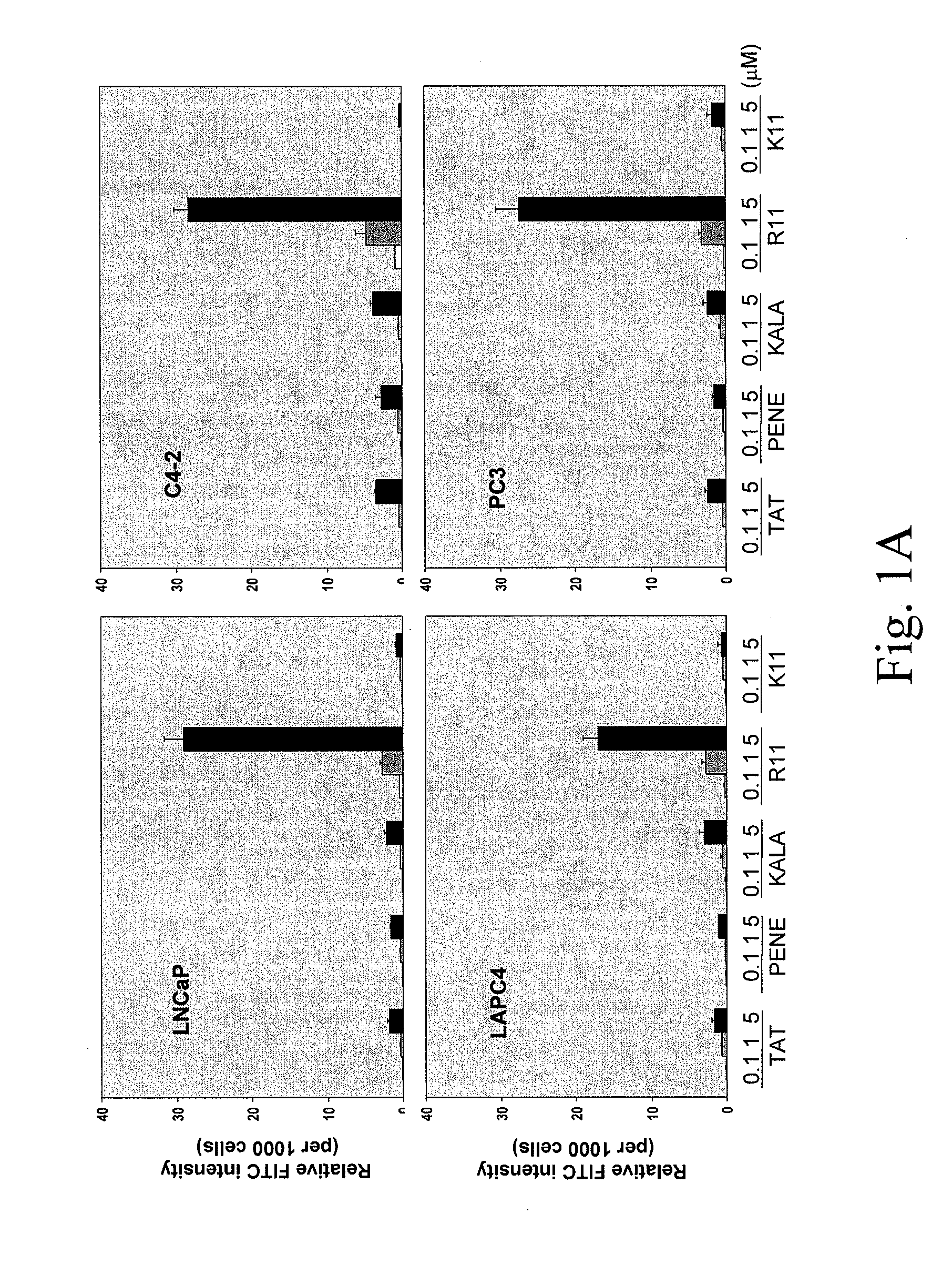
[0256]Screening of the most efficient CPP uptake by prostate cancer (PCa) cell lines. To deliver small peptide into target cells, CPP was employed as a delivery system. Five different CPPs were synthesized including TAT (amino acid 49-57), Penetratin (Antennapedia 43-58) (PENE), KALA peptide, poly-arginine (R11) and poly-lysine (K11). All the CPPs were incorporated with a FITC-labeled glycine at their N-terminal ends. From initial observation under fluorescence microscope, the inventors found that all five CPPs exhibited positive staining on cells without fixation and R11 appeared to have stronger staining than others. To determine the uptake quantitatively, fluorometer was used to measure the intensity of each CPP per cell basis. To rule out the possible artifacts such as CPP only associated with the cell surface and the pH variation from different organelles, cells were trypsinized and washed extensively with PBS, and a buffer with neutral pH was...
example 3
PCA Cell Internalization of the CPPS as Determined by Fluorescence Microscopy
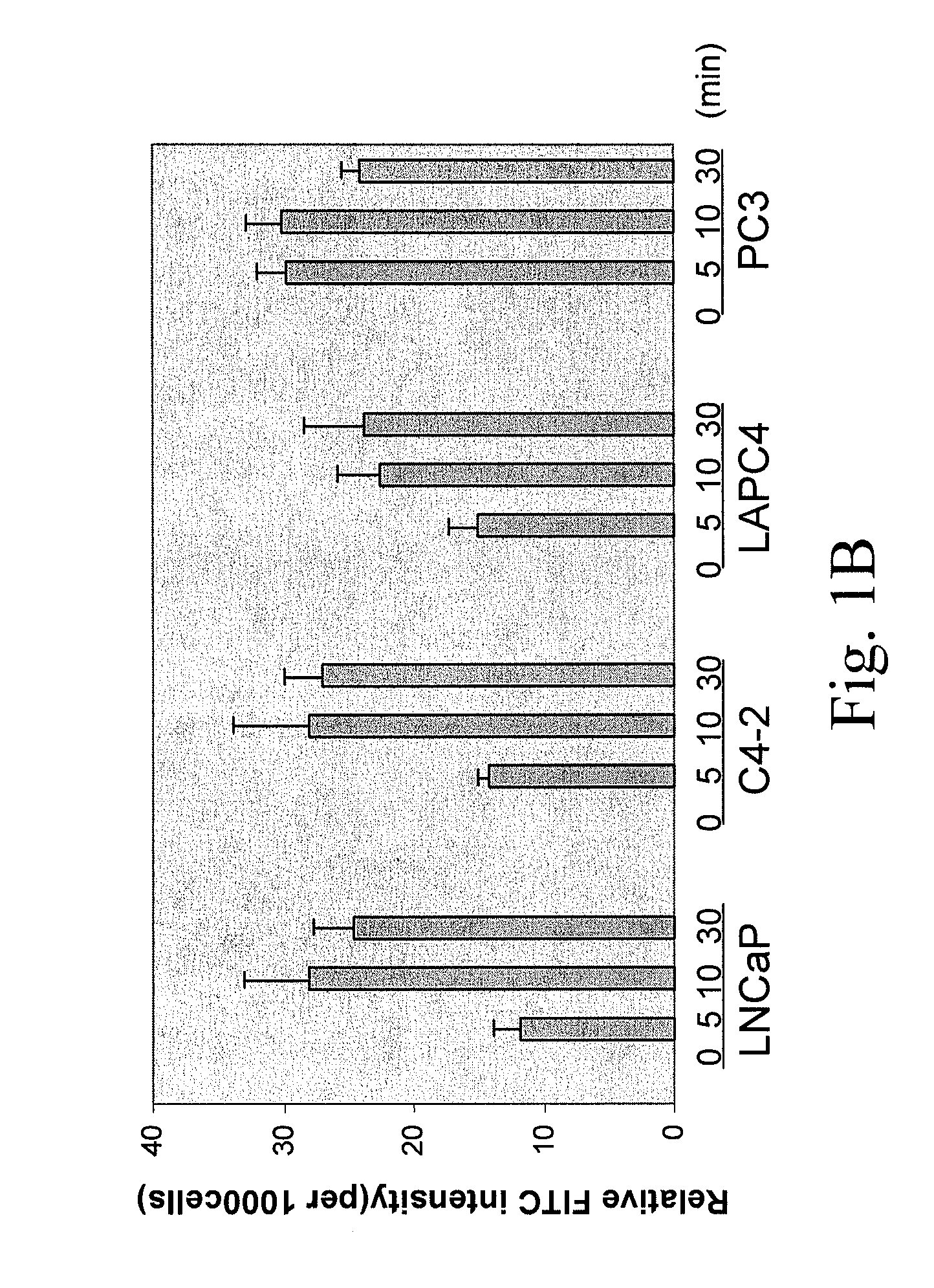
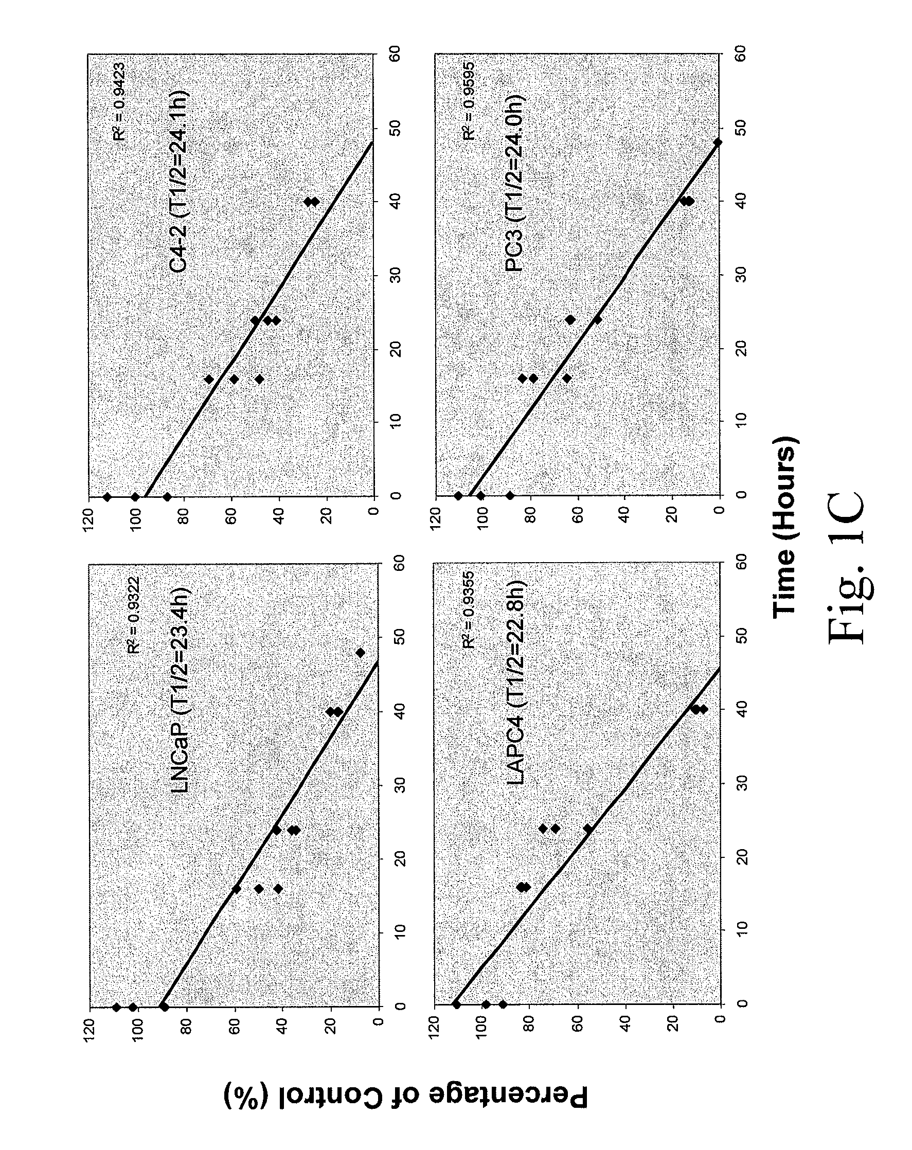
[0269]To determine the uptake efficiency of each CPP, different concentrations (0.1, 1, 5 μM) of FITC-tagged peptides were incubated with cells for 30 minutes (or different time for the time course study) then cells were washed twice with PBS and trypsinized. After washing with PBS twice, the total cell number was determined and cells were lysed in Tris Buffer (50 mM Tris-HCl, pH 7.5, 150 mM NaCl, 5 mM EDTA, 1% Triton X100). The fluorescence intensity was examined by fluorometer (excitation 490-500 nm; emission 515-525 nm). To determine the cellular localization of CPPs, after 30-minute incubation, cells were fixed with 4% paraformaldehyde in PBS plus DAPI (1 μg / ml) counterstaining (Sigma, St. Louis, Mo.) and cells were examined under fluorescence microscope. As shown in FIG. 1, a dose-dependent CPP uptake by four different PCa cells (LNCaP, C4-2, LAPC4 and PC3) was observed. Among the five CPPs, R11 exhibi...
PUM
| Property | Measurement | Unit |
|---|---|---|
| volume | aaaaa | aaaaa |
| time | aaaaa | aaaaa |
| time | aaaaa | aaaaa |
Abstract
Description
Claims
Application Information
 - R&D
- Intellectual Property
- Life Sciences
- Materials
- Tech Scout
- Unparalleled Data Quality
- Higher Quality Content
- 60% Fewer Hallucinations
Browse by: Latest US Patents, China's latest patents, Technical Efficacy Thesaurus, Application Domain, Technology Topic, Popular Technical Reports.
© 2025 PatSnap. All rights reserved.Legal|Privacy policy|Modern Slavery Act Transparency Statement|Sitemap|About US| Contact US: help@patsnap.com


