Sonographically guided transvaginal or transrectal pelvic abscess drainage using trocar method and biopsy guide attachment

a transvaginal or transrectal pelvic abscess, ultrasound guided technology, applied in the field of needle guides, can solve the problems of long recovery time, high surgical and anesthetic risk, long recovery time, and patients infertile and devoid of ovarian hormones

Active Publication Date: 2011-07-19
RGT UNIV OF CALIFORNIA
View PDF10 Cites 57 Cited by
  • Summary
  • Abstract
  • Description
  • Claims
  • Application Information

AI Technical Summary

Benefits of technology

[0021]In another embodiment, the guide body has a proximal end and a distal end, wherein the slot is configured such that the retainer may be installed in the needle guide by positioning the retainer in a slot opening at the proximal end of the guide body, and sliding the retainer in the slot longitudinally along the guide body toward the distal end of the guide body. The slot may preferably terminate at a stop at the distal end of the guide body such that the retainer is restrained from forward motion past the stop and the distal end.

Problems solved by technology

Although many pelvic abscesses respond to antibiotic therapy, surgery is often needed when antibiotics fail.
However, laparotomy for pelvic abscesses is one of the more technically difficult gynecologic procedures and has associated surgical and anesthetic risks, hospitalization costs, and prolonged recovery times. The definitive surgical treatment of a total hysterectomy and bilateral salpingo-oophorectomy is highly effective.
However, the surgical treatment leaves patients infertile and devoid of ovarian hormones, which are major problems in the young premenopausual women who tend to be affected by pelvic abscesses.
However, laparotomy is associated with both anesthesia and surgical risks, and prolonged recovery time.
However, for sonographically guided transrectal or transvaginal trocar method of needle placement, the use of a punctured channel that is not open to allow the catheter to be free is a technical limitation for all current endorectal and endovaginal transducers.
However, there may be difficulty in catheter placement via the Seldinger technique when using the endovaginal route.
This is because of the difficulty with buckling of the guidewire when dilators or catheters are placed through the thick vaginal musculature.

Method used


Image

  • Sonographically guided transvaginal or transrectal pelvic abscess drainage using trocar method and biopsy guide attachment
  • Sonographically guided transvaginal or transrectal pelvic abscess drainage using trocar method and biopsy guide attachment
  • Sonographically guided transvaginal or transrectal pelvic abscess drainage using trocar method and biopsy guide attachment

Examples


Embodiment Construction

[0040]Referring more specifically to the drawings, for illustrative purposes the present invention is embodied in the apparatus generally shown in FIG. 1 through FIG. 9D. It will be appreciated that the apparatus may vary as to configuration and as to details of the parts, and that the method may vary as to the specific steps and sequence, without departing from the basic concepts as disclosed herein.

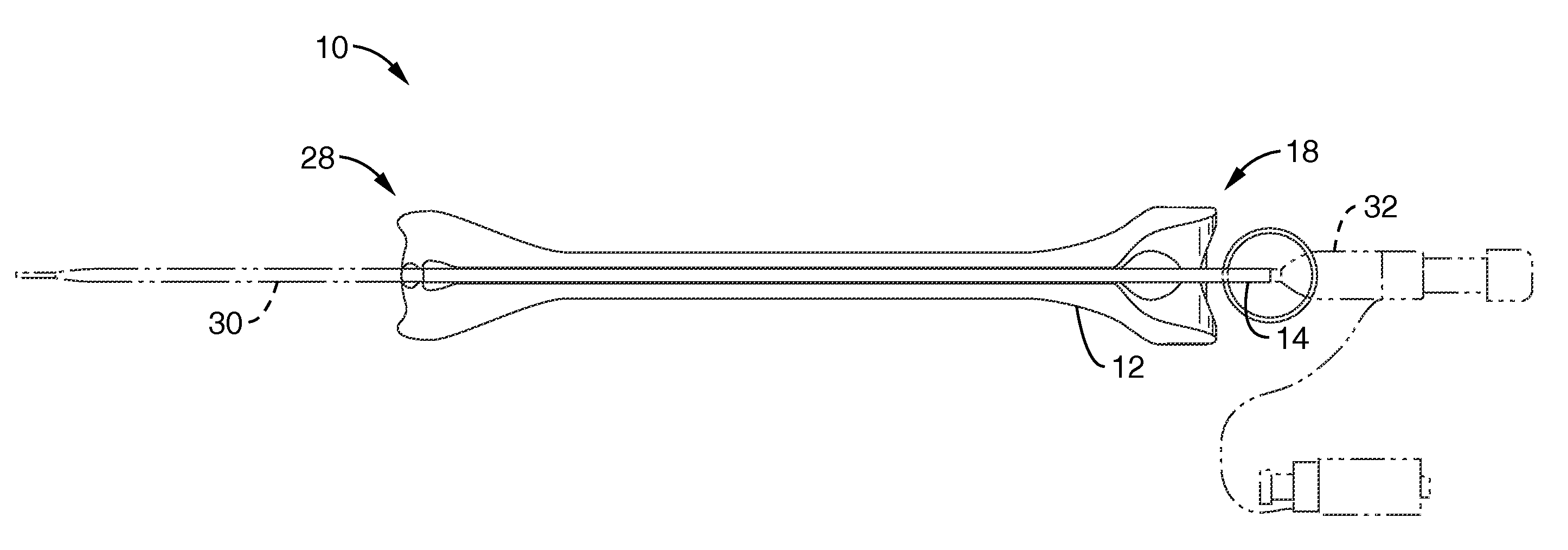
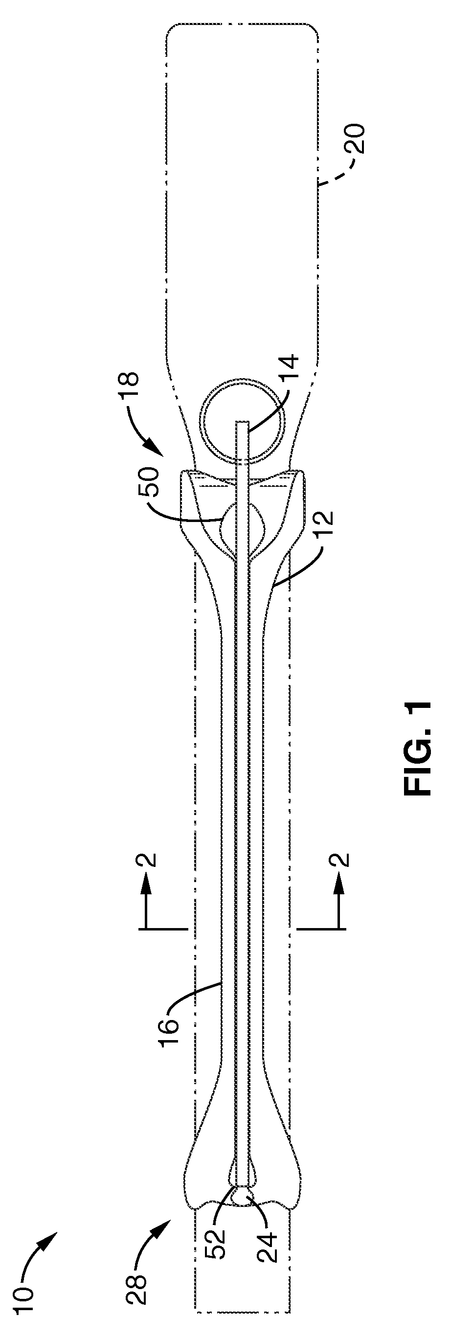
[0041]FIG. 1 illustrates a top view of a needle guide 10 configured to attach to a diagnostic instrument 20, such as an ultrasound probe (transducer) or the like. Needle guide 10 primarily includes a guide body 12 and a retainer 14 that is configured to slide longitudinally, i.e. along the length of the guide body 12, into a slot 16 that runs along the top of guide body 12.

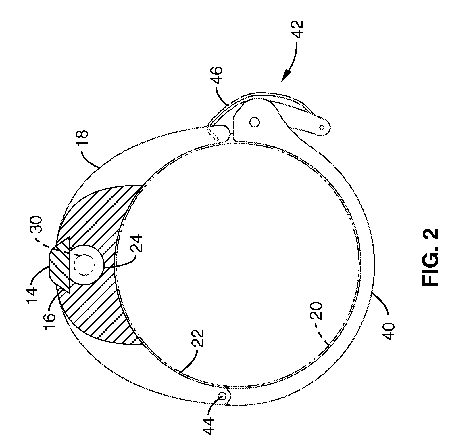
[0042]FIG. 2 illustrates further detail of the guide body 12 in cross-sectional view with the retainer 14 inserted. As shown in FIGS. 2 and 4, the slot 16 is preferably configured to have sidewalls 34 that that are sl...


PUM


Abstract

A needle biopsy guide system is disclosed for attachment to an endoluminal ultrasound probe or like sonographic instrument. The device includes a biopsy-guide attachment that allows for trocar catheter placement for abscess drainage or like procedures, using the transvaginal or transrectal route under sonographic control. The device has a base portion, which is attachable to an ultrasound probe. A removable retainer is provided that slides into the base unit to hold a biopsy needle in place. A physician may locate the target area in the body with the ultrasound probe, insert the biopsy needle into the target area, and then remove the insert (retainer) from the base unit and ultrasound probe, and leave the biopsy needle in place in the body.

Description

CROSS-REFERENCE TO RELATED APPLICATIONS[0001]This application claims priority from U.S. provisional application Ser. No. 60 / 885,342 filed on Jan. 17, 2007, incorporated herein by reference in its entirety.STATEMENT REGARDING FEDERALLY SPONSORED RESEARCH OR DEVELOPMENT[0002]Not ApplicableINCORPORATION-BY-REFERENCE OF MATERIAL SUBMITTED ON A COMPACT DISC[0003]Not ApplicableNOTICE OF MATERIAL SUBJECT TO COPYRIGHT PROTECTION[0004]A portion of the material in this patent document is subject to copyright protection under the copyright laws of the United States and of other countries. The owner of the copyright rights has no objection to the facsimile reproduction by anyone of the patent document or the patent disclosure, as it appears in the United States Patent and Trademark Office publicly available file or records, but otherwise reserves all copyright rights whatsoever. The copyright owner does not hereby waive any of its rights to have this patent document maintained in secrecy, inclu...

Claims


Application Information

Patent Timeline
Patent Type & Authority Patents(United States)
IPC IPC(8): A61B8/00A61B8/14A61B17/32A61M5/00
CPCA61B17/3403A61B2017/00296A61B2017/3413
Inventor MCGAHAN, JOHN P.
Owner RGT UNIV OF CALIFORNIA
Why Eureka
Try Eureka