Tissue oximetry apparatus and method

a technology of tissue oximetry and oximetry, which is applied in the field of tissue oximetry apparatus and method, can solve the problems of complicated and expensive calibration of optical instruments, insufficient pulse oximetry to monitor critically ill patients, and insufficient diagnostic information for critically ill patients, so as to improve the precision of measured output variables and eliminate influences on calibration

Active Publication Date: 2011-11-08
BERNREUTER PETER
View PDF112 Cites 53 Cited by
  • Summary
  • Abstract
  • Description
  • Claims
  • Application Information

AI Technical Summary

Benefits of technology

[0011]The present invention is directed to an apparatus and method which eliminate influences on calibration by subtracting and adding measured light attenuations, and a model-based calibration calculation to improve precision of measured output variables. In one embodiment, an apparatus utilizes a combination of light emitters and detectors with a light wavelength combination of more than two wavelengths, where the peak spectrum of a third wavelength is about the geometric mean value of the first and second wavelengths.
[0012]As a result, influences on the calibration of different tissue properties can be minimized in order to measure arterial or venous or the combination of arterial and venous oxygenation. It has been discovered that by choosing one of the wavelengths as a geometric mean value of two other wavelengths, variations due to scattering can be reduced. Additional determination of light attenuation can reduce measurement errors because of variations of light absorption due to different tissue composition, i.e., variations of relative amounts of muscle, skin, fat, bone, etc.

Problems solved by technology

Another problem is that pulse oximetry alone does not provide sufficient diagnostic information to monitor critically ill patients (See: When Pulse Oximetry Monitoring of the Critically Ill is Not Enough by Brian F. Keogh in Anesth Analg (2002), 94:96-99).
A problem of those disclosed solutions is that hair, dirt or other optically non-transparent material on the surface of tissue can influence the measured results for SvO2.
A disadvantage of this solution is that the calibration of the optical instruments is complicated and expensive, which makes it impractical to use such devices for sports activity applications, where light weight wearable devices would be of interest.

Method used


Image

  • Tissue oximetry apparatus and method
  • Tissue oximetry apparatus and method
  • Tissue oximetry apparatus and method

Examples


example 1

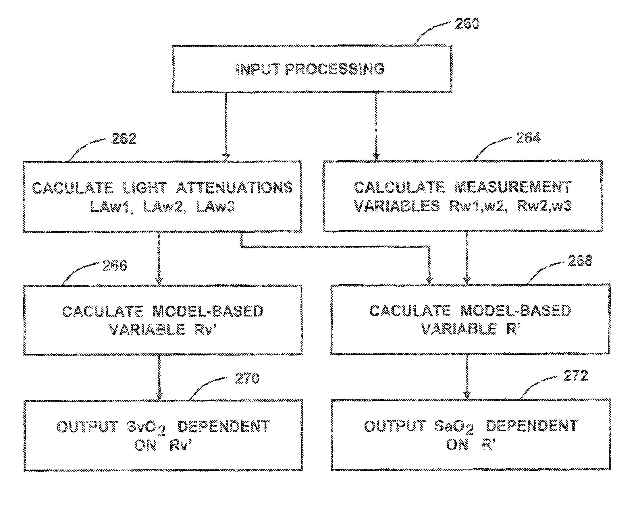
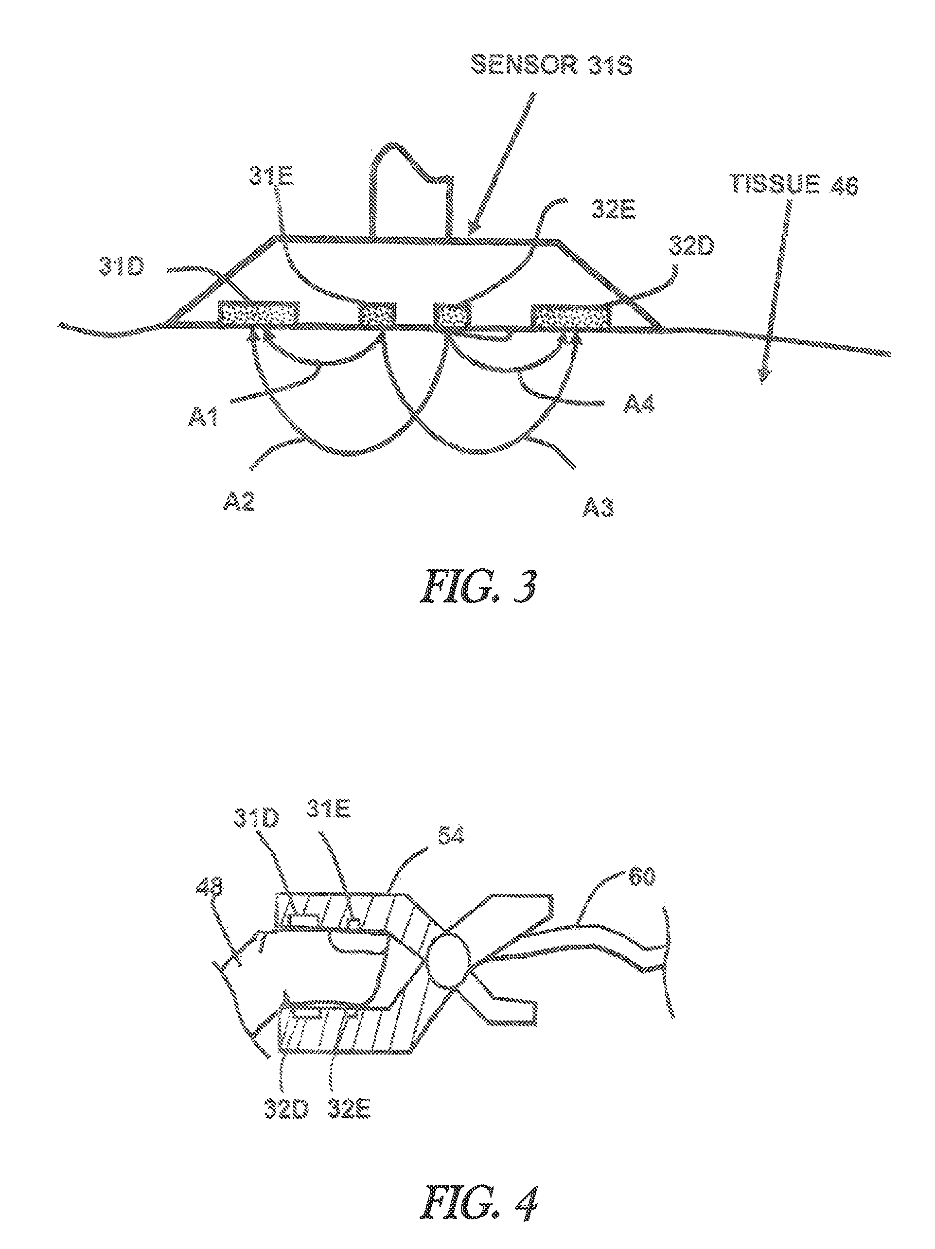
[0058]The sensor 31S shown in FIG. 3 is used to determine the arterial oxygenation and the mixed venous blood oxygenation of tissue with improved precision. Equation (2) is used to provide a measurement variable R′ for the arterial oxygenation. For each of the emitters 31E and 32E, three wavelengths are defined. Initially, two measurement wavelengths w0=940 nm and w2=660 nm are selected. Using equation (4) the third wavelengths w1 is about 788 nm. Wavelength w1=805 nm is chosen because it is close to the calculated third wavelength and is additionally at an isobestic point of the blood absorption spectrum. The next step is to determine the resulting light attenuation LA for each of the three wavelengths w0, w1 and w3:

LAw1=LA(A3w1)+LA(A2w1)−LA(A1w1)−LA(A4w1)  eq. (5)

LAw2=LA(A3w2)+LA(A2w2)−LA(A1w2)−LA(A4w2)  eq. (6)

LAw3=LA(A3w3)+LA(A2w3)−LA(A1w3)−LA(A4w3)  eq. (7)[0059]where LA(Axwy) is the logarithm of received light intensity in the detector related to light arrow Ax at wavelength w...

example 2

[0072]In FIG. 4 finger clip sensor 54 is shown with the two emitters 31E, 32E and the two detectors 31D and 32D. The benefit of the finger clip sensor is that it is easy to apply. Equivalent to sensor 31S in FIG. 3, four representative light paths between the two emitters and the two detectors are possible so that all calculations according example 1 can be performed in order to calculate the output variables R′ and Rv′ as a measure for mixed venous and arterial oxygenation in the finger 48. The corresponding calculations can also be performed using sensor of FIG. 9. The difference here is the alternative form of detectors 35D and 36D, which are able to increase detected light intensity due to an enlarged, concentric detector area.

example 3

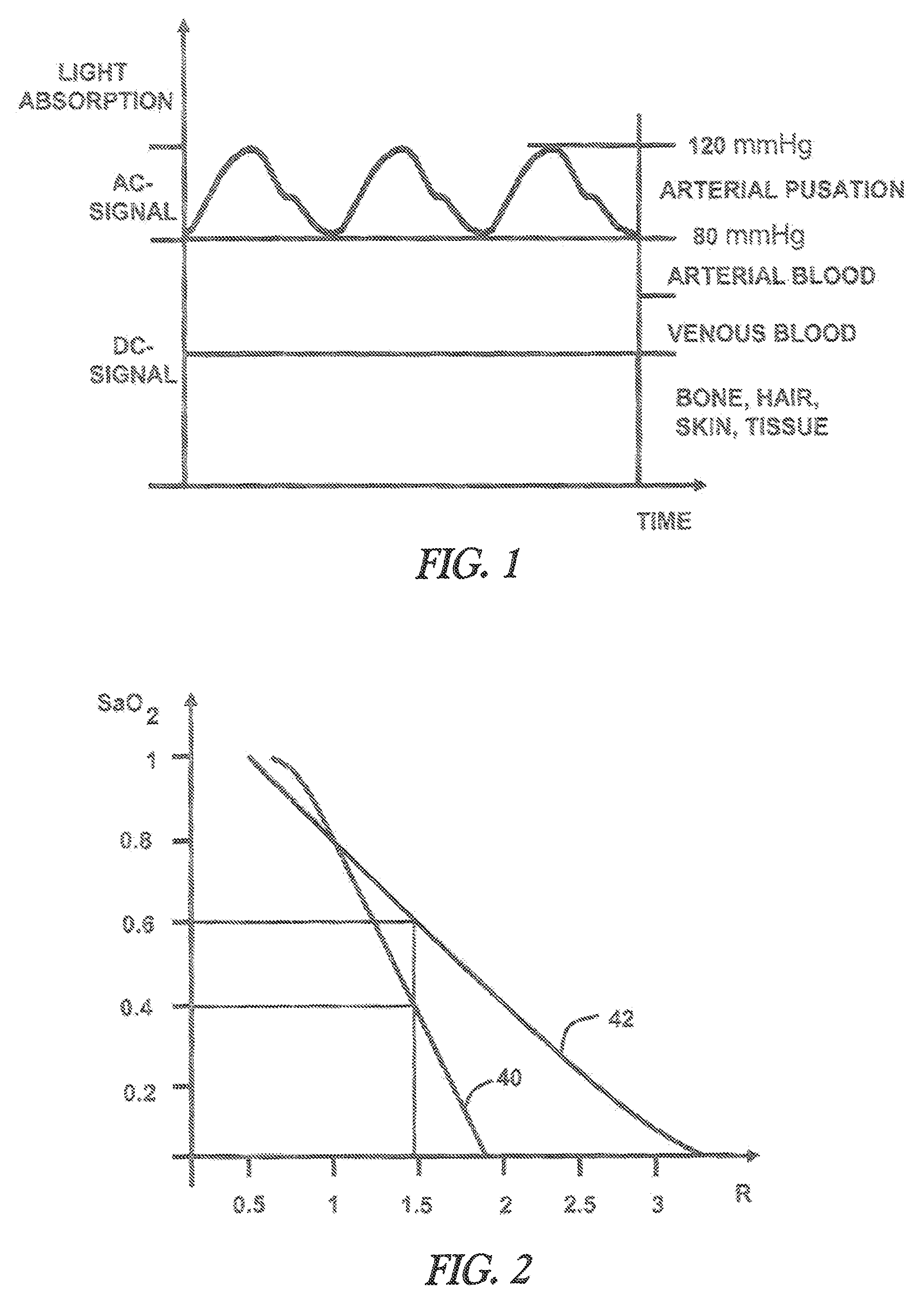
[0073]FIG. 5 shows a multidimensional calibration of SaO2 vs. R1 and R2. R1 and R2 can be calculated according (1) by selecting two wavelengths pairs where for the first wavelengths pair the wavelengths wm1=660 nm and wm2=910 nm is chosen and for the second wavelengths pair wm3=810 nm and wm2=910 nm. The second wavelengths pair is less sensitive towards arterial oxygenation and is used to compensate errors due to optical tissue parameter variations. In order to guarantee that the multidimensional calibration delivers improved precision in presence of varying tissue parameters, it is important to select exactly the correspondent calibration which is specified for a distinct wavelengths set and a distinct detector emitter distance. Therefore additional information has to be coded to the selected sensor. The tissue oximeter device can read out this information and use the appropriate calibration. The coding of information can be achieved for example by a resistor implemented in the LED...


PUM


Abstract

An apparatus and method for determining tissue oxygenation such as arterial and venous oxygenation and cerebral oxygenation. In one embodiment, the optical properties of tissue are determined using measured light attenuations at a set of wavelengths. By choosing distinct wavelengths and using light attenuation information, the influence of variables such as light scattering, absorption and other optical tissue properties can be minimized.

Description

RELATED APPLICATIONS[0001]This application is a continuation-in-part of pending U.S. Ser. No. 11 / 078,399 filed on Mar. 14, 2005, and incorporated by reference herein.TECHNICAL FIELD[0002]The present invention relates to a process and apparatus for improving accuracy of optical measurements of oxygenation of blood in tissue.BACKGROUND OF THE INVENTION[0003]A standard method to measure the arterial oxygenation of blood is known as pulse oximetry. Pulse oximeters function on the basis that at differing wavelengths, blood attenuates light very differently depending upon the level of oxygenation. Pulse waves starting from the heart cause in the arterial blood vessel system a periodic fluctuation in the arterial blood content in the tissue. As a consequence, a periodic change in the light absorption (FIG. 1) can be registered between the light transmitter, whose radiation passes through the tissue, and the receivers, which are integrated in a pulse oximetry sensor. The evaluation of the s...

Claims


Application Information

Patent Timeline
Patent Type & Authority Patents(United States)
IPC IPC(8): A61B5/1455
CPCA61B5/14551A61B5/14552A61B5/14553A61B2562/0242A61B5/1495A61B5/72A61B5/7203A61B5/742A61B2560/0238A61B2562/0238
Inventor BERNREUTER, PETER
Owner BERNREUTER PETER
Why Eureka
Try Eureka