

Methods for amyloid removal using anti-amyloid antibodies
an amyloid and anti-amyloid technology, applied in the field of amyloid related diseases, can solve the problems that no therapeutic antibody has been shown to stop or reverse the deposition of amyloid fibrils in patients with clinically proven amyloidosis spontaneously achieving complete remission
- Summary
- Abstract
- Description
- Claims
- Application Information
AI Technical Summary
Benefits of technology
Problems solved by technology
Method used
Image

Examples
example 1
Unassisted Resolution of Human IgLC Amyloid in Murine Host
[0068]Human IgLC amyloid was extracted and purified from infected organs obtained during an autopsy. The first experiments involved transplanting 50-200 mg of this amyloid material into a Balb / c mouse. The amyloid mass, or “amyloidoma,” was prepared in sterile PBS by serial sonication and grinding steps in order to produce a fine suspension of amyloid fibrils complete with the accessory molecules found in vivo. This procedure was performed to allow the amyloid to be injected into the mice through a wide-gauge hypodermic needle.
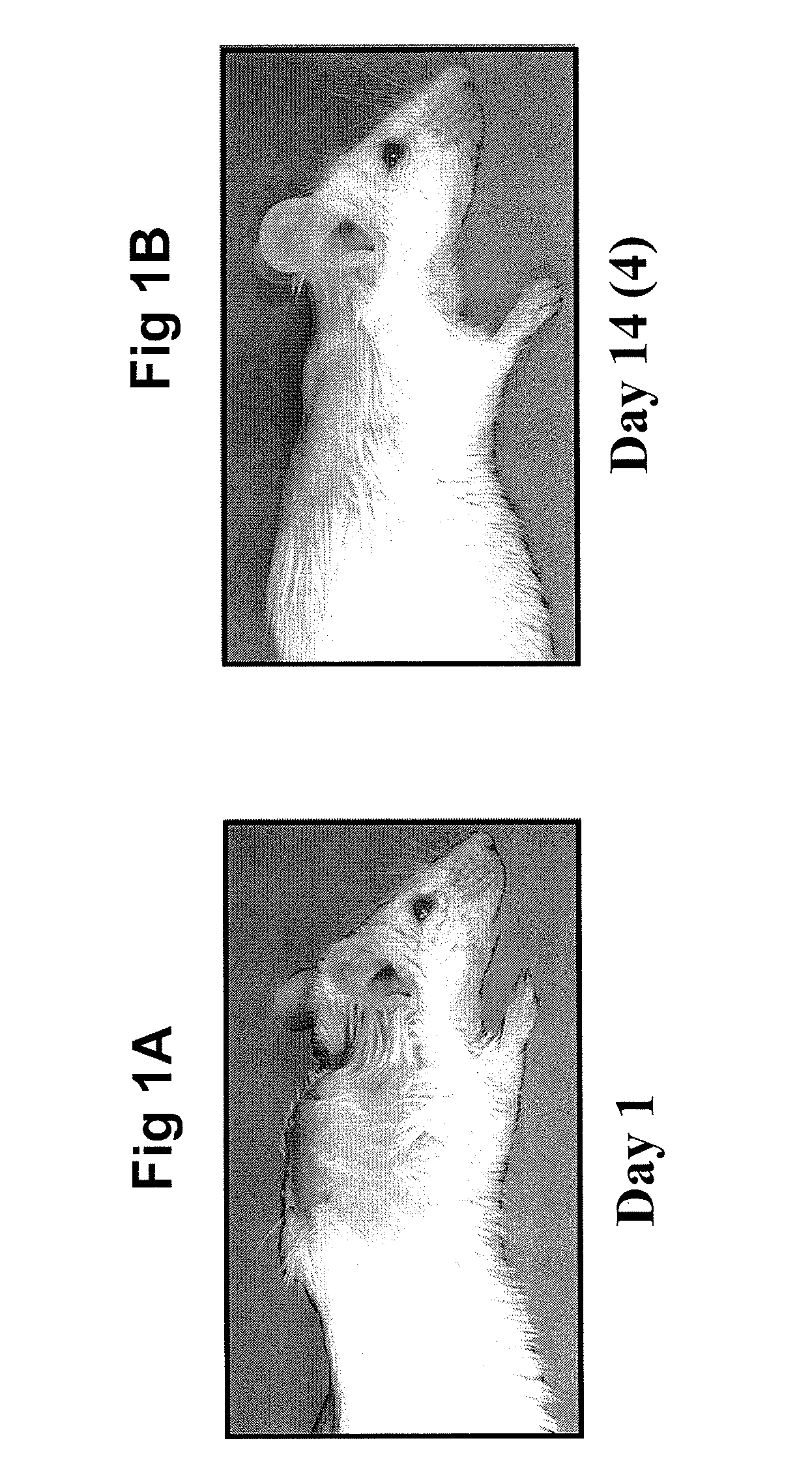
[0069]The amyloid material, equivalent to 10% of the body weight of the animal, was injected into mice (under anesthetic) between the scapula, which resulted in a large mass being visible (see FIG. 1A). The mouse required 15-18 days to achieve the complete removal of the amyloidoma (see FIG. 1B), after which the animal appeared healthy and lived a normal life span. The removal of the amyloidoma was dete...
example 2
Involvement of Both Antibody-Mediated and Cellular Immunity in the Removal of Amyloidomas
[0070]The involvement of anti-amyloid antibodies in the removal of amyloidomas was shown by screening serum from a mouse previously injected with amyloid material against a sample of the injected material. This was done by Western blot analysis using suitable dilutions of the mouse serum as the primary antibody. It was shown that there were antibodies to every component of the amyloid matrix, i.e., every band on the gel was stained by the mouse serum, even at a 10,000-fold serum dilution (data not shown).
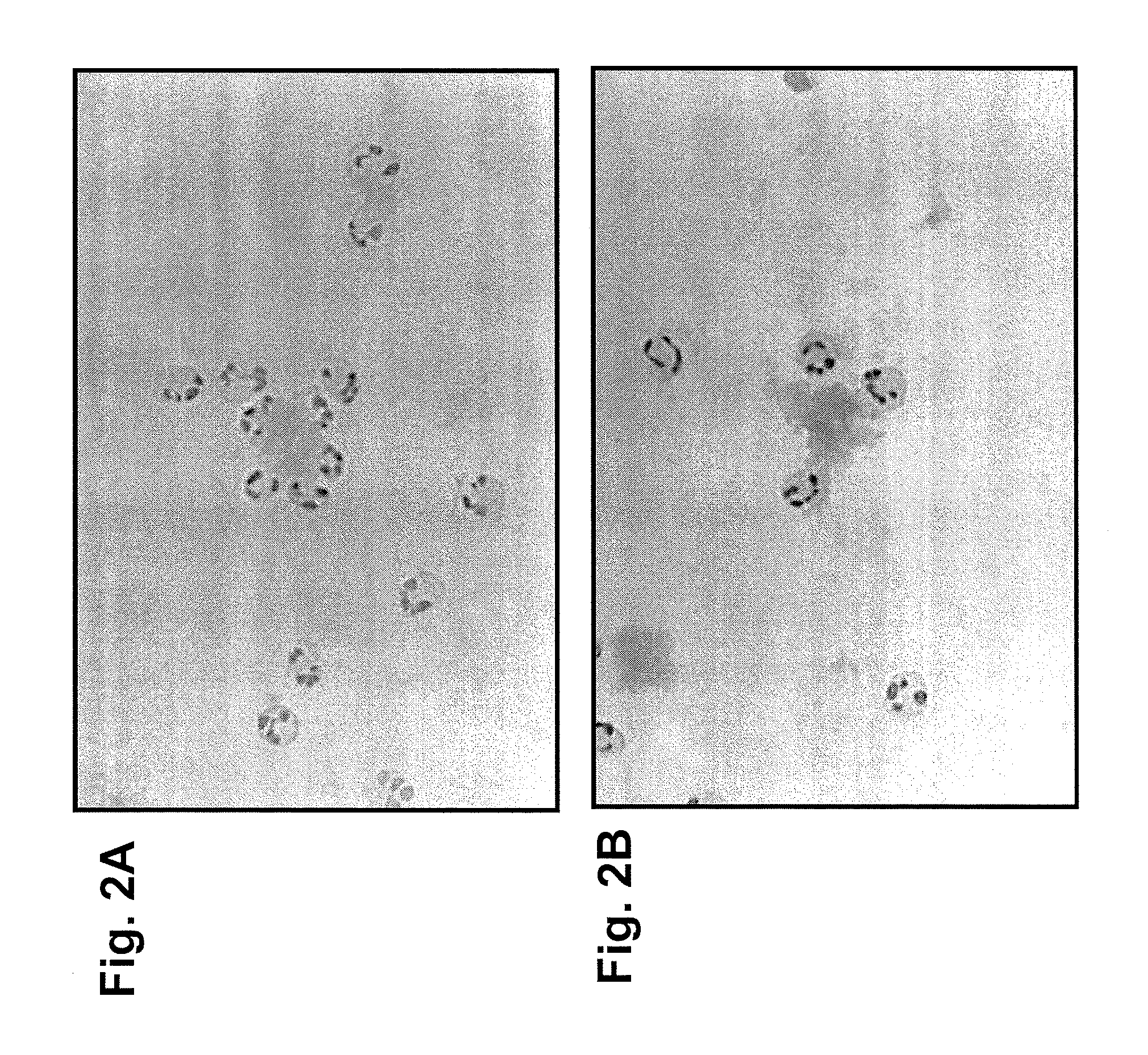
[0071]The involvement of a cellular component was demonstrated by in vitro neutrophil binding assays (see FIGS. 2A and 2B) and by using knockout-mutant mouse strains (data not shown). FIGS. 2A and 2B show human neutrophils adhering to human amyloid after the amyloid was treated with mouse anti-human IgLC mAbs. This shows that the mouse mAb can bind to human amyloid as well as attract human neutr...
example 3
ELISA Screening of IgLC Subsets
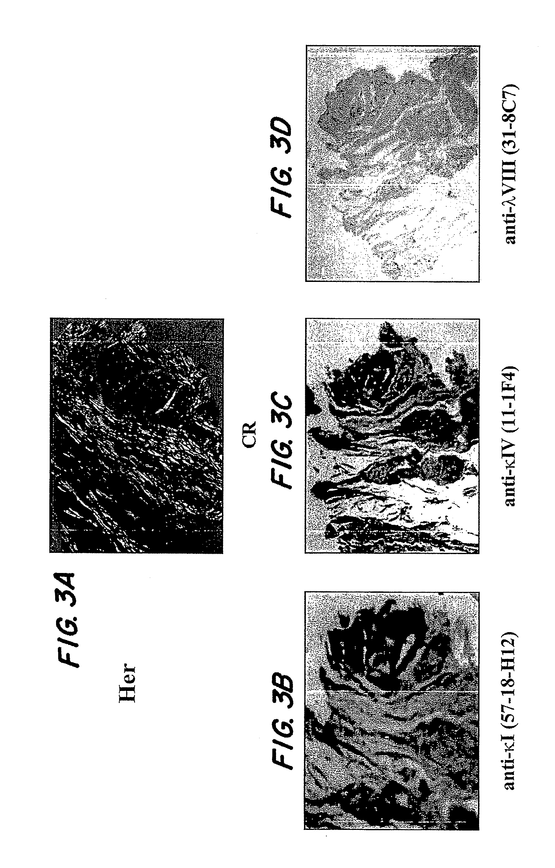
[0074]A systematic study was performed using ELISA techniques to screen a large number of human extracted amyloid samples using mAbs raised against the IgLC subsets (λ1, λ2, λ3, λ4, λ5, λ6, κ1, κ2, κ3, κ4, free κ and λ and total κ and λ). Interestingly, it was found that more often than not, the amyloids tested positive with mAbs specific for their own subtype, the total κ or λ antibodies and a κ1 (57-18H12), κ4 (11-1F4) and λ8(31-8C7) mAb. These latter three reagents were found to react in a non-subgroup specific manner, i.e., κ1 reacted with amyloids comprised of IgLCs other than κ1; and the other two mAbs exhibit the same quality. This shows that the epitope recognized by these antibodies may be a general feature of amyloid fibrils, indicating the possibility of a shared amyloid epitope that can be targeted.
PUM
| Property | Measurement | Unit |
|---|---|---|
| cross-isotype reactivity | aaaaa | aaaaa |
| epifluorescence microscope | aaaaa | aaaaa |
| purity | aaaaa | aaaaa |
Abstract
Description
Claims
Application Information