
Sutureless methods for laceration closure

a sutureless, laceration technology, applied in the field of sutureless laceration closure, can solve the problems of corneal trauma, current suturing techniques present limitations, insufficient tissue to effect suture repair,

Inactive Publication Date: 2012-06-12
SRI INTERNATIONAL
View PDF20 Cites 6 Cited by
  • Summary
  • Abstract
  • Description
  • Claims
  • Application Information

AI Technical Summary

Problems solved by technology

Trauma to the cornea and sclera is common, but current suturing techniques present limitations.
In the case of lacerations that extend to the posterior part of the eye, surgeons have no choice but to leave the wounds open because of difficulty in accessing and closing them with sutures.
In other cases, injuries may involve loss of tissue due to avulsion or necrosis, leaving insufficient tissue to effect a suture repair.
Likewise, in modern cataract surgery where sutures are often not used the risk of infection remains.
At present there are few alternatives to suture closure of scleral defects, and each has drawbacks.
While cyanoacrylate glue has been used, problems such as ease and uniformity of application exist.
[2, 3] Fibrin glues suffer from poor tensile strength.
[2] Laser tissue welding uses laser energy to cure solder commonly composed of albumin, but its application remains limited due to the lack of consistency in the outcomes, solubility in physiologic fluids, and thermal damage to the underlying tissue.

Method used


Image

  • Sutureless methods for laceration closure
  • Sutureless methods for laceration closure
  • Sutureless methods for laceration closure

Examples


example

Robotic Laser Tissue Welding of Sclera Using Chitosan Films

[0030]In this study we evaluated scleral wound closure using chitosan film with and without the application of near-infrared laser irradiation using a robotic manipulator and a laser scanner. We incorporated the laser tissue welding into a surgical robotic platform in an attempt to provide greater control over some of the parameters that are critical for a successful outcome of the bonds, such as the scanning uniformity and consistency of the energy delivery.

[0031]The experiments were performed on porcine eyeballs with radial lacerations in the sclera. To compare the different methods, we evaluated their ability to effect a watertight closure, measured leak pressures and times to complete the closures using manual microsuturing as a baseline.

[0032]Preparation of Chitosan Film: In a series of preliminary experiments we explored several previously reported methods to produce the chitosan films:

[0033]Method 1: Cast Chitosan dis...


PUM


Abstract

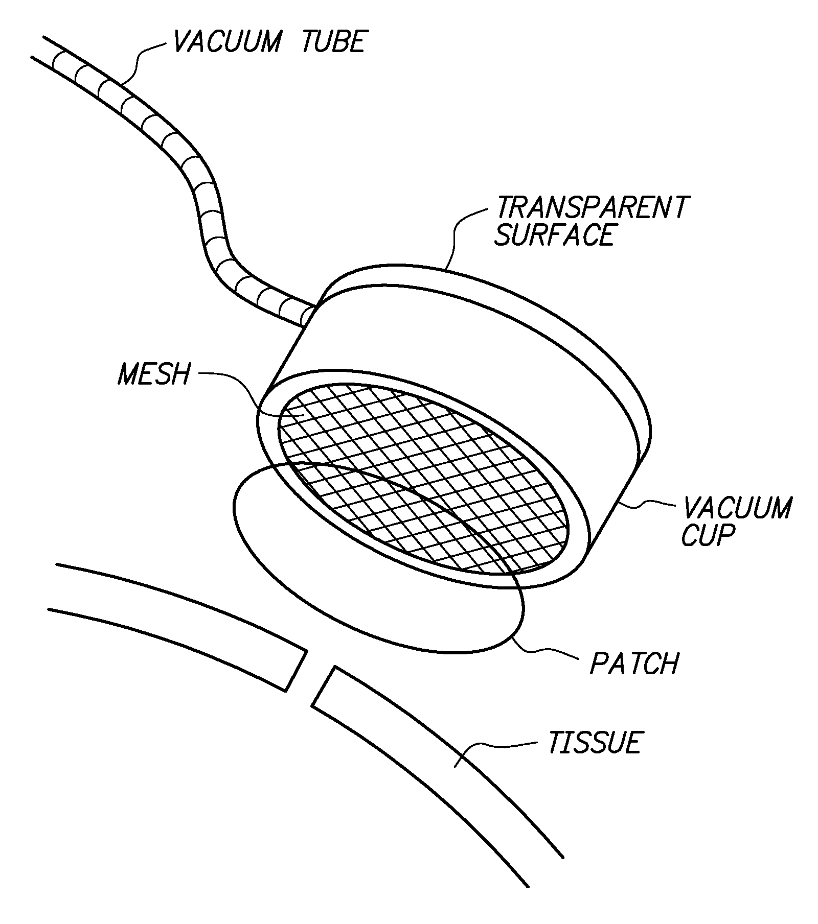
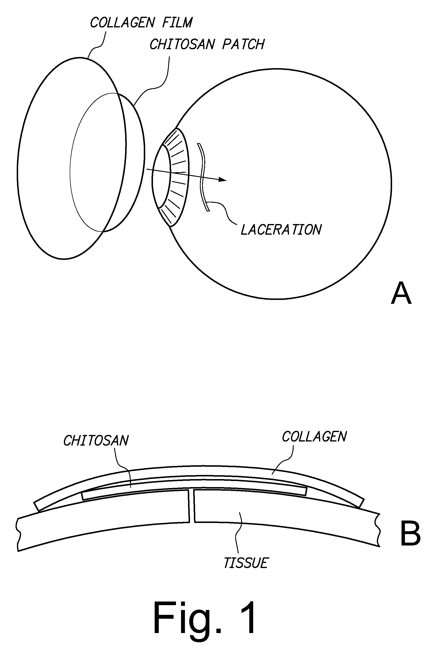
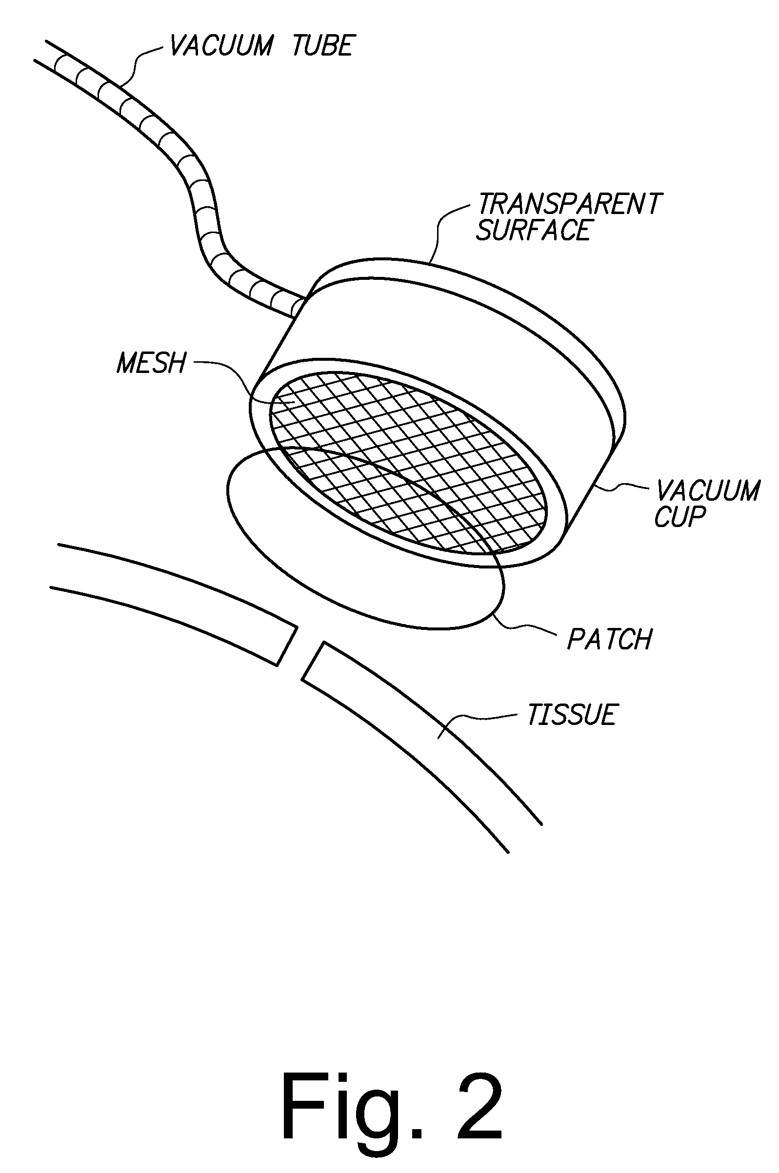
Tissue lacerations are closed using a vacuum cup applied to the tissue surface having a tissue-abutting, optically transparent mesh surface that under vacuum conforms with the tissue surface, apposing edges of the wound, and is optionally loaded with a bandage comprising a chitosan film and a collagen backing. An eye tissue surface wound is closed without sutures by closing the wound with a bioadhesive, biocompatible sclera or cornea wound patch comprising a chitosan film and a collagen backing, wherein the backing is bonded to the film without adhesive and protects the film against dissociation when the patch is exposed to a physiological fluid, and the film adheres to the sclera sufficient to retain apposed edges of the wound.

Description

[0001]This application claims priority to U.S. Ser. No. 61 / 020,979, filed Jan. 14, 2008, and U.S. Ser. No. 61 / 142,837, filed Jan. 6, 2009, both by the same inventors.[0002]This work was funded in part by US Army Medical Research and Materiel Command, Telemedicine and Advanced Technology Research Center Contract Number W81XWH-05-C-0117; the Government has certain rights in this invention.FIELD OF THE INVENTION[0003]The field of the invention is sutureless methods for laceration closure.BACKGROUND OF THE INVENTION[0004]Trauma to the cornea and sclera is common, but current suturing techniques present limitations. In the case of lacerations that extend to the posterior part of the eye, surgeons have no choice but to leave the wounds open because of difficulty in accessing and closing them with sutures. In other cases, injuries may involve loss of tissue due to avulsion or necrosis, leaving insufficient tissue to effect a suture repair. Likewise, in modern cataract surgery where sutures...

Claims


Application Information

Patent Timeline
Patent Type & Authority Patents(United States)
IPC IPC(8): A61B17/08
CPCA61B17/085A61F9/04
Inventor GARCIA, PABLO E.CHAVEZ, BRYANHILL, JOMAYON
Owner SRI INTERNATIONAL
Learn More