Continuous X-ray image screening examination device, program, and recording medium

a technology of x-ray image and examination device, applied in the field of continuous x-ray image screening examination technology, can solve the problems of insufficient examination, significant burden on the examinee, abnormal pulmonary blood flow, etc., and achieve the effect of simple and convenient generation of information effective for diagnosis

Inactive Publication Date: 2012-10-30
KANAZAWA UNIV
View PDF12 Cites 7 Cited by
  • Summary
  • Abstract
  • Description
  • Claims
  • Application Information

AI Technical Summary

Benefits of technology

[0011]Therefore, the present invention is attained in view of such a circumstance and an object thereof is to provide a continuous X-ray image screening examination device, program and recording medium which do not impose a considerable burden on an examinee in an examination such as of a lung embolism or a heart disease causing abnormality in blood flow such as pulmonary blood flow and cardiac blood flow and allows simple and convenient generation of information effectively utilizable for a diagnosis on these diseases.
[0031]Without imposing a considerable burden on an examinee in examination such as of a lung embolism or a heart disease causing abnormality in blood flow such as pulmonary blood flow and cardiac blood flow, the present invention will enable simple and convenient generation of information effectively utilizable for a diagnosis on these diseases.

Problems solved by technology

Such a lung embolism causes an embolus to close the pulmonary artery and will, therefore, give rise to abnormality in pulmonary blood flow.
However, those examinations are not sufficient as the last evidence for a diagnosis of a lung embolism.
However, the technology in the Patent Document 1 requires supplying stable xenon and the like, which operate to absorb X-ray, to an examinee and, therefore, is not simple and convenient for realizing imaging of an affected area and, moreover, the burden on the examinee is significant, giving rise to a problem.
In addition, the technology in the Patent Document 2 only uses the ultrasonic echo sensor and, therefore, cannot obtain sufficient information for a diagnosis on a blood clot in the pulmonary artery and the like.Patent Document 1: Japanese Patent Application Laid-Open No. 5-279268Patent Document 2: Japanese Patent Application Laid-Open No. 2003-235846

Method used


Image

  • Continuous X-ray image screening examination device, program, and recording medium
  • Continuous X-ray image screening examination device, program, and recording medium
  • Continuous X-ray image screening examination device, program, and recording medium

Examples


Embodiment Construction

[0075]An embodiment of the present invention will be described below in detail with the drawings.

[Summary of the Present Invention]

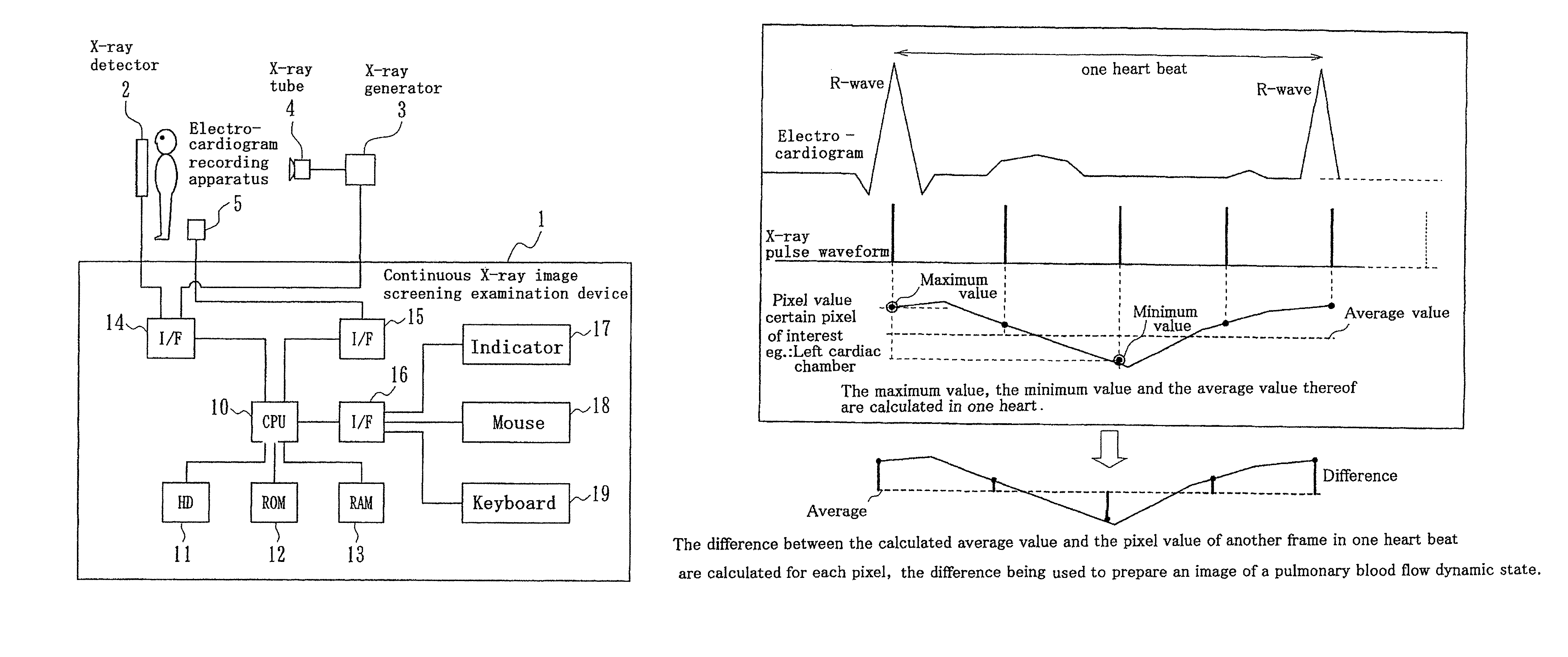
[0076]At first, the summary of the present invention will be described. The present invention uses the nature that the pixel value in a lung and a mediastinal part of a chest X-ray moving image varies due to heart beat. That is, focusing attention on the pixel value which increases and decreases according to blood flow such as pulmonary blood flow and cardiac blood flow due to heart beat, the variation information on the pixel value is effectively used for diagnosis such as of a lung embolism or a heart disease, considering this variation information of the pixel as information on blood flow such as pulmonary blood flow and cardiac blood flow. A chest X-ray moving image can be obtained from an X-ray detector and phase information on heart beat can be obtained from an electrocardiogram recording apparatus. In addition, the heart dynamic state during the c...


PUM

PropertyMeasurementUnit
strengthaaaaaaaaaa
blood velocityaaaaaaaaaa
velocityaaaaaaaaaa

Abstract

Using nature that a pixel value in a lung of a chest X-ray moving image varies due to heart beat, the variation information on the pixel value is effectively used for diagnosis such as of a lung embolism or a heart disease, considering the variation information as lung blood flow information. A continuous X-ray image screening examination device receives a chest X-ray moving image from an X-ray detector and receives an electrocardiogram to become original information on a heart beat variation from an electrocardiogram recording apparatus. From the dynamic state of the heart wall measured by the electrocardiograph or the chest X-ray moving image, the heart dynamic state during the cardiac chamber systolic and diastolic phases is grasped, and information such as the variation of the pixel value of the chest X-ray moving image due to increase (lung blood flow increase) of the blood flow from the heart to the lung during the cardiac chamber systolic phase is generated.

Description

CROSS REFERENCE OF RELATED APPLICATIONS[0001]The present application demands for priority on Japanese Patent Application No. 2006-587 (applied on Jan. 5, 200) and Japanese Patent Application No. 2006-172760 (Jun. 22, 2006) and these applications are incorporated to the present specification with reference thereto.TECHNICAL FIELD[0002]The present invention relates to continuous X-ray image screening examination technology carrying out computer-analysis with an X-ray moving image and an electrocardiogram and generates information for assessing blood flow such as pulmonary blood flow and cardiac blood flow.RELATED ART[0003]In general, a lung embolism refers to a circulatory disorder caused by an embolus being carried on venous blood flow to close a pulmonary artery (a blood vessel transmitting venous blood for getting oxidized to the lung). For example, an economy-class syndrome being one of the lung embolism is a disease caused by maintaining the state of sitting on a seat with the sa...

Claims


Application Information

Patent Timeline
Patent Type & Authority Patents(United States)
IPC IPC(8): G06K9/70G06T7/20G06K9/78G01N23/12A61B5/352
CPCA61B5/0205A61B6/507A61B5/0456A61B5/08A61B5/415A61B5/418A61B6/4233A61B6/463A61B6/469A61B6/503A61B6/504G06T7/0012G06T7/20A61B5/026A61B5/029A61B6/481G06T2207/30048G06T2207/30061G06T7/0016A61B5/352
Inventor SANADA, SHIGERUTANAKA, RIEOKAZAKI, NOBUO
Owner KANAZAWA UNIV
Why Eureka
Try Eureka